Chest X-Ray Portable — Dictation, Appropriateness, and Dose for Residents
1. The ICU Stat Read: Portable Chest X-Ray for Line Placement
It’s a familiar scene: the ICU calls for a stat portable chest film to confirm endotracheal tube placement. The patient is unstable, the clinical team is waiting, and your attending wants a confident read on that tube tip’s location relative to the carina—yesterday. You also need to systematically check every other line, look for iatrogenic pneumothorax, and evaluate for acute cardiopulmonary changes. This isn’t just a “check film”; it’s a critical data point in a sick patient’s care.
When I was a resident, the pressure was all about speed and accuracy on these common studies. You need a bulletproof checklist so you don’t miss a malpositioned PICC while you’re focused on the ETT. Having a solid template isn’t just about efficiency; it’s about patient safety and looking sharp for your attending. Let’s build that process.
2. What an X-Ray Chest Portable (AP Single-View) Covers and What Attendings Look For
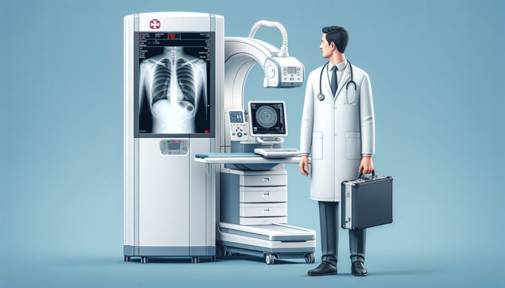
The portable anteroposterior (AP) chest X-ray is the undisputed workhorse of inpatient and critical care imaging for any patient who can’t make it to the department for a standard upright PA/lateral study. The most common indications are procedural follow-ups or evaluating acute changes in clinical status.
Your attending expects a systematic, prioritized report. The first priority is always answering the specific clinical question—usually the position of a newly placed tube or line. After that, a comprehensive survey is mandatory. Don’t be the resident who misses the new pneumothorax because you only looked at the CVC tip.
A solid report on a portable chest film will always address:
- Tubes, Lines, and Devices: The primary reason for most portable studies. Confirm position and check for complications.
- Lungs and Pleura: Evaluate for pneumothorax, pleural effusion, edema, atelectasis, or new focal opacity suggesting pneumonia. Remember to compare to the prior study, as interval change is key.
- Cardiomediastinal Silhouette: Assess for gross changes, but be cautious. The AP view magnifies the heart, so don’t overcall cardiomegaly.
- Bones and Soft Tissues: A quick check for fractures (especially in trauma) or subcutaneous emphysema.
3. Radiology Report Template for X-Ray Chest Portable (AP Single-View)
This template is designed for efficiency and completeness. Use it as a starting point for your personal macros in PowerScribe or your PACS dictation system. The key is to have a structured search pattern that you follow every single time.
Technique
Single portable AP view of the chest. Patient is [supine/semi-upright]. The exam is limited by [patient rotation, incomplete inspiration, overlying lines/leads]. Comparison is made to the prior study from [Date].
Findings
Support Devices:
- Endotracheal tube: The tip is approximately [X] cm superior to the carina. The cuff is inflated.
- Central venous catheter ([side] [type]): The tip terminates at the [cavoatrial junction / lower SVC / right atrium].
- PICC line ([side]): The tip terminates in the [lower SVC].
- Nasogastric/Orogastric tube: The tip is below the diaphragm in the expected location of the stomach.
- Chest tube ([side]): The tip is in the [location] pleural space. The side port is within the thoracic cavity.
Lungs: Lungs are [clear/demonstrate…] Opacities concerning for [atelectasis, pneumonia, edema] are seen at the [location].
Pleura: No pneumothorax. No pleural effusion. [OR] There is a [small/moderate/large] [left/right] pneumothorax. [OR] There are bilateral pleural effusions, [right greater than left / left greater than right]. Note the deep sulcus sign on the [left/right] concerning for pneumothorax in the supine position.
Cardiomediastinal Silhouette: The cardiomediastinal silhouette is [normal/enlarged], though limited by AP technique. The aortic knob is [normal/prominent].
Bones and Soft Tissues: No acute fracture. No subcutaneous emphysema.
Impression
- [Semi-upright/supine] portable chest radiograph.
- Endotracheal tube with tip approximately [X] cm above the carina, in adequate position.
- [Name of line/tube] with tip at the [location], in adequate position. [OR] Malpositioned [Name of line/tube] with tip at [location], which is too [deep/shallow].
- [Stable/Improved/Worsened] [pulmonary edema/atelectasis/pleural effusions].
- [No pneumothorax / Size and location of pneumothorax].
4. Free Radiology Template Sources
Building your own templates is a great way to learn, but you don’t have to start from scratch. Two great free repositories exist that are curated by radiologists and serve as an excellent reference for nearly any study you’ll encounter on call.
- RadReport.org: Maintained by the RSNA, this is a comprehensive library of peer-reviewed templates covering all subspecialties. It’s a go-to for standardized, best-practice reporting language.
- Radiology Templates (AU): This Australian-maintained site offers a fantastic collection of practical, user-friendly templates that are easy to adapt for your own use.
Bookmark them. They’re invaluable resources, especially in your first couple of years.
5. The Next-Level Move: AI-Assisted Structured Reporting
The classic workflow is dictating into a macro, then tabbing through the fields to fill in the blanks. It works, but it’s clunky. A more modern approach separates the act of finding from the act of formatting. You can simply dictate your positive findings in free form—”ETT tip is 4 cm above the carina, right IJ CVC tip is at the cavoatrial junction, new small right pneumothorax”—and let the software handle the rest.
Tools like GigHz Precision AI are designed for this exact workflow. It takes your free-form dictation of positive findings and automatically generates a clean, structured report based on pre-loaded ACR and SIR templates. It also helps surface relevant Clinical Decision Support (CDS) guidance where appropriate, ensuring your reports meet current standards without you having to stop and look things up. This approach streamlines the reporting process, letting you focus on the images, not the template fields.
6. When Should You Order an X-Ray Chest Portable (AP Single-View)? ACR Appropriateness Criteria
The American College of Radiology (ACR) provides evidence-based guidelines to help clinicians choose the right imaging study. For patients in the intensive care unit, the guidance is clear.
According to the ACR Appropriateness Criteria for Routine Chest Radiography in ICU, the portable chest X-ray is Usually Appropriate for monitoring ICU patients as clinically indicated. This includes common scenarios like post-procedure evaluation for lines and tubes, or assessing for acute cardiopulmonary changes like edema, pneumonia, or barotrauma.
While it’s the go-to study for non-ambulatory patients, it’s important to know the alternatives and their roles:
- Upright PA and lateral chest X-ray: This is the preferred study for any patient who can stand. It provides a more accurate assessment of heart size, pleural effusions, and pneumothorax.
- Bedside ultrasound: Increasingly used in the ICU, ultrasound is excellent for detecting pneumothorax and pleural effusions with high sensitivity, often exceeding that of a supine chest film. It’s also useful for assessing for pulmonary edema (B-lines).
- CT chest: Reserved for cases where more anatomical detail is required to solve a specific clinical problem that the portable radiograph cannot, such as characterizing a complex opacity, evaluating for pulmonary embolism, or assessing for aortic injury.
7. How Much Radiation Does an X-Ray Chest Portable (AP Single-View) Deliver?
Patients and clinical teams are increasingly aware of radiation dose. Fortunately, the portable chest X-ray is one of the lowest-dose imaging studies performed in the hospital. This is a key reason why it’s used so frequently for serial monitoring in the ICU.
The estimated effective dose from a single AP portable chest X-ray is approximately 0.05-0.1 mSv. To put that in perspective, the average person in the U.S. receives about 3 mSv per year from natural background radiation. This means a single portable chest film delivers a dose equivalent to only a few days of background radiation.
| Imaging Study | Typical Effective Dose (mSv) | Equivalent Background Radiation |
|---|---|---|
| Portable Chest X-Ray (AP) | 0.05 – 0.1 mSv | ~10 days |
| Standard 2-View Chest X-Ray (PA/Lat) | 0.1 mSv | ~10 days |
| CT Chest (Standard Dose) | 7 mSv | ~2 years |
Modern digital detectors are highly efficient, allowing for diagnostic images at very low mAs settings, which helps keep the dose for these frequent follow-up exams as low as reasonably achievable (ALARA).
8. X-Ray Chest Portable (AP Single-View) Imaging Protocol — Technique and Common Pitfalls
While residents don’t perform the exam, understanding the protocol is crucial for interpreting the images correctly and knowing their limitations. The portable technique is a compromise driven by the patient’s inability to cooperate or move to the radiology department.
The goal is always to get the patient as upright as possible. A semi-upright (45°) position is far superior to a fully supine one for detecting pleural fluid and pneumothorax. The technologist uses a portable X-ray machine with the source tube positioned anteriorly, about 40 inches from the digital detector plate placed behind the patient.
| Parameter | Typical Setting | Clinical Note |
|---|---|---|
| Projection | Anteroposterior (AP) | X-ray tube is in front, detector is behind the patient. |
| kVp | 75-90 kVp | May be increased to 100+ kVp for larger patients to ensure adequate penetration. |
| mAs | 4-10 mAs | Digital detectors are very dose-efficient. |
| Source-to-Image Distance (SID) | ~40 inches (100 cm) | Shorter distance than standard PA chest (72 inches), causing cardiac magnification. |
| Grid | Often omitted | Anti-scatter grids are difficult to align properly on portable exams, and misalignment can degrade the image more than scatter. |
Common Protocol Pitfalls: The biggest pitfall is patient positioning. A fully supine patient makes it difficult to detect a small pneumothorax (which will collect anteriorly) or a pleural effusion (which will layer posteriorly, causing diffuse haziness). Rotation can also significantly distort the appearance of the cardiomediastinal silhouette, mimicking pathology that isn’t there. Always assess the technique before diving into the findings.
9. The 3-Months-Free Offer for Radiology Residents and Fellows
Look like a rockstar on your reports — get 3+ months of GigHz Precision AI free.
We built this tool to solve the exact workflow headaches we had during training. You can dictate your positive findings in free form, and the AI instantly generates a perfectly structured report using the latest ACR and SIR templates, with the appropriate clinical decision support firing automatically. It helps you be faster, more accurate, and more consistent.
All we ask in return is your feedback so we can keep improving the product for trainees. There’s no credit card required and no long forms. To get set up, just reply to the application with three items:
- Your PGY year (e.g., PGY-2, PGY-4)
- Your training type (radiology residency or fellowship specialty)
- Your training program / hospital name
Ready to give it a try? You can apply for the residents free-access program here. And for more high-yield content, check out the residents and fellows resource hub.
10. Frequently Asked Questions
Is GigHz Precision AI HIPAA-compliant?
Yes. The platform is designed for de-identified workflows by default. It processes the clinical content of your dictation to structure the report, but no patient-identifying information (PHI) is required or stored.
Does this require a complex IT setup at my hospital?
No. It’s a secure, browser-based tool. There’s no software to install on hospital machines. You can use it on any computer, including the call-room PC or your personal laptop or iPad.
How does this work with PowerScribe or other dictation systems?
It works alongside your existing system. You can dictate your free-form findings, let the AI generate the structured report, and then copy/paste the final text into your hospital’s dictation software. It streamlines the “thinking and formatting” part of the job.
Can I use this on my phone or iPad?
Yes, the interface is fully responsive and works well on mobile devices and tablets, making it easy to use in any reading room or on the go.
Can I customize the templates?
Yes. While the system comes pre-loaded with ACR and society-recommended templates, you can create, modify, and save your own templates to match your personal style or your institution’s specific requirements.
What happens after my residency or fellowship ends?
The free access program is specifically for trainees. After you graduate, you can choose to transition to a paid plan for practicing radiologists. We offer discounts for recent graduates to help with the transition into practice.
Free GigHz Tools That Pair With This Article
Three free tools that complement the material above:
- Imaging Appropriateness Selector — Type an indication or clinical scenario in plain language and get the imaging studies the ACR rates for it, with adult and pediatric radiation levels. Built directly from 297 ACR topics, 1,336 clinical variants, and 15,823 procedure ratings.
- GigHz Imaging Protocol Library — A searchable library of 131 imaging protocols with the physics specs surfaced and the matching ACR Appropriateness Criteria alongside. Plain-English narratives readable in 60 seconds, organized by modality.
- GigHz Radiation Dose Calculator — Pick the imaging studies a patient has had and see total dose in millisieverts (mSv) with comparisons to natural background radiation, transatlantic flights, and chest X-rays. Useful for shared decision-making.
Reviewed by Pouyan Golshani, MD, Interventional Radiologist — August 4, 2026