Hand/Wrist X-Ray — Dictation, Appropriateness, and Dose for Residents
1. The Most Common Read on Your MSK Rotation
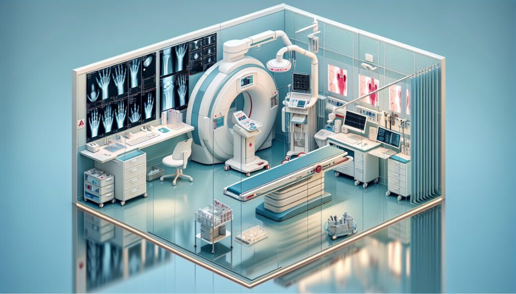
It’s a busy shift. The ED sends over a patient with wrist pain after a fall on an outstretched hand — a classic FOOSH. The images pop up: a standard 3-view wrist series. Your eyes immediately go to the distal radius. Is it angulated dorsally? Volarly? Is the fracture intra-articular? Is there a subtle scaphoid waist fracture you’re missing? The attending is going to want a clear, decisive read that covers all the bases, and they’ll want it five minutes ago. This isn’t an esoteric MR; it’s the bread-and-butter of MSK, and getting it right, fast, is table stakes. When you’re just starting out, having a solid framework is key. We’ve built out a ton of these frameworks and other high-yield tools in the residents and fellows resource hub to help you through shifts just like this one.
2. What an X-Ray of the Hand and Wrist Covers and What Attendings Look For
The hand and wrist X-ray is the workhorse of musculoskeletal imaging, ordered for everything from acute trauma to chronic arthritic pain. Your primary job is to systematically evaluate the bones and joints for abnormalities. Attendings expect a structured report that moves methodically through the anatomy, clearly answering the clinical question.
A comprehensive report on a hand or wrist series should address:
- Alignment: Assess the carpal arcs and the relationship between the radius, lunate, and capitate, especially on the lateral view. A break in these lines can signal a dislocation.
- Bones: Scrutinize all 27 bones for fractures, paying special attention to high-yield areas like the distal radius, scaphoid waist, and metacarpal necks. Describe fracture location, displacement, angulation, and any intra-articular extension.
- Cartilage/Joints: Evaluate joint spaces for narrowing, which suggests osteoarthritis, or for erosions, which can indicate an inflammatory arthropathy like rheumatoid arthritis.
- Soft Tissues: Look for swelling, radiopaque foreign bodies, or soft tissue calcifications like tophi in gout.
This study is the definitive first step for evaluating fractures, dislocations, arthritis, radiopaque foreign bodies, and the position of surgical hardware. For subtle injuries like an occult scaphoid fracture or soft tissue pathology, it serves as the critical starting point before advanced imaging like MRI is considered.
3. Radiology Report Template for X-Ray Hand and Wrist (Standard Series)
Use this template as a starting point for your dictations. You can adapt it into a macro in your speech recognition software. The key is to be systematic so you don’t miss subtle but critical findings.
Technique
PA, oblique, and lateral radiographs of the [right/left] [hand/wrist] were obtained. [Comparison is made to a prior study dated YYYY-MM-DD].
Findings
Alignment: The carpal bones are in anatomic alignment. The radiocapitellar and radiolunate alignment is preserved on the lateral view. No evidence of dislocation or subluxation.
Bones: The visualized bones, including the distal radius and ulna, carpal bones, metacarpals, and phalanges, are intact. There is no acute fracture. Bone mineralization is normal for the patient’s age. No aggressive osseous lesions are identified.
[If fracture is present, replace the above with a specific description:]
Example 1 (Colles): There is a transverse fracture of the distal radius with [XX] mm of dorsal displacement and [XX] degrees of dorsal angulation. The fracture extends to the articular surface. The distal radioulnar joint is intact.
Example 2 (Scaphoid): There is a nondisplaced transverse fracture through the waist of the scaphoid.
Example 3 (Boxer’s): There is a fracture of the fifth metacarpal neck with approximately [XX] degrees of volar angulation of the distal fracture fragment.
Joints: The joint spaces are preserved. There are no erosions, osteophytes, or chondrocalcinosis.
[If arthritis is present, replace the above with a specific description:]
Example 1 (OA): There is joint space narrowing and marginal osteophyte formation at the first carpometacarpal joint and the distal interphalangeal joints, consistent with osteoarthritis.
Example 2 (RA): There are marginal erosions and symmetric joint space narrowing involving the metacarpophalangeal and proximal interphalangeal joints, with associated periarticular osteopenia.
Soft Tissues: The soft tissues are unremarkable. No radiopaque foreign body is seen.
Impression
1. No acute fracture or dislocation.
[OR]
1. Acute, [displaced/nondisplaced] fracture of the [e.g., distal radius, scaphoid waist, fifth metacarpal neck] as described above.
2. [Mild/Moderate/Severe] degenerative changes, as described.
4. Free Radiology Template Sources
Building a personal library of templates is a rite of passage in residency. But you don’t have to start from scratch. Two great free repositories exist that are worth bookmarking. The Radiological Society of North America (RSNA) curates a comprehensive library at RadReport.org, which covers nearly every modality and subspecialty. Another excellent resource, maintained by Australian radiologists, is Radiology Templates (AU), which often has well-structured MSK and trauma templates.
5. The Next-Level Move: Free-Form Dictation to Structured Report
Macros are great, but they can feel rigid. You see the finding, and you just want to dictate it naturally: “transverse fracture of the fifth metacarpal neck with 30 degrees volar angulation.” You don’t want to tab through a dozen fields in a clunky template. This is where AI-powered tools can streamline your workflow. Instead of forcing you into a template, they let you dictate the positive findings in free form. The AI then parses your language and generates a complete, structured report in the background, using vetted templates from sources like the American College of Radiology (ACR). This approach combines the speed of natural dictation with the clarity and consistency of structured reporting. Tools like GigHz Precision AI are designed to do exactly this, helping you create attending-level reports without the friction of traditional templates.
6. When Should You Order an X-Ray for Hand and Wrist Trauma? ACR Appropriateness Criteria
The American College of Radiology (ACR) provides evidence-based guidelines to help clinicians choose the right test for the right reason. For a patient presenting with acute hand or wrist trauma, the guidance is straightforward.
According to the ACR Appropriateness Criteria for Acute Hand and Wrist Trauma, plain radiographs (X-rays) are Usually Appropriate as the first-line imaging study. This is the initial test for nearly all cases of suspected fracture or dislocation following an injury.
If the initial X-rays are negative but clinical suspicion for a fracture remains high (e.g., persistent snuffbox tenderness suggesting an occult scaphoid fracture), the ACR outlines several appropriate next steps. An MRI without contrast is considered a highly sensitive alternative for detecting occult fractures, ligamentous injuries, or other soft tissue damage. CT without contrast is also an excellent alternative for characterizing complex fractures that are already visible on X-ray or for identifying occult fractures. A radionuclide bone scan can also be used but is less specific and involves more radiation.
7. How Much Radiation Does a Hand or Wrist X-Ray Deliver?
Patients often ask about radiation, and being able to give a clear, confident answer is part of our job. A standard 3-view hand or wrist X-ray series delivers an extremely low effective radiation dose, estimated to be between 0.001 and 0.005 mSv.
To put that in perspective, this is a negligible dose, far less than the radiation from a single cross-country flight or the natural background radiation we all receive over a few days. The ACR’s Relative Radiation Level (RRL) categorizes this exam as ☢ (less than 0.1 mSv), the lowest possible category.
| Imaging Study | Estimated Effective Dose | ACR Relative Radiation Level |
|---|---|---|
| Hand/Wrist X-Ray (3 views) | 0.001 – 0.005 mSv | ☢ (<0.1 mSv) |
| Natural Background Radiation (per day) | ~0.008 mSv | N/A |
| Chest X-Ray (2 views) | ~0.1 mSv | ☢☢ (0.1 – 1.0 mSv) |
| CT Head (non-contrast) | ~2.0 mSv | ☢☢☢ (1.0 – 10 mSv) |
Because the dose is already so low, specific dose-reduction techniques are less of a focus compared to higher-dose studies like CT, but proper collimation (restricting the beam to the area of interest) is always standard practice.
8. X-Ray Hand and Wrist Imaging Protocol — Views and Technique
A standard hand or wrist series consists of three views, each designed to profile different anatomical structures. While simple, precise positioning is critical for an accurate diagnosis. The optional scaphoid view is added when there is specific clinical concern for a scaphoid fracture.
| View | Positioning | Key Technical Parameters | Purpose |
|---|---|---|---|
| PA (Posteroanterior) | Hand pronated and flat on the detector, fingers slightly spread. | kVp: 60-65; SID: 40 inches; Collimation: Distal radius/ulna to fingertips. | Provides a general overview of all 27 bones and joints. |
| Oblique | Hand pronated, with the ulnar side rotated up 45 degrees. | Same as PA. | Profiles the metacarpals and phalanges without overlap, useful for subtle fractures. |
| Lateral | Ulnar side of the hand/wrist placed on the detector, fingers fanned out. | Same as PA. | Critical for assessing carpal alignment (e.g., perilunate dislocation) and distal radius angulation (e.g., Colles’ vs. Smith fractures). Profiles the thumb. |
| Scaphoid Views (Optional) | PA view with ulnar deviation. | Same as PA. | Elongates the scaphoid, providing a clear view of the waist, where most fractures occur. |
A common pitfall to be aware of is institutional variation. Some hospitals or orthopedic services may routinely perform a 4-view series as their standard protocol, often including the ulnar-deviated PA view by default. Always be sure you know your institution’s specific protocol.
9. The 3-Months-Free Offer for Radiology Residents and Fellows
3+ months free for radiology residents and fellows
Look like a rockstar on your reports — dictate positive findings in free form, and the AI generates a structured report using ACR + SIR templates with the appropriate Clinical Decision Support (CDS) firing automatically. All we ask in return is your feedback so we can keep improving the product for trainees.
Signup is simple. No credit card, no long forms. To apply, just provide these three items:
- Your PGY year (e.g., PGY-2, PGY-4)
- Your training type (radiology residency or specific fellowship — IR, body, MSK, neuro, peds, breast, nucs)
- Your training program / hospital name
Send us your details and we’ll get you set up. You can apply for the residents free-access program here.
10. Frequently Asked Questions
Is this HIPAA-compliant?
Yes. The platform is designed for de-identified workflows by default. You dictate findings, not Protected Health Information (PHI). It operates securely without needing access to your PACS or EMR.
Do I need my hospital’s IT department to set this up?
No. It’s a browser-based tool that requires no local installation or special permissions. It works on any computer, including the call-room PC or your personal iPad, without any IT involvement.
Does it work with PowerScribe or other speech recognition software?
Yes, it works alongside your existing dictation software. You can dictate into the GigHz platform to generate the structured report, then copy-paste the final, clean text into your official reporting system like PowerScribe, Fluency, or Epic.
Can I customize the templates?
Yes. While the system comes pre-loaded with ACR and other society-vetted templates, you can create, modify, and save your own templates to match your personal style or your attending’s specific preferences.
What happens after my residency or fellowship ends?
The free access is for trainees. After you graduate, you can choose to transition to a paid plan for practicing radiologists. There’s no automatic-billing or obligation to continue.
Free GigHz Tools That Pair With This Article
Three free tools that complement the material above:
- ACR Appropriateness Criteria Lookup — Type an indication or clinical scenario in plain language and get the imaging studies the ACR rates for it, with adult and pediatric radiation levels. Built directly from 297 ACR topics, 1,336 clinical variants, and 15,823 procedure ratings.
- GigHz Imaging Protocol Library — A searchable library of 131 imaging protocols with the physics specs surfaced and the matching ACR Appropriateness Criteria alongside. Plain-English narratives readable in 60 seconds, organized by modality.
- GigHz Radiation Dose Calculator — Pick the imaging studies a patient has had and see total dose in millisieverts (mSv) with comparisons to natural background radiation, transatlantic flights, and chest X-rays. Useful for shared decision-making.
Reviewed by Pouyan Golshani, MD, Interventional Radiologist — May 7, 2026