How Should You Assess Cervical Cancer Response After Chemoradiation? An ACR-Guided Workflow
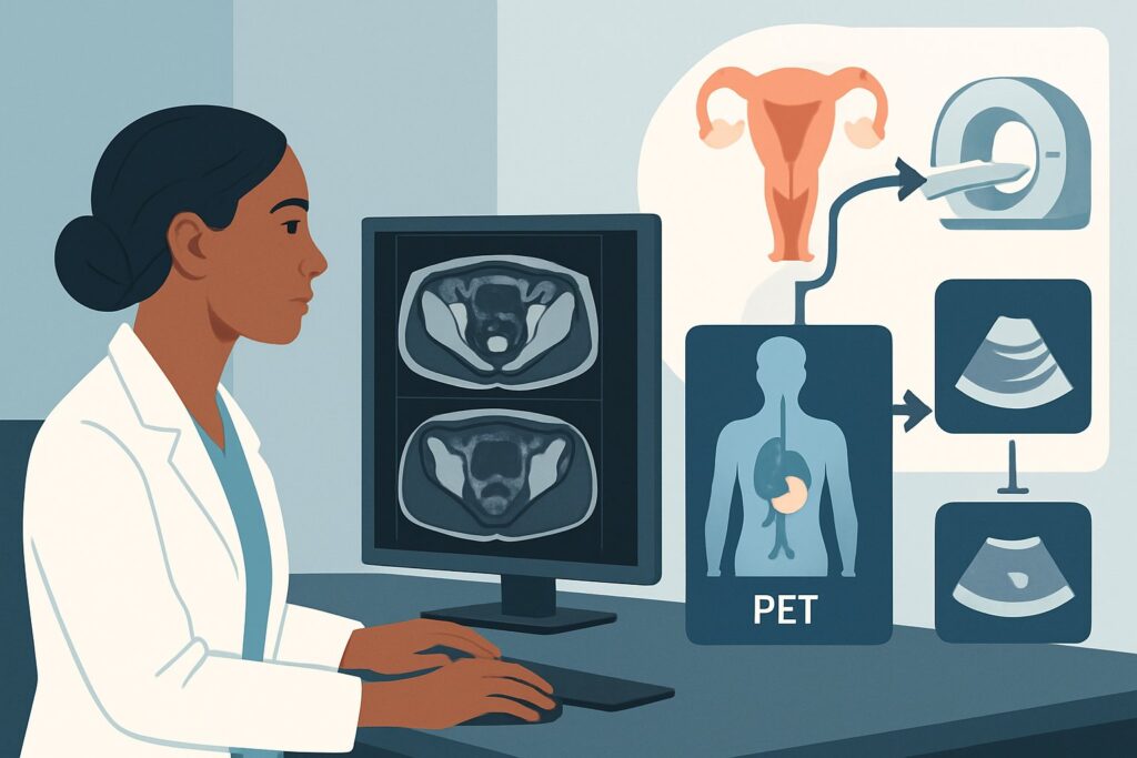
A 48-year-old woman with FIGO stage IIB squamous cell carcinoma of the cervix is in your clinic for her first follow-up visit, three months after completing definitive chemoradiation. Her physical exam is reassuring, with a non-palpable cervical mass and no new symptoms. Now, you must decide on the optimal imaging study to assess her initial treatment response, a critical step that will guide her future surveillance and management. This article provides a detailed clinical workflow for this specific scenario, explaining why the American College of Radiology (ACR) rates MRI pelvis without and with IV contrast as Usually appropriate for the initial post-treatment evaluation.
Who Fits This Clinical Scenario for Cervical Cancer Response Assessment?
This guidance is specifically for patients undergoing their initial imaging-based treatment response assessment after completing a course of definitive chemoradiation for invasive cervical cancer. Typically, this evaluation occurs approximately 3 to 6 months after the conclusion of therapy. The timing is crucial: imaging too early can be confounded by acute post-radiation inflammation, leading to false-positive results, while waiting too long may delay the detection of residual or progressive disease.
This workflow applies to patients who have received concurrent chemotherapy and radiation with curative intent. It is distinct from several related but different clinical situations:
- Initial Staging: This article does not cover the initial workup of a newly diagnosed cervical cancer for local (T stage) or systemic (N/M stage) disease.
- Asymptomatic Surveillance: This workflow is for the first major post-treatment assessment, not for routine, long-term surveillance in a patient who has already been deemed to have a complete response.
- Symptomatic Recurrence: This guidance is not for a patient presenting with new symptoms concerning for recurrence (e.g., pelvic pain, vaginal bleeding, leg edema) long after treatment has been completed. That scenario represents an evaluation of known or suspected recurrence, a separate ACR variant.
What Diagnoses Are You Working Up in This Post-Treatment Scenario?
The primary goal of imaging at this 3-to-6-month juncture is to differentiate between a successful treatment outcome and persistent disease. The differential diagnosis is narrow but has profound prognostic implications.
Complete Response: This is the desired outcome, where imaging demonstrates the resolution of the primary tumor and any involved lymph nodes. The expected findings include a significant decrease in the size of the cervical mass, replacement by low-signal-intensity fibrotic tissue on T2-weighted MRI, and resolution of abnormal FDG avidity on PET/CT. Differentiating this from other possibilities is the central task of the imaging study.
Residual Viable Tumor: This represents an incomplete response to therapy. The imaging goal is to detect any remaining tumor tissue that is still metabolically active or demonstrates imaging characteristics of malignancy, such as persistent enhancement, restricted diffusion on MRI, or FDG avidity on PET. Identifying residual disease early is critical for considering salvage therapy options, such as pelvic exenteration or additional systemic treatments.
Post-Radiation Changes Mimicking Recurrence: This is a significant diagnostic challenge. The inflammatory response to radiation can cause tissue edema, enhancement, and increased metabolic activity that can be difficult to distinguish from residual tumor. These changes are most pronounced in the first few months after treatment, which is why imaging is typically delayed until at least 3 months post-therapy. The superior soft-tissue resolution of MRI is particularly valuable in distinguishing organized, linear post-radiation fibrosis from a nodular, disorganized residual mass.
Why Is Pelvic MRI the Recommended Study for Initial Response Assessment?
The ACR Appropriateness Criteria panel designates MRI pelvis without and with IV contrast as Usually appropriate for the initial assessment of treatment response. This recommendation is based on MRI’s exceptional ability to characterize pelvic soft tissues and differentiate post-treatment changes from residual cancer.
The strength of MRI lies in its multi-parametric approach. High-resolution T2-weighted imaging is excellent for evaluating cervical anatomy and identifying the low-signal scar tissue characteristic of a complete response. Conversely, residual tumor often appears as an intermediate-to-high signal intensity mass on T2-weighted images.
The addition of functional sequences provides further diagnostic confidence. Diffusion-weighted imaging (DWI) is highly sensitive for detecting viable, hypercellular tumor tissue, which typically demonstrates restricted diffusion (appearing bright on DWI and dark on corresponding ADC maps). Intravenous gadolinium-based contrast helps delineate enhancing viable tumor from non-enhancing necrotic or fibrotic tissue. This combination of anatomic and functional information makes MRI a powerful tool for this specific clinical question.
While MRI is a primary recommendation, other modalities are also highly rated:
- FDG-PET/CT is also rated Usually appropriate. It provides crucial metabolic information, assessing for FDG-avid residual disease in the pelvis and simultaneously surveying for distant metastases. However, it carries a significant radiation dose (☢☢☢☢ 10-30 mSv) and is more susceptible to false positives from post-radiation inflammation. It is often reserved for cases where MRI findings are equivocal or when there is a high pretest probability of metastatic disease.
- CT abdomen and pelvis with IV contrast is rated May be appropriate. While useful for detecting bulky nodal disease or hydronephrosis, its inferior soft-tissue contrast makes it significantly less sensitive than MRI for evaluating the primary tumor site within the cervix.
Ultimately, MRI provides the best balance of high diagnostic accuracy for local pelvic disease assessment with no ionizing radiation (0 mSv), making it the preferred initial imaging test in this scenario.
What’s Next After the Post-Treatment Pelvic MRI? Downstream Workflow
The results of the initial post-treatment MRI will direct the subsequent clinical pathway. The interpretation should be integrated with the findings of a thorough physical and pelvic examination.
- If the MRI shows a complete response: This is an excellent prognostic sign. The patient can transition to a standard surveillance protocol. This typically involves regular clinical follow-up with physical exams. The next ACR variant, Surveillance of treated cervical cancer in asymptomatic patients, would then guide any future imaging decisions, which are often not performed routinely in the absence of symptoms.
- If the MRI is negative but clinical exam is suspicious: A biopsy of any suspicious area on pelvic exam is warranted, regardless of the reassuring imaging. Clinical findings should always be correlated with imaging results, and tissue sampling remains the gold standard for confirming recurrence.
- If the MRI shows unequivocal residual or recurrent disease: This finding necessitates a biopsy to confirm viable cancer. If confirmed, the patient should be evaluated by a multidisciplinary tumor board to discuss options for salvage therapy. This may include radical surgery (e.g., pelvic exenteration), further radiation, or systemic therapy, depending on the location and extent of the disease.
- If the MRI is indeterminate or equivocal: This is a common and challenging situation. The findings may represent post-radiation inflammation or a small volume of residual disease. In this case, FDG-PET/CT is often the next best step to provide metabolic information that can help differentiate between these possibilities. If still indeterminate, a short-interval follow-up MRI in 2-3 months or an examination under anesthesia with directed biopsies may be necessary.
Pitfalls to Avoid (and When to Get Help)
Navigating the post-treatment assessment requires careful attention to timing and technique to avoid common errors.
- Imaging Too Early: Ordering the first response assessment MRI less than 3 months after completing chemoradiation significantly increases the risk of false-positive results due to intense post-treatment inflammation.
- Omitting Key MRI Sequences: A standard pelvic MRI protocol may be insufficient. Ensure the order specifies the inclusion of high-resolution T2-weighted images in multiple planes and diffusion-weighted imaging (DWI/ADC), as these are critical for distinguishing tumor from fibrosis.
- Relying on Imaging Alone: Never disregard the physical examination. A palpable nodule or abnormal finding on speculum exam warrants a biopsy even if the MRI report is negative.
- Misinterpreting Inflammatory FDG Avidity: Be aware that post-radiation inflammation can cause intense FDG uptake on a PET/CT, mimicking malignancy. Correlating the PET findings with the anatomic detail from CT or MRI is essential.
If imaging findings are equivocal or conflict with the clinical picture, escalation to a multidisciplinary tumor board including gynecologic oncology, radiation oncology, and diagnostic radiology is the most appropriate next step.
Related ACR Topics and Tools
This article focuses on a single clinical scenario. For a comprehensive overview of all related variants and for tools to assist in your clinical practice, please refer to the following resources.
For breadth across all scenarios in Pretreatment Evaluation and Follow-up of Invasive Cancer of the Cervix, see our parent guide: Pretreatment Evaluation and Follow-up of Invasive Cancer of the Cervix: ACR Appropriateness Decoded.
- ACR Appropriateness Criteria Lookup — for adjacent scenarios
- Imaging Protocol Library — for technique on the recommended study
- Radiation Dose Calculator — for cumulative dose conversations
Frequently Asked Questions
Why is imaging delayed until 3 months after completing chemoradiation for cervical cancer?
Imaging is typically delayed for at least 3 months to allow acute post-radiation inflammation and edema to subside. Performing imaging, especially FDG-PET/CT, too early can lead to false-positive results, as inflammation can mimic the appearance and metabolic activity of residual cancer. This waiting period provides a more accurate assessment of the true treatment response.
If the initial MRI is negative, is any further imaging needed for surveillance?
If the initial 3-6 month MRI and clinical exam both indicate a complete response, routine surveillance imaging is generally not recommended for asymptomatic patients. Follow-up typically consists of regular physical and pelvic exams. Imaging is reserved for patients who develop new signs or symptoms concerning for recurrence, which falls under a different ACR clinical scenario.
Can I order an MRI without contrast for this assessment?
While an MRI of the pelvis without IV contrast is also rated as *Usually appropriate* by the ACR, the addition of a gadolinium-based contrast agent is generally preferred. Contrast enhancement helps to better delineate viable tumor tissue from post-treatment fibrosis or necrosis, increasing diagnostic confidence, especially in equivocal cases.
When should I choose FDG-PET/CT instead of MRI for the initial response assessment?
FDG-PET/CT is also rated *Usually appropriate* and is an excellent alternative or complementary study. It may be preferred over MRI if there is a high clinical suspicion for distant metastatic disease, as it provides whole-body staging. It is also a valuable problem-solving tool when MRI findings are indeterminate, as the metabolic information from PET can help clarify whether an abnormality represents inflammation or viable tumor.
What specific MRI sequences should I ensure are included in the protocol?
For optimal evaluation, the MRI protocol should include high-resolution T2-weighted sequences in at least two planes (e.g., sagittal and axial oblique, perpendicular to the long axis of the cervix). Crucially, it must also include diffusion-weighted imaging (DWI) with an apparent diffusion coefficient (ADC) map, and T1-weighted sequences before and after the administration of IV gadolinium contrast. These sequences provide the necessary anatomic and functional detail to assess for residual tumor.
Reviewed by Pouyan Golshani, MD, Interventional Radiologist — May 30, 2026