How Should You Image DCIS in Patients on Active Surveillance? An ACR Workflow
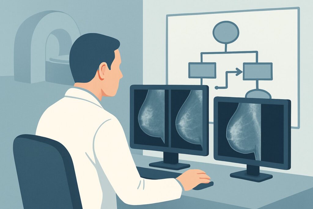
A 52-year-old patient sits in your clinic for a follow-up visit. Three months ago, a stereotactic biopsy of suspicious microcalcifications revealed low-grade ductal carcinoma in situ (DCIS). After a thorough discussion with the multidisciplinary breast team, she elected to pursue active surveillance rather than immediate surgical intervention. Now, her first surveillance imaging study is due, and you need to decide on the most appropriate order. What is the optimal imaging modality to monitor her known DCIS for stability or signs of progression, ensuring both efficacy and patient safety?
This clinical workflow article addresses this specific decision point. Based on the American College of Radiology (ACR) Appropriateness Criteria, for a patient with newly diagnosed DCIS on active surveillance, a Digital breast tomosynthesis diagnostic study is rated Usually Appropriate.
Who Fits This Clinical Scenario for DCIS Active Surveillance?
This guidance is specifically for adult patients with a recent, biopsy-proven diagnosis of Ductal Carcinoma in Situ (DCIS) who have made a conscious and informed decision to forego immediate surgical treatment (lumpectomy or mastectomy) in favor of an active surveillance protocol. The key inclusion criteria are a confirmed DCIS diagnosis and a management plan that involves serial imaging to monitor the lesion over time.
It is crucial to distinguish this scenario from similar but distinct clinical situations that require different imaging pathways:
- Initial Diagnostic Workup: This article does not apply to the initial evaluation of suspicious findings (e.g., calcifications) before a DCIS diagnosis is established. That workup falls under the “Newly diagnosed DCIS. Initial imaging” variant.
- Post-Surgical Surveillance: This guidance is not for patients who have already undergone breast conservation therapy (lumpectomy with or without radiation) or mastectomy for DCIS. Those patients follow different routine surveillance protocols for detecting local recurrence.
- Symptomatic Patients: If a patient on active surveillance develops new symptoms, such as a palpable lump, nipple discharge, or skin changes, the workup shifts from routine surveillance to a diagnostic evaluation of a new problem, which is covered in the “Suspected local recurrence based on symptoms” variant.
- DCIS with Microinvasion: If the initial biopsy revealed DCIS with microinvasion, the management and imaging strategy would differ, as this finding changes the risk profile and staging.
What Are You Monitoring For During DCIS Active Surveillance?
Unlike a typical diagnostic workup aimed at identifying an unknown disease, imaging in active surveillance is focused on monitoring a known, low-risk malignancy. The primary goals are to detect any signs of change that might warrant a shift from surveillance to active treatment. The differential considerations are centered on stability versus progression.
Stability of Known DCIS: The most desired outcome is demonstrating that the known area of DCIS is unchanged. For DCIS that presents as microcalcifications, this means the extent, morphology, and distribution of the calcifications remain stable compared to prior examinations. This finding supports continuing with the active surveillance protocol.
Progression to Invasive Ductal Carcinoma (IDC): This is the most critical concern that active surveillance aims to detect at the earliest possible stage. Imaging signs of progression from DCIS to invasive cancer can be subtle. Radiologists are looking for an increase in the number or extent of microcalcifications, a change to a more suspicious morphology (e.g., fine linear or branching), or the development of an associated soft tissue mass or architectural distortion that was not previously present.
Development of New Disease: The surveillance study also serves as a screening tool for the remainder of the ipsilateral breast and the entire contralateral breast. A patient with DCIS has a higher risk of developing a new breast cancer, so the examination must be comprehensive enough to detect any new, unrelated suspicious findings that would require their own diagnostic workup.
Why Is Diagnostic Digital Breast Tomosynthesis the Recommended Study?
The ACR Appropriateness Criteria rate Digital breast tomosynthesis diagnostic as Usually Appropriate for monitoring DCIS in patients on active surveillance. This recommendation is based on the modality’s superior ability to characterize the key features of DCIS, particularly microcalcifications.
Digital Breast Tomosynthesis (DBT), or 3D mammography, acquires images of the breast from multiple angles, which are then reconstructed into thin, one-millimeter slices. This technique significantly reduces the effect of overlapping breast tissue, a common limitation of standard 2D mammography. For DCIS surveillance, this is paramount for accurately assessing the extent, distribution, and morphology of calcifications. The improved clarity helps radiologists detect subtle changes that might indicate progression to invasive disease. While standard 2D Mammography diagnostic is also rated Usually Appropriate, DBT is often preferred where available due to its enhanced visualization.
The radiation dose for DBT is low, with a relative radiation level (RRL) of ☢☢ (0.1-1 mSv), making it safe and suitable for the serial imaging required in a long-term surveillance program.
Why are alternative studies rated lower for this specific scenario?
- MRI breast without and with IV contrast: This study is rated May be appropriate. While breast MRI has very high sensitivity for detecting invasive cancer, its specificity is lower. This can lead to a higher rate of false-positive findings, resulting in patient anxiety and potentially unnecessary biopsies. Its use may be considered in select cases, such as for patients with extremely dense breasts where mammography is limited, or if there is a clinical or mammographic concern for a larger extent of disease than is visible.
- US breast: Ultrasound is rated Usually not appropriate as a primary surveillance tool for DCIS. The vast majority of DCIS presents as microcalcifications, which are the primary target of surveillance. Ultrasound has poor sensitivity for visualizing and characterizing these calcifications. Its role is reserved for evaluating palpable lumps or mammographically identified masses, not for monitoring calcific DCIS.
When ordering the study, it is essential to specify “diagnostic” rather than “screening.” This designation signals to the radiology department that this is a targeted examination, allowing the radiologist to request additional views, such as magnification, to meticulously evaluate the area of known DCIS and compare it to prior studies.
Downstream Workflow: Interpreting Results in DCIS Surveillance
The results of the diagnostic DBT will direct the next steps in the patient’s management plan. The workflow is a clear decision tree based on the imaging findings.
- If the study is stable: When the radiologist reports no change in the known area of DCIS and no new suspicious findings, the patient continues on the active surveillance protocol. The next imaging study is typically scheduled in 6 to 12 months, according to the institution’s established protocol for DCIS surveillance.
- If the study shows suspicious changes (progression): This is the most significant outcome. Findings suggesting progression include an increased extent of calcifications, development of a new mass, or architectural distortion in the area of the known DCIS. This result should prompt an immediate recommendation for a repeat image-guided biopsy of the area to histologically assess for an invasive component. If invasive carcinoma is confirmed, the patient would be counseled to transition from surveillance to definitive surgical treatment.
- If the study shows a new, indeterminate finding elsewhere: If a new, unrelated finding is discovered in the ipsilateral or contralateral breast, it is worked up independently according to standard BI-RADS guidelines. This may involve further imaging with spot compression views, targeted ultrasound, and potentially a biopsy of the new lesion.
Pitfalls to Avoid (and When to Get Help)
Careful execution of the imaging protocol is critical to the success of an active surveillance strategy for DCIS. Avoiding common pitfalls can prevent missed diagnoses or unnecessary interventions.
- Using Screening Instead of Diagnostic Mammography: Ordering a “screening” study is insufficient. A “diagnostic” mammogram is required to ensure the radiologist can legally and procedurally perform the necessary additional views, like magnification, to properly assess the known DCIS.
- Under-characterizing Calcifications: Failing to obtain high-quality magnification views can obscure subtle but critical changes in calcification morphology that may signal progression to invasive cancer.
- Inappropriate Reliance on Ultrasound: Do not use ultrasound as the primary or sole imaging tool for following calcific DCIS. It is not sensitive for the key features being monitored.
- Poor Communication of Biopsy Location: The ordering clinician must ensure the radiologist is aware of the precise location of the original biopsy-proven DCIS. The presence of a post-biopsy marker is essential for accurate year-over-year comparison.
If imaging findings are equivocal or if there is a discrepancy between imaging and a new clinical finding, escalation to a multidisciplinary breast tumor board discussion is the appropriate next step.
Related ACR Topics and Tools
For a comprehensive overview of all clinical variants related to Ductal Carcinoma in Situ, please refer to our parent topic hub article. Additional GigHz tools can help you navigate adjacent scenarios, understand imaging techniques, and discuss radiation dose with your patients.
- For breadth across all scenarios in Imaging of Ductal Carcinoma in Situ (DCIS), see our parent guide: Imaging of Ductal Carcinoma in Situ (DCIS): ACR Appropriateness Decoded.
- To explore other clinical presentations, use the ACR Appropriateness Criteria Lookup.
- For details on imaging techniques, consult the Imaging Protocol Library.
- To help frame conversations about cumulative exposure, see the Radiation Dose Calculator.
Frequently Asked Questions
How often should imaging be performed for DCIS on active surveillance?
While protocols can vary by institution, diagnostic mammography or digital breast tomosynthesis is typically performed every 6 to 12 months. The exact interval is determined by the multidisciplinary breast care team based on the patient’s specific risk factors and lesion characteristics.
Is breast MRI better than mammography for monitoring DCIS?
Not necessarily for routine surveillance. The ACR rates breast MRI as ‘May be appropriate’ because while it is highly sensitive, it has lower specificity, which can lead to more false positives and unnecessary biopsies. Digital breast tomosynthesis is rated ‘Usually appropriate’ and remains the primary recommended tool for most patients on active surveillance.
What should be done if a patient on active surveillance for DCIS develops a new lump?
The development of a new symptom like a palpable lump changes the clinical situation from routine surveillance to a diagnostic workup. The patient should undergo a diagnostic mammogram/DBT and a targeted ultrasound of the new lump to characterize it, which may lead to a biopsy.
Does the grade of DCIS (low, intermediate, high) affect the choice of imaging for surveillance?
The choice of imaging modality itself—diagnostic DBT—is generally the same regardless of the DCIS grade. However, the decision to offer active surveillance in the first place is heavily influenced by grade. Active surveillance is most commonly considered for low-grade DCIS, as higher-grade DCIS has a greater risk of progression to invasive cancer and is more often treated surgically upfront.
Why is ultrasound ‘Usually not appropriate’ for routine DCIS surveillance?
The vast majority of DCIS cases, especially those selected for active surveillance, present as microcalcifications on mammography. Ultrasound is not a sensitive tool for detecting or characterizing these microcalcifications. Its role is primarily for evaluating masses or other non-calcific findings, making it unsuitable as the main imaging tool for monitoring calcific DCIS.
Reviewed by Pouyan Golshani, MD, Interventional Radiologist — May 30, 2026