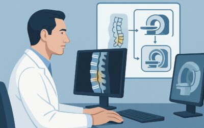
How Should You Manage Hilar Cholangiocarcinoma with Vascular Invasion? ACR Insights

A 68-year-old male presents with progressive jaundice, pruritus, and unintentional weight loss. His initial workup included a multiphase CT of the abdomen, and the report is now on your screen. It describes a 4 cm, ill-defined mass at the hepatic duct confluence, encasing the main portal vein and involving regional lymph nodes. The biliary ducts are markedly dilated upstream. You are now faced with a complex management decision for what appears to be a locally advanced hilar cholangiocarcinoma. This article provides a detailed clinical workflow for this specific scenario, explaining why the American College of Radiology (ACR) Appropriateness Criteria designate Systemic therapies as Usually appropriate as the primary management step.
Who Fits This Clinical Scenario for Advanced Hilar Cholangiocarcinoma?
This guidance is specifically for patients whose diagnostic imaging and biopsy results confirm a hilar cholangiocarcinoma (also known as a Klatskin tumor) with features indicating locally advanced, likely unresectable disease. The key inclusion criteria are:
- Tumor Location: Centered at the hepatic hilum (biliary confluence).
- Tumor Size: Greater than 3 cm in its largest dimension.
- Tumor Margins: Poorly defined or infiltrative, suggesting invasion into surrounding liver parenchyma.
- Vascular Invasion: Clear evidence of tumor encasement or invasion of major portal or hepatic arterial branches.
- Lymphadenopathy: Presence of suspicious periportal or other regional lymph nodes.
It is critical to distinguish this scenario from others that may present similarly but follow different management pathways. This guidance does not apply to:
- Small, well-defined peripheral intrahepatic cholangiocarcinoma: These tumors, located away from the hilum and without vascular invasion, may be candidates for surgical resection or ablation.
- Hepatocellular carcinoma (HCC) with vascular invasion: While also a primary liver cancer, HCC has a different biology and responds differently to locoregional therapies. Its management is covered under a separate ACR variant.
- Metastatic disease to the liver hilum: Tumors from the gallbladder, pancreas, or gastrointestinal tract can metastasize to the porta hepatis and mimic a primary cholangiocarcinoma. The management is dictated by the primary cancer type.
What Diagnoses Are You Working Up in This Scenario?
While the imaging findings in this scenario are highly suggestive of a specific diagnosis, a concise differential remains essential for confirming the treatment plan and considering rare mimics. The primary goal of the workup is to confirm unresectability and obtain tissue for pathologic and molecular analysis to guide systemic therapy.
Hilar Cholangiocarcinoma (Klatskin Tumor): This is the leading diagnosis. The combination of a mass at the biliary confluence causing obstructive jaundice, coupled with vascular encasement and regional adenopathy, is the classic presentation of a Bismuth-Corlette type III or IV hilar cholangiocarcinoma that is beyond the criteria for surgical cure.
Hepatocellular Carcinoma (HCC) with Biliary Invasion: Less commonly, an aggressive HCC can infiltrate the biliary tree and present as a hilar mass. This is an important distinction, as the systemic therapies for HCC and cholangiocarcinoma differ significantly. Serum tumor markers like Alpha-Fetoprotein (AFP) and CA 19-9, alongside a biopsy, are crucial for differentiation.
Metastatic Disease to the Porta Hepatis: The liver hilum is a common site for nodal metastases from other abdominal cancers, particularly gallbladder, pancreatic, and gastric adenocarcinomas. These can form a confluent mass that encases vessels and bile ducts, creating an identical imaging picture. A biopsy is essential, and a search for an alternative primary tumor may be warranted if the pathology is not definitive for cholangiocarcinoma.
IgG4-Related Sclerosing Cholangitis: This is a rare but critical inflammatory mimic. It can produce a mass-like inflammatory infiltrate in the porta hepatis that causes biliary stricturing and can be mistaken for malignancy. While vascular invasion is less common than in cancer, it can occur. Elevated serum IgG4 levels and a biopsy showing a characteristic lymphoplasmacytic infiltrate can confirm this diagnosis, which is treated with steroids, not chemotherapy.
Why Are Systemic Therapies the Recommended First Step for This Presentation?
For a hilar cholangiocarcinoma with features of advanced disease—large size, vascular invasion, and nodal involvement—the likelihood of achieving a complete (R0) surgical resection is exceedingly low. The clinical goal shifts from curative-intent local treatment to systemic disease control, management of symptoms like biliary obstruction, and extension of survival. This is why the ACR panel rates Systemic therapies as Usually appropriate.
The rationale for prioritizing systemic therapy is based on a multidisciplinary assessment that the cancer has extended beyond the boundaries of safe and effective local removal. Attempting aggressive local treatments first in this setting often leads to high morbidity without a survival benefit. Instead, initiating chemotherapy (often a gemcitabine and cisplatin-based regimen, potentially with immunotherapy depending on molecular testing) addresses both the primary tumor and any potential micrometastatic disease.
Understanding why other options are rated lower is key to decision-making:
- Surgical Liver Resection and Liver Transplantation: Both are rated Usually not appropriate. The presence of major vascular invasion and confirmed nodal metastasis are widely accepted contraindications for both resection and transplantation due to the near-certainty of early and widespread recurrence.
- Percutaneous Ablation: This is rated Usually not appropriate. Ablative energies (like radiofrequency or microwave) cannot safely or effectively treat a large tumor (>3 cm) that is wrapped around critical structures like the portal vein, hepatic artery, and major bile ducts. The risk of catastrophic injury is unacceptably high.
- Transarterial Chemoembolization (TACE) or Bland Embolization: These are also Usually not appropriate. Unlike HCC, which is typically hyperarterial, cholangiocarcinomas are often hypovascular. They derive a significant blood supply from the portal system, making them poor targets for therapies delivered solely via the hepatic artery.
Therapies rated as May be appropriate, such as Transarterial radioembolization (Y-90) and External beam radiation therapy (EBRT), are generally considered as potential adjuncts or second-line options. They may be used in combination with systemic therapy or for consolidation if there is a good response to initial chemotherapy, but this is determined on a case-by-case basis by a multidisciplinary tumor board.
Note that the ACR does not assign a relative radiation level (RRL) to these therapeutic interventions, as the concept is designed for diagnostic imaging. The decision to proceed with radiation-based treatments is based on a complex risk-benefit analysis of therapeutic efficacy versus potential toxicity to the liver and surrounding organs.
What’s Next After Systemic Therapies? Downstream Workflow
Initiating systemic therapy is the first step in a longer management journey. The downstream workflow is guided by treatment response, symptom control, and ongoing multidisciplinary evaluation.
Biliary Decompression: A critical, often concurrent, first step is relieving the biliary obstruction. This is typically achieved via percutaneous transhepatic biliary drainage (PTBD) or an endoscopic retrograde cholangiopancreatography (ERCP) with stent placement. Decompressing the biliary system is vital to prevent cholangitis, improve liver function (which is necessary to tolerate chemotherapy), and alleviate debilitating symptoms like jaundice and pruritus.
Monitoring Treatment Response: The patient will undergo restaging imaging (typically CT or MRI) after a set number of chemotherapy cycles (e.g., 2-3 months).
- If the tumor responds or is stable: Systemic therapy is typically continued. In select cases of exceptional response, the patient may be re-evaluated by the tumor board for potential conversion to a locoregional therapy or even surgery, though this is uncommon.
- If the tumor progresses: The next step is to consider second-line systemic agents, guided by molecular profiling of the tumor tissue (e.g., testing for FGFR2 fusions, IDH1 mutations). Enrollment in a clinical trial is often a preferred option at this stage.
Palliative Care and Symptom Management: Throughout the treatment course, a strong focus on palliative care is essential. This includes managing pain, nutritional support, and addressing the psychological and social needs of the patient and their family. This is not just end-of-life care but an integrated part of managing a complex cancer diagnosis from the outset.
Pitfalls to Avoid (and When to Get Help)
Navigating this clinical scenario requires careful coordination and avoidance of common pitfalls.
- Delaying Biliary Drainage: Failing to promptly decompress an obstructed biliary system can lead to life-threatening cholangitis and delay the start of essential systemic therapy.
- Inadequate Biopsy Sample: Obtaining only a cytology sample (e.g., from biliary brushings) may be insufficient. A core needle biopsy is often necessary to provide enough tissue for both histologic confirmation and comprehensive next-generation sequencing to identify targetable mutations.
- Attempting Futile Surgery: Proceeding with a high-risk resection in a patient with clear contraindications (vascular and nodal involvement) can cause significant harm and delay the initiation of more appropriate systemic treatment.
- Neglecting Multidisciplinary Input: Management of advanced cholangiocarcinoma is not the responsibility of a single specialist. Regular discussion among medical oncology, interventional radiology, surgery, radiation oncology, and gastroenterology is the standard of care.
If liver function deteriorates rapidly or signs of sepsis (fever, hypotension) develop, this constitutes a medical emergency requiring immediate escalation for biliary drainage and supportive care.
Related ACR Topics and Tools
This article focuses on a single, complex scenario. For a broader overview of imaging and intervention for all types of liver cancer, or to explore the tools used in the diagnostic process, the following resources are valuable.
- For breadth across all scenarios in Management of Liver Cancer, see our parent guide: Management of Liver Cancer: ACR Appropriateness Decoded.
- To search for other clinical presentations, consult the ACR Appropriateness Criteria Lookup.
- For details on imaging techniques, explore the Imaging Protocol Library.
- To discuss cumulative radiation exposure from diagnostic scans with patients, use the Radiation Dose Calculator.
Frequently Asked Questions
Why isn’t surgery the first choice for a large hilar cholangiocarcinoma with vascular invasion?
Surgery is rated ‘Usually not appropriate’ because the involvement of major blood vessels (like the portal vein or hepatic artery) and regional lymph nodes means the cancer has spread beyond the area that can be safely and completely removed. Attempting surgery in this setting has a very high risk of leaving cancer behind (an R1 or R2 resection), leading to rapid recurrence and significant surgical morbidity without a survival benefit.
What is the role of a biopsy if the imaging is classic for unresectable cholangiocarcinoma?
A biopsy is essential for two reasons. First, it confirms the diagnosis and rules out rare mimics like IgG4-related disease or metastatic cancer from another source. Second, and critically, it provides tissue for molecular and genomic testing. Identifying specific mutations (e.g., FGFR2 fusions, IDH1 mutations) can open the door to targeted therapies that may be more effective than standard chemotherapy.
Can radiation therapy be used as the primary treatment for this condition?
External beam radiation therapy (EBRT) is rated ‘May be appropriate’ but is not typically used as the sole primary treatment for this advanced stage. It is more often considered as an adjunct to systemic therapy to improve local control of the tumor, or as a palliative measure to relieve symptoms like pain or obstruction, especially after initial chemotherapy.
What is the purpose of biliary drainage before starting chemotherapy?
Biliary drainage (via ERCP or a percutaneous drain) is crucial to relieve the obstruction caused by the tumor. This accomplishes three goals: 1) It prevents life-threatening infection (cholangitis). 2) It improves liver function by reducing bilirubin levels, which is often a prerequisite for safely administering chemotherapy. 3) It alleviates severe symptoms like jaundice and itching, improving the patient’s quality of life.
If the tumor is larger than 3 cm but has no vascular invasion or adenopathy, does this guidance still apply?
No, that would represent a different clinical scenario. The absence of vascular invasion and lymphadenopathy might make the patient a candidate for surgical resection, even with a larger tumor. That presentation would fall under a different set of considerations and would not align with this specific ACR variant, which is defined by features of unresectability.
Reviewed by Pouyan Golshani, MD, Interventional Radiologist — May 30, 2026