How Should You Manage Multiple Splenic Abscesses in an Intravenous Drug User with Sepsis?
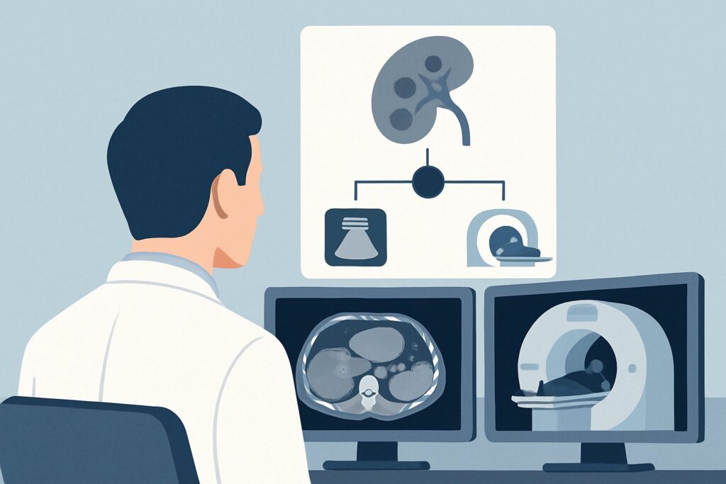
A 28-year-old man with a known history of intravenous drug use is brought to the emergency department with a high fever, rigors, and a heart rate of 130 bpm. He is lethargic and appears acutely ill. Blood cultures are drawn, and broad-spectrum antibiotics are started. A contrast-enhanced computed tomography (CT) scan of the abdomen and pelvis is performed to identify a source of sepsis, revealing two distinct, rim-enhancing fluid collections in the spleen, measuring 4 cm and 5 cm. They are noncommunicating, and each appears accessible through at least 1 cm of normal splenic parenchyma. You are now faced with a critical management decision: escalate to surgical consultation for splenectomy or consult interventional radiology for drainage. This article details the clinical workflow for this specific scenario, where the American College of Radiology (ACR) rates **Percutaneous catheter drainage only** as **Usually appropriate**.
Who Fits This Clinical Scenario for Splenic Abscess Management?
This guidance is tailored for a specific, high-acuity patient presentation. The key inclusion criteria are a patient who is systemically unwell (e.g., fever, tachycardia, leukocytosis) with a clear risk factor for hematogenous infection, such as intravenous drug abuse, and has confirmed imaging findings of one or more splenic abscesses. Critically, the abscesses must be technically amenable to percutaneous intervention, meaning there is a safe and accessible path for a needle and catheter that avoids transgressing other organs, major vessels, or the pleural space. The presence of a 1 cm rim of normal splenic tissue is a classic indicator of a safe percutaneous window.
It is crucial to distinguish this scenario from other presentations involving infected fluid collections:
- This workflow does not apply to patients with infected pancreatic pseudocysts, which often require specialized endoscopic or surgical management due to their enzymatic content and proximity to other structures.
- This is distinct from a patient with a contained appendiceal abscess, where management is closely tied to the underlying appendicitis and may involve interval appendectomy.
- This guidance is not intended for a patient with a tubo-ovarian abscess, where the anatomical location and typical pathogens necessitate a different gynecologic-focused treatment algorithm.
Applying this workflow to the wrong clinical context can lead to suboptimal outcomes. This article is strictly for the septic patient with confirmed, accessible splenic abscesses.
What Is the Underlying Cause of Splenic Abscesses in This Scenario?
While imaging has identified the splenic abscesses, understanding the likely underlying etiology is critical for comprehensive patient management, as it dictates further workup and long-term treatment. In a patient with a history of intravenous drug use, several diagnoses must be considered.
The leading consideration is infective endocarditis with septic emboli. This is the most common pathway for splenic abscess formation in this population. Bacteria, often Staphylococcus aureus, form vegetations on heart valves (typically right-sided, but left-sided involvement is also common) that can break off and travel through the arterial system, lodging in end-organs like the spleen. The presence of splenic abscesses should immediately trigger a high suspicion for endocarditis and prompt an urgent echocardiogram and multiple sets of blood cultures.
A related possibility is hematogenous seeding from bacteremia without endocarditis. The patient may have transient or persistent bacteremia from non-sterile injections that directly seeds the spleen. This is particularly relevant if the spleen has pre-existing damage, such as a small, otherwise asymptomatic infarct, which can act as a nidus for infection.
Less commonly, a splenic abscess can arise from contiguous spread from an adjacent infection, such as a perforated gastric or colonic ulcer or a severe pancreatic infection. However, in this scenario, the primary source of infection would typically be evident on the initial CT scan, which is not the case here.
Finally, consider an infected splenic infarct from a non-bacterial cause. While septic emboli are most likely, other causes of emboli or hypercoagulability can lead to splenic infarction, which can then become secondarily infected during an episode of bacteremia.
Why Is Percutaneous Catheter Drainage a Recommended First-Line Treatment?
For a septic patient with accessible splenic abscesses, percutaneous catheter drainage is rated as Usually appropriate by the ACR because it offers effective source control with significantly lower morbidity and mortality compared to open surgery. The primary goal is to evacuate the purulent material, which reduces the bacterial load, alleviates systemic toxicity, and allows antibiotics to penetrate the abscess cavity more effectively. As a minimally invasive procedure, it avoids the risks of general anesthesia, laparotomy, and the long-term immunological consequences of splenectomy.
The ACR evaluates other potential management strategies, which receive different ratings for this specific scenario:
- Splenectomy: While also rated as Usually appropriate, splenectomy is a major abdominal surgery with substantial risks, including bleeding, injury to adjacent organs (like the pancreatic tail), and overwhelming post-splenectomy infection (OPSI). It is generally reserved for cases where percutaneous drainage fails, the abscesses are inaccessible, the spleen is ruptured, or there is extensive splenic necrosis. Starting with the less invasive option is the preferred modern approach.
- Needle aspiration: This is rated as May be appropriate. Aspiration alone can be diagnostic and may be sufficient for small, unilocular abscesses. However, for larger collections like those in this scenario (4-5 cm), simple aspiration often fails to provide complete evacuation, leading to rapid re-accumulation and treatment failure. Catheter drainage provides continuous, dependent drainage, which is generally more effective for definitive treatment.
- Conservative management only: Relying solely on antibiotics is rated as Usually not appropriate. While antibiotics are a critical component of treatment, they have poor penetration into the avascular, necrotic core of an established abscess. Without source control via drainage or surgery, antibiotic therapy alone has a high failure rate and is associated with prolonged illness and increased mortality.
What’s Next After Percutaneous Catheter Drainage? Downstream Workflow
Placing a drainage catheter is the beginning, not the end, of the management pathway. The subsequent steps depend on the patient’s clinical response and the catheter’s output.
If the procedure is successful and the patient improves: You should see a rapid defervescence and improvement in tachycardia and leukocytosis within 24-48 hours. The catheter will be left in place and flushed regularly to maintain patency. The purulent fluid should be sent for culture and sensitivity testing to tailor antibiotic therapy. The catheter remains until drainage diminishes to a minimal amount (e.g., <10-20 mL/day), the fluid becomes serous, and follow-up imaging (CT or ultrasound) confirms resolution or near-resolution of the collection. This typically takes 7-14 days.
If the patient fails to improve: If fever and sepsis persist 48-72 hours after catheter placement, you must troubleshoot. First, obtain a CT scan with contrast injected through the catheter (a “drainogram”) to confirm proper catheter position and assess for any undrained loculations or new collections. If the catheter is malpositioned or the abscess is multiloculated, repositioning the existing catheter or placing an additional one may be necessary. If percutaneous options are exhausted and the patient remains septic, this constitutes a failure of minimally invasive therapy. At this point, you must escalate to surgical consultation for splenectomy.
If the initial workup for endocarditis is positive: A positive echocardiogram for valve vegetation solidifies the diagnosis and requires a multidisciplinary approach involving infectious disease and potentially cardiothoracic surgery. The patient will require a prolonged course of intravenous antibiotics (typically 4-6 weeks), and the splenic abscesses are managed as a metastatic complication of the primary cardiac infection.
Pitfalls to Avoid (and When to Get Help)
Several common pitfalls can complicate the management of splenic abscesses. First, avoid delaying source control; if a patient with a drainable abscess remains septic on antibiotics, prompt consultation with interventional radiology is critical. Second, do not prematurely remove the drainage catheter. Removing it before the cavity has fully collapsed and drainage has ceased can lead to recurrence. Third, never overlook the heart. In an IV drug user with splenic abscesses, failing to perform an echocardiogram to rule out endocarditis is a significant clinical error. Finally, be aware of the risk of hemorrhage during and after percutaneous drainage, as the spleen is a highly vascular organ. If the patient develops sudden hypotension, tachycardia, or a drop in hematocrit after the procedure, or if the catheter begins draining frank blood, this is a medical emergency requiring immediate imaging and potential escalation to angiography with embolization or emergent splenectomy.
Related ACR Topics and Tools
This article focuses on a single, specific clinical scenario. For a comprehensive overview of managing other types of infected fluid collections, or to explore the tools used in making these decisions, the following resources are valuable:
- For breadth across all scenarios in Radiologic Management of Infected Fluid Collections, see our parent guide: Radiologic Management of Infected Fluid Collections: ACR Appropriateness Decoded.
- To explore other clinical presentations and their corresponding ACR recommendations, use the ACR Appropriateness Criteria Lookup.
- For details on the techniques used in various imaging and interventional procedures, consult the Imaging Protocol Library.
- To help discuss radiation exposure from CT scans with your patients, the Radiation Dose Calculator can be a useful aid.
Frequently Asked Questions
Why is splenectomy also rated ‘Usually Appropriate’ if percutaneous drainage is the preferred first step?
Splenectomy is rated ‘Usually Appropriate’ because it is a definitive and effective treatment for splenic abscesses. However, it is a major surgery with higher risks of complications, including bleeding, pancreatic injury, and long-term immunodeficiency (overwhelming post-splenectomy infection). Modern practice favors a minimally invasive-first approach. Percutaneous drainage is preferred as the initial step due to its lower morbidity, reserving surgery for cases where drainage fails, is not technically possible, or if the spleen has ruptured.
What if there is only a very small rim of splenic tissue to traverse for percutaneous access?
The presence of a 1 cm rim of normal splenic tissue is a guideline for a safe percutaneous window. A smaller window increases the risk of complications like hemorrhage or capsular violation leading to peritonitis. The decision to proceed with a smaller window is up to the discretion of the interventional radiologist, who will weigh the risk of the procedure against the risk of uncontrolled sepsis. In some cases, an alternative approach (e.g., a retroperitoneal route) may be possible, or the risk may be deemed too high, making surgery the safer option.
Does the presence of two noncommunicating abscesses change the management plan?
Yes, it requires placing two separate drainage catheters, one into each collection, during the same procedure. Attempting to drain both with a single catheter is ineffective and will lead to treatment failure of the undrained collection. The technical feasibility of safely placing two catheters is a key consideration for the interventional radiologist.
What are the most common organisms cultured from splenic abscesses in IV drug users?
In patients with a history of intravenous drug use, the most common causative organism is *Staphylococcus aureus*, often methicillin-resistant (MRSA). Other common organisms include streptococcal species and gram-negative bacteria. Polymicrobial infections are also frequent. Sending the drained fluid for Gram stain, culture, and sensitivity is essential to guide definitive antibiotic therapy.
Is an ultrasound-guided approach as good as a CT-guided approach for the drainage?
Both CT and ultrasound can be used for guidance. CT provides excellent anatomical detail, clearly delineates the abscess and surrounding structures, and is often preferred for initial placement, especially for deep collections or complex anatomy. Ultrasound offers real-time visualization without ionizing radiation and can be very effective, particularly for more superficial abscesses or for follow-up catheter checks at the bedside. The choice of modality depends on the specific anatomy, institutional preference, and the expertise of the interventional radiologist.
Reviewed by Pouyan Golshani, MD, Interventional Radiologist — May 21, 2026