Initial Diagnosis of Varicose Veins: Why Duplex Ultrasound Is the Right First Step
A 48-year-old high school teacher presents to your primary care clinic with a three-year history of worsening, aching, and visibly tortuous veins in her left leg. The discomfort is most pronounced at the end of the day after standing for long periods. On examination, you note prominent varicosities along the distribution of the great saphenous vein. You suspect chronic venous insufficiency, but before referring her to a vascular specialist for potential treatment, you need to confirm the diagnosis and map the extent of the underlying venous reflux. This article details the appropriate imaging workflow for this exact scenario: the initial diagnosis of varicose veins. Based on the American College of Radiology (ACR) Appropriateness Criteria, the definitive first step is a study rated as Usually appropriate: US duplex Doppler of the lower extremity.
Who Fits This Clinical Scenario?
This guidance applies to adult patients presenting for the initial evaluation of visible or symptomatic varicose veins. The classic presentation includes dilated, tortuous superficial veins, often accompanied by symptoms like leg aching, heaviness, swelling, fatigue, or itching that worsens with prolonged standing and improves with elevation. This workflow is intended for the first diagnostic imaging study in a patient who has not had prior imaging or treatment for this condition.
It is crucial to distinguish this presentation from related but distinct clinical scenarios that require a different diagnostic approach. This guidance does not apply if:
- The patient has an active venous leg ulcer. This represents a more advanced stage of chronic venous disease (C6 according to the CEAP classification) and is addressed in a separate ACR variant, Venous leg ulcer. Initial diagnosis.
- There is suspicion of acute deep vein thrombosis (DVT). A patient with acute onset of unilateral leg swelling, pain, and warmth requires an urgent DVT study, which has a different protocol and clinical urgency.
- Varicose veins are suspected to originate from the pelvis. In female patients with atypical varicosities (e.g., involving the vulva, perineum, or upper medial thigh) or symptoms of pelvic congestion syndrome, the workup should follow the ACR variant for Suspected pelvic-origin lower extremity varicose veins.
What Diagnoses Are You Working Up in This Scenario?
When ordering an initial imaging study for varicose veins, you are primarily investigating the underlying cause of superficial venous hypertension. The differential diagnosis includes several key conditions that a comprehensive ultrasound can evaluate.
Superficial Venous Insufficiency (SVI): This is by far the most common cause of varicose veins. The imaging study aims to identify and map incompetent valves leading to reflux (reversed flow) in the superficial venous system. The primary targets are the great saphenous vein (GSV), small saphenous vein (SSV), and their major tributaries, as well as key junctions like the saphenofemoral and saphenopopliteal junctions.
Deep Venous Insufficiency (DVI): While less commonly the sole cause of isolated varicose veins, reflux in the deep system (e.g., femoral or popliteal veins) is a consequential finding. It can contribute to the overall severity of venous hypertension and may alter the treatment plan. A thorough initial evaluation must assess the deep veins for competency.
Chronic Venous Obstruction: In some cases, varicose veins are a secondary manifestation of a more proximal obstruction in the deep venous system, such as chronic post-thrombotic changes or non-thrombotic iliac vein compression (e.g., May-Thurner syndrome). While not the most common presentation, duplex ultrasound can reveal monophasic flow patterns in the common femoral vein, raising suspicion for a proximal issue that requires further investigation.
Congenital Vascular Malformations: Rarely, prominent leg veins can be part of a low-flow arteriovenous malformation (AVM) or a congenital venous malformation (e.g., Klippel-Trenaunay syndrome). Duplex ultrasound can often identify the abnormal vascular connections and flow patterns characteristic of these less common but important diagnoses.
Why Is US Duplex Doppler the Recommended Study for This Presentation?
The ACR designates US duplex Doppler lower extremity as Usually appropriate for the initial diagnosis of varicose veins because it provides a comprehensive, non-invasive, and dynamic assessment of both the anatomy and physiology of the venous system without using ionizing radiation.
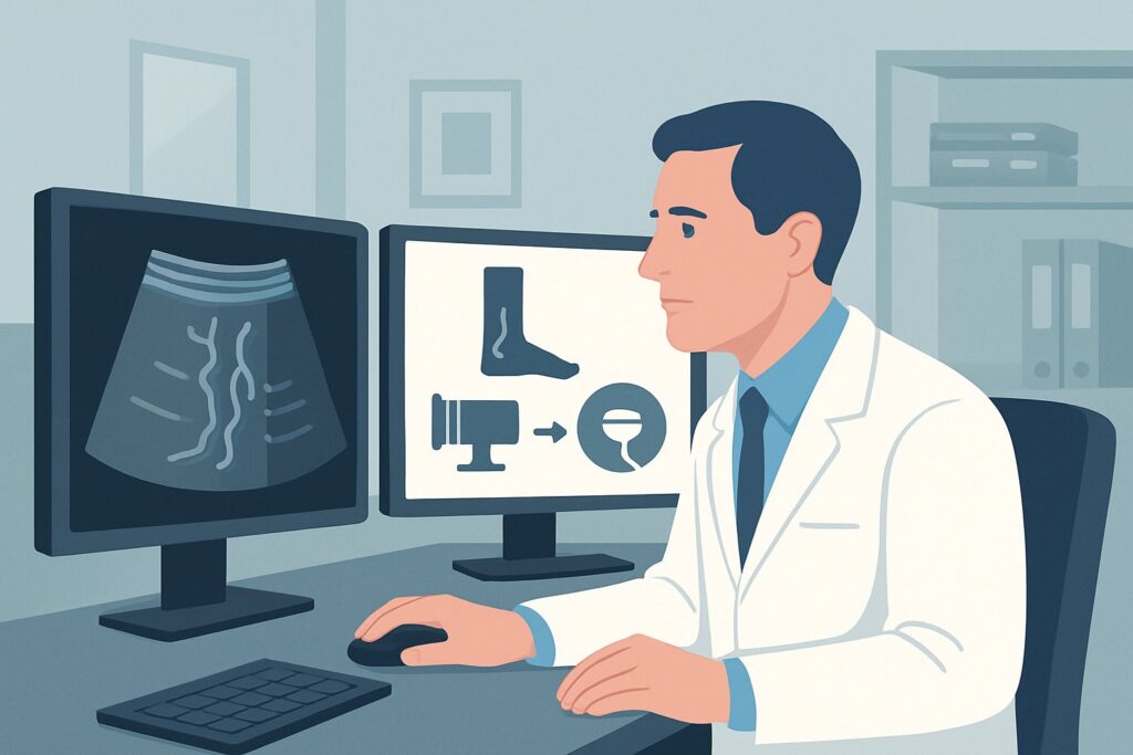
The “duplex” nature of the study is key. It combines two forms of ultrasound: B-mode imaging, which creates a grayscale anatomical map of the superficial and deep veins, and Doppler ultrasound, which assesses the direction and velocity of blood flow. This combination allows the sonographer to directly visualize venous reflux. During a venous insufficiency or reflux study, the patient is examined while standing to maximize gravitational stress on the venous valves. The sonographer then uses maneuvers like calf compression/release or Valsalva to challenge the valves and measure the duration of any reversed flow. A reflux time greater than 0.5 seconds in the superficial system is generally considered pathologic.
This detailed mapping of reflux sources and pathways is essential for planning subsequent minimally invasive treatments like endovenous thermal ablation or sclerotherapy. The study provides the proceduralist with a precise roadmap of which veins to treat.
Alternative imaging modalities are rated lower for this specific scenario for clear reasons:
- MR Venography (MRV) and CT Venography (CTV) are both rated Usually not appropriate. While these cross-sectional techniques provide excellent anatomical detail, they are static examinations and cannot perform the dynamic reflux assessment that is central to the diagnosis. Furthermore, CTV exposes the patient to significant ionizing radiation (adult RRL ☢☢☢☢ 10-30 mSv) and iodinated contrast, which is not justified for an initial workup of a benign condition. MRV avoids radiation but is more costly, less available, and still lacks the functional reflux data of ultrasound.
- Catheter Venography is also Usually not appropriate. This invasive procedure requires venous access and contrast injection, carrying risks of bleeding, infection, and contrast reaction. It has been almost entirely replaced by duplex ultrasound for the initial diagnosis of venous insufficiency.
When ordering this study, it is critical to specify “lower extremity venous insufficiency/reflux evaluation” rather than simply “lower extremity venous Doppler.” This ensures the technologist performs the correct protocol with the patient in the upright position and includes the necessary reflux-provoking maneuvers. Once you’ve decided on this study, our protocol guide covers the technique, contrast, and reading principles: US Lower Extremity Doppler (DVT).
What’s Next After US Duplex Doppler? Downstream Workflow
The results of the duplex ultrasound directly guide the next steps in patient management. The report should provide a clear map of venous incompetence, which forms the basis for a referral to a vascular surgeon, interventional radiologist, or other vein specialist.
- If the study is positive for significant superficial venous reflux (e.g., in the GSV or SSV), the patient is a candidate for treatment. The detailed ultrasound findings—including vein diameters, depth, and the location of the reflux origin—allow the specialist to determine the most appropriate therapeutic option, such as endovenous laser ablation (EVLA), radiofrequency ablation (RFA), mechanochemical ablation (MOCA), or ultrasound-guided sclerotherapy.
- If the study is negative for any venous reflux or obstruction, the patient’s symptoms are unlikely to be caused by chronic venous disease. The diagnostic focus should shift to other potential causes of leg pain and swelling, such as lymphedema, musculoskeletal conditions (e.g., arthritis, radiculopathy), peripheral artery disease, or peripheral neuropathy. A negative, high-quality venous reflux study effectively rules out a significant venous etiology.
- If the study is positive for deep venous reflux or suggests proximal iliac vein obstruction, the management becomes more complex. The patient should be referred to a specialist with expertise in deep venous disease. While superficial vein treatments may still be part of the plan, addressing the underlying deep system pathology is critical and may involve compression therapy, and in select cases, more advanced interventions.
Pitfalls to Avoid (and When to Get Help)
Navigating the initial workup for varicose veins is generally straightforward, but several common pitfalls can lead to diagnostic errors or suboptimal management. Be mindful of the following:
- Ordering the Wrong Test: Requesting a “DVT study” is the most common mistake. This protocol is designed to find acute thrombus and is typically performed with the patient supine, which will not elicit valvular reflux. Always specify a “venous insufficiency” or “reflux” study.
- Incomplete Examination: A proper study must evaluate the deep system, the superficial system (GSV and SSV from junction to ankle), and major perforator veins. An incomplete map can lead to treatment failure and recurrent varicosities.
- Ignoring Atypical Patterns: If varicosities appear in unusual locations (buttocks, perineum, posterior thigh) or if the duplex scan is negative despite high clinical suspicion, consider a pelvic origin. Escalate to a specialist or consider pelvic-focused imaging if symptoms like pelvic pain or dyspareunia are present.
- Attributing All Leg Symptoms to Veins: Do not fall into the trap of confirmation bias. If a patient has mild reflux but severe symptoms, continue to investigate other potential causes, particularly peripheral artery disease or neurologic conditions.
Related ACR Topics and Tools
This article focuses on a single, common clinical scenario. For a comprehensive overview of all variants within this topic, see our parent guide. For other tools to help with imaging decisions, use the resources below.
- Parent Topic Hub: For breadth across all scenarios in Lower Extremity Chronic Venous Disease, see our parent guide: Lower Extremity Chronic Venous Disease: ACR Appropriateness Decoded.
- ACR Appropriateness Criteria Lookup: For adjacent or different clinical presentations, consult the full criteria directly: ACR Appropriateness Criteria Lookup.
- Imaging Protocol Library: For detailed techniques on recommended studies: Imaging Protocol Library.
- Radiation Dose Calculator: For discussing cumulative dose with patients when considering studies that use radiation: Radiation Dose Calculator.
Frequently Asked Questions
What is the difference between a DVT ultrasound and a venous insufficiency ultrasound?
A DVT (deep vein thrombosis) study is an urgent exam performed with the patient lying down, focused on compressing the deep veins to rule out an acute blood clot. A venous insufficiency (or reflux) study is an elective, detailed mapping exam performed with the patient standing up. It uses specific maneuvers like calf squeezes to measure backward blood flow (reflux) in both the superficial and deep veins, which is the cause of varicose veins.
If my patient’s varicose veins are only in one leg, should I order a bilateral or unilateral ultrasound?
It is standard practice to order a bilateral lower extremity venous insufficiency study, even if symptoms are unilateral. This is because venous reflux is often a bilateral process, even if not yet symptomatic on the other side. A bilateral exam also provides a normal contralateral leg for comparison and ensures the saphenofemoral junction on the asymptomatic side is competent.
Is a duplex ultrasound sufficient for pre-procedural planning, or will the specialist need another study?
For the vast majority of patients undergoing standard treatments like endovenous ablation or sclerotherapy, a high-quality, comprehensive duplex ultrasound is the only imaging study required. It provides all the necessary anatomical and physiological information to plan the procedure. More advanced imaging like MRV or CTV is only reserved for complex cases, such as suspected deep vein obstruction or congenital malformations.
What if I suspect the varicose veins are coming from the pelvis?
If a female patient has varicose veins in an atypical distribution (e.g., vulva, perineum, upper medial or posterior thigh) or has symptoms of pelvic congestion syndrome (e.g., chronic pelvic pain, dyspareunia), you should suspect a pelvic origin. This is a distinct clinical scenario. The standard leg duplex may be negative or inconclusive. The workup should be tailored to evaluating the pelvic veins, which may involve a dedicated pelvic ultrasound or, more commonly, an MRV of the pelvis.
Does the patient need to stop any medications before a venous duplex scan?
No, patients generally do not need to stop any medications, including blood thinners, before a venous insufficiency ultrasound. The study is non-invasive and does not carry a risk of bleeding. Patients should be well-hydrated, as dehydration can make the veins harder to visualize.
Reviewed by Pouyan Golshani, MD, Interventional Radiologist — May 30, 2026