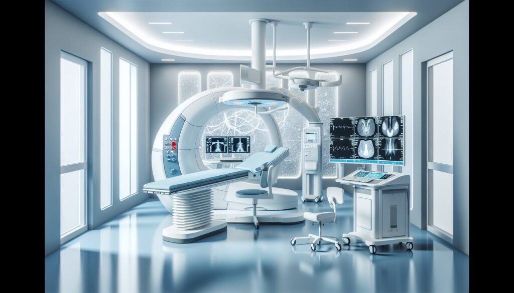
IR Management of Acute PE (CDT, Embolectomy) — Dictation, Appropriateness, and Dose for Residents
Stat call from the Pulmonary Critical Care fellow. You just read the CTA on a patient from the ED: saddle pulmonary embolism with a right ventricle to left ventricle (RV/LV) ratio of 1.2 and a bowed septum. Troponins are elevated. The patient is tachycardic and tachypneic but holding a blood pressure for now. The PE Response Team (PERT) is activated, and your attending, the on-call IR, is asking for your assessment and recommendation. This isn’t just about seeing the clot; it’s about stratifying risk and knowing the interventional options cold. When I was a fellow, this was the exact scenario where you either step up or get sidelined. Let’s make sure you’re ready to step up. For more high-yield guides like this, check out the residents free-reference hub.
What an Interventional Radiology Pulmonary Embolism Procedure Covers and What Attendings Look For
The IR management of acute Pulmonary Embolism (PE) is a high-stakes procedure focused on rapidly reducing right heart strain and restoring pulmonary perfusion in patients with massive or intermediate-high risk (submassive) PE. It’s an alternative or adjunct to systemic thrombolysis, especially when bleeding risk is high. Your attending expects a procedure note that is clear, concise, and justifies the intervention based on established risk criteria.
Your report must clearly document:
- Pre-procedure Assessment: Confirmation of PE severity (massive vs. submassive), including key metrics like RV/LV ratio, biomarkers (troponin, BNP), and hemodynamic status. Mention coordination with the PERT.
- Vascular Access: Site of access (typically right common femoral vein) and sheath size.
- Pulmonary Angiography: Initial findings confirming the location and extent of the thrombus burden.
- Intervention Details: The specific technique used—catheter-directed thrombolysis (CDT), ultrasound-assisted thrombolysis (USAT), or mechanical thrombectomy—and the device(s) employed (e.g., EKOS, FlowTriever, Penumbra). For lytics, document the drug, dose, and infusion rate.
- Post-intervention Angiography: A clear statement on the degree of improvement in pulmonary perfusion and resolution of thrombus.
- Hemostasis and Plan: Method of sheath removal/hemostasis and the post-procedure plan, including anticoagulation and ICU monitoring.
Radiology Report Template for IR Management of Acute Pulmonary Embolism
This template provides a solid foundation. Dictate the specifics of your case, focusing on the pre-procedure justification and post-procedure outcome.
Technique
Procedure: Pulmonary Angiography and Catheter-Directed Therapy for Acute Pulmonary Embolism.
Indication: [e.g., 55-year-old male with acute submassive pulmonary embolism, presenting with tachycardia, an RV/LV ratio of 1.3 on CTA, and elevated troponin].
Operators: [Attending Name, MD], [Fellow/Resident Name, MD].
Consent: Informed consent was obtained after explanation of the risks, benefits, and alternatives, including major bleeding, vessel injury, and death. The patient was able to participate and all questions were answered.
Access Site: [Right/Left] common femoral vein.
Catheters and Devices: [e.g., 8-French sheath, 5-French pigtail catheter, Inari FlowTriever 24-French aspiration catheter, EKOS acoustic lysis catheters].
Contrast: [e.g., 80 mL of Iohexol 350].
Fluoroscopy Time: [e.g., 12.5 minutes].
Procedure Summary: After sterile prep and local anesthesia, the [right] common femoral vein was accessed under ultrasound guidance. A pigtail catheter was advanced into the main pulmonary artery. Initial pulmonary angiography was performed. Based on the findings and the patient’s clinical status, a decision was made to proceed with [mechanical thrombectomy / catheter-directed thrombolysis].
[For Mechanical Thrombectomy: A large-bore aspiration catheter was advanced into the [right/left] pulmonary artery and multiple aspirations were performed, removing a significant volume of thrombus.]
[For Catheter-Directed Thrombolysis: A [e.g., 50 cm EKOS] catheter was advanced over a wire and positioned across the thrombus in the [right/left] pulmonary artery. A continuous infusion of alteplase at [e.g., 1 mg/hr] was initiated.]
Completion pulmonary angiography was performed, demonstrating the post-intervention result. The access sheath was [removed and hemostasis was achieved with a figure-of-eight suture / left in place for subsequent thrombolysis infusion]. The patient tolerated the procedure well and was transferred to the ICU in stable condition.
Findings
Pre-Intervention Pulmonary Angiography: Large filling defects consistent with extensive thrombus are identified in the [e.g., main, right, and left pulmonary arteries, with diminished flow to the right lower lobe]. Mean pulmonary artery pressure was measured at [e.g., 45 mmHg].
Post-Intervention Pulmonary Angiography: Following [mechanical thrombectomy / thrombolysis], there is significant improvement in perfusion to the pulmonary arteries. There is near-complete resolution of thrombus in the [e.g., main and left pulmonary arteries] with residual non-occlusive thrombus in the [e.g., right interlobar artery]. Mean pulmonary artery pressure decreased to [e.g., 28 mmHg].
Impression
Successful [mechanical thrombectomy / catheter-directed thrombolysis] for acute [massive/submassive] pulmonary embolism.
1. Pre-procedure angiography demonstrated extensive thrombus burden in the [location].
2. Post-procedure angiography shows significant improvement in pulmonary perfusion and reduction in thrombus burden.
3. Plan: Continue anticoagulation as per the PERT protocol. Monitor in the ICU for hemodynamic improvement and any signs of bleeding.
Free Radiology Template Sources
Building a personal macro library is a key part of residency. While you’re building it, two great free repositories exist that can provide excellent starting points. The Radiological Society of North America (RSNA) curates a broad library at RadReport.org, covering nearly every modality. Another excellent, independently maintained resource is Radiology Templates from Australia.
The Next-Level Move: AI-Powered Structuring
The challenge on call isn’t just knowing what to do, but documenting it quickly and accurately. This is where AI tools can streamline your workflow. Instead of clicking through a structured template or remembering every field, you can dictate your positive findings in free form—”Successful mechanical thrombectomy of a large saddle PE using a FlowTriever device, with post-procedure angio showing restored flow to both lungs”—and let the software handle the rest. GigHz Precision AI is designed to do exactly this. It takes your free-form dictation of key findings and organizes them into a clean, comprehensive report using pre-loaded templates from the American College of Radiology (ACR) and Society of Interventional Radiology (SIR). This approach helps ensure your reports are consistently complete and structured the way attendings expect, without slowing you down. It also supports the integration of radiology reporting and clinical decision support, automatically flagging when frameworks like LI-RADS or Bosniak should be applied.
When Should You Order IR Management for Acute Pulmonary Embolism? ACR Appropriateness Criteria
The decision to intervene on a PE is a critical one, guided by risk stratification. The American College of Radiology (ACR) provides evidence-based guidelines to help determine the right course of action.
For a patient with extensive acute bilateral central PEs and sustained hypotension (massive PE), catheter-directed therapy is considered Usually Appropriate. This is an emergent situation where rapid intervention is needed to prevent cardiovascular collapse.
In the more common scenario of an adult with acute bilateral PEs who is normotensive but shows signs of right heart strain—such as an RV/LV ratio greater than 0.9 on CTA, right heart strain on echo, and an elevated troponin (submassive or intermediate-high risk PE)—catheter-directed therapy is also Usually Appropriate. This intervention aims to prevent hemodynamic decompensation.
Conversely, for a patient with acute bilateral PEs but no signs of right heart strain (RV/LV < 0.9, normal echo, normal troponin, no hypotension), catheter-directed therapy is Usually Not Appropriate. This includes patients with a saddle PE who are otherwise stable. In these low-risk cases, anticoagulation alone is the standard of care.
A particularly challenging case is a patient with a massive or submassive PE who also has a contraindication to thrombolytics, such as an acute intracranial hemorrhage from a syncopal event. In this scenario, mechanical thrombectomy (which avoids lytics) is Usually Appropriate as a life-saving measure.
How Much Radiation Does an IR Pulmonary Embolism Procedure Deliver?
An interventional procedure for PE management involves fluoroscopy, which imparts an ionizing radiation dose. The estimated effective dose for this procedure is typically in the range of 5-15 mSv. This dose is highly variable depending on the complexity of the case, fluoroscopy time, and patient size.
To put this in perspective, this dose is comparable to several months to a few years of natural background radiation. While not insignificant, the radiation exposure is justified by the life-threatening nature of massive and submassive PE. The IR team always uses dose-reduction techniques, such as pulsed fluoroscopy, collimation, and minimizing magnification, to adhere to the ALARA (As Low As Reasonably Achievable) principle.
| Procedure / Exposure | Typical Effective Dose (mSv) |
|---|---|
| IR PE Intervention | 5 – 15 mSv |
| CT Pulmonary Angiogram | 3 – 10 mSv |
| Annual Natural Background Radiation (U.S.) | ~3 mSv |
IR Pulmonary Embolism Intervention Protocol — Phases, Contrast, and Key Steps
The protocol for IR management of PE is a multi-step process focused on safe access, accurate diagnosis via angiography, and effective treatment delivery. The core of the procedure involves navigating catheters to the pulmonary arteries to either aspirate clot or deliver thrombolytic medication directly.
The table below outlines the key phases of the procedure. Contrast is iodinated (Iohexol or Iopamidol 350), with volumes typically ranging from 60-100 mL for the entire case, used sparingly for angiographic runs.
| Phase / Step | Description | Key Considerations |
|---|---|---|
| Pre-procedure Imaging | Review diagnostic CTA and often a baseline echocardiogram. | Confirm RV/LV ratio, clot location, and RV strain. |
| Venous Access | Ultrasound-guided access to the common femoral vein (right preferred). | Upsize to a large-bore sheath (e.g., 16-24 French for mechanical thrombectomy). |
| Initial Pulmonary Angiography | A pigtail catheter is placed in the main pulmonary artery. Angiography confirms CTA findings and establishes a baseline. | Measure pulmonary artery pressures. |
| Intervention | Either mechanical thrombectomy (e.g., FlowTriever, Penumbra) or catheter-directed thrombolysis (e.g., EKOS) is performed. | Device choice depends on clot burden, patient stability, and bleeding risk. |
| Post-procedure Angiography | Repeat angiogram to assess for improvement in perfusion and reduction of thrombus. | Re-measure pulmonary artery pressures to document hemodynamic improvement. |
| Sheath Removal | Hemostasis is achieved, often with a figure-of-eight suture for large sheaths. | If continuing lytic infusion, sheath is secured and patient goes to ICU. |
Common protocol pitfalls: A key point of discussion is the thrombolytic dose for CDT; trials like OPTALYSE-PE suggest that lower total doses of alteplase (e.g., 8-24 mg) over shorter durations can be as effective with potentially lower bleeding risk. Also, remember that the single most important “protocol” step is multidisciplinary consultation. The involvement of a PE Response Team (PERT) is increasingly the standard of care and is proven to improve patient outcomes.
3+ months free for radiology residents and fellows
Look like a rockstar on your reports. With the GigHz Radiology Report Assistant, you can dictate your positive findings in free form, and our AI will generate a perfectly structured report using the latest ACR and SIR templates. The appropriate Clinical Decision Support (CDS) frameworks fire automatically, so you never miss a key classification. All we ask in return is your feedback so we can keep improving the product for trainees.
Signup is simple. No credit card, no long forms. To apply, just provide these three items:
- Your PGY year (e.g., PGY-2, PGY-4)
- Your training type (radiology residency or a specific fellowship like IR, Body, MSK, etc.)
- Your training program or hospital name
- (Optional) Your institutional email
Ready to give it a try? Send us your details and apply for the residents free-access program.
Free GigHz Tools That Pair With This Article
Three free tools that complement the material above:
- ACR Appropriateness Criteria Lookup — Type an indication or clinical scenario in plain language and get the imaging studies the ACR rates for it, with adult and pediatric radiation levels. Built directly from 297 ACR topics, 1,336 clinical variants, and 15,823 procedure ratings.
- GigHz Imaging Protocol Library — A searchable library of 131 imaging protocols with the physics specs surfaced and the matching ACR Appropriateness Criteria alongside. Plain-English narratives readable in 60 seconds, organized by modality.
- GigHz Radiation Dose Calculator — Pick the imaging studies a patient has had and see total dose in millisieverts (mSv) with comparisons to natural background radiation, transatlantic flights, and chest X-rays. Useful for shared decision-making.
Frequently Asked Questions
Is it HIPAA-compliant?
Yes. The platform is designed for de-identified workflows by default. You dictate only the clinical findings, not patient-identifying information. All data is handled within a secure, HIPAA-compliant environment.
Do I need IT to set it up?
No. GigHz Precision AI is browser-based and requires no local software installation or special permissions from your IT department. It works on any hospital computer, your personal laptop, or even the call-room iPad.
Does it work with PowerScribe or other dictation systems?
Yes. It works alongside your existing dictation system. You can dictate into our web interface, let the AI structure the report, and then copy/paste the final, clean text into PowerScribe, Fluency, or any other PACS or EMR field.
Can I use my own templates?
Yes. While the system comes pre-loaded with ACR and other society-endorsed templates, you can easily create, modify, and save your own custom templates to match your personal style or your institution’s specific requirements.
What happens after my residency or fellowship ends?
We offer continuity plans for graduating residents and fellows who want to continue using the platform in their practice. Your templates and account settings carry over seamlessly.
Reviewed by Pouyan Golshani, MD, Interventional Radiologist — May 7, 2026