Mammography – Implant Evaluation — Dictation, Appropriateness, and Dose for Residents
1. The Read You Can’t Rush: Mammography with Implants
The next case on your list is a screening mammogram for a 45-year-old with bilateral implants. The tech messages you: “Patient is asking if we really need the extra pictures, they’re uncomfortable.” You know the answer is an emphatic yes. Your attending expects a full read—not just of the implants, but of every millimeter of native breast tissue you can visualize. Getting the Eklund views right is non-negotiable, and your report needs to systematically address both the device and the patient.
When I was a resident, this was the kind of study that could slow the whole list down. It’s not just a screening; it’s a diagnostic evaluation of the implant layered on top of a screening of the native tissue. There’s no shortcut. For more high-yield guides and tools like this, check out the residents and fellows resource hub we’ve put together.
2. What a Mammogram for Implant Evaluation Covers and What Attendings Look For
A mammogram for a patient with breast implants is a dual-purpose exam. It evaluates the integrity of the implants themselves while also serving as a primary screening tool for breast cancer. The challenge is that implants, particularly older subglandular ones, can obscure a significant amount of native breast tissue on standard views. This is why the protocol is expanded beyond a typical screening mammogram.
Your attending will expect a comprehensive report that methodically addresses:
- Implant Assessment: Type (saline vs. silicone), position (subglandular vs. subpectoral), shape, and integrity.
- Capsular Contracture: Any signs of fibrous encapsulation causing distortion or asymmetry, often graded on the Baker scale.
- Peri-implant Changes: Look for fluid collections, masses, or other abnormalities immediately surrounding the implant.
- Native Breast Tissue Evaluation: This is where the Eklund (implant-displaced) views are critical. You must meticulously search for masses, calcifications, asymmetries, and architectural distortion in the tissue pulled forward, away from the implant.
- Axillary Evaluation: The axilla must always be checked for adenopathy. Remember, free silicone in axillary lymph nodes is a strong indicator of implant rupture.
- Comparison: Note any changes from prior studies.
- Final BI-RADS Assessment: A conclusive BI-RADS category and recommendation.
3. Radiology Report Template for Mammography – Implant Evaluation
Use this template as a starting point for your dictations. The key is to be systematic, addressing the implants first, then the native tissue, and concluding with a clear impression and recommendation.
Technique
Bilateral full-field digital mammography with computer-aided detection was performed, including standard craniocaudal (CC) and mediolateral oblique (MLO) views, as well as implant-displaced (Eklund) CC and MLO views. When available, comparison is made to the prior study from [DATE].
Findings
Implants: The patient has bilateral [saline/silicone] breast implants, which appear to be in a [subglandular/subpectoral] position. The implants are [intact/deflated/ruptured]. There is no mammographic evidence of extracapsular rupture. There is [no/mild/moderate/severe] capsular contracture. No suspicious peri-implant fluid collections or masses are identified.
Breast Parenchyma: The native breast tissue is [predominantly fatty/scattered fibroglandular/heterogeneously dense/extremely dense]. On the implant-displaced views, the visualized breast tissue is unremarkable. No suspicious masses, calcifications, or areas of architectural distortion are seen.
(If findings are present, describe them here using standard BI-RADS lexicon, e.g., “In the right breast at the 2 o’clock position, middle depth, there is a [shape] mass measuring [size] cm with [margin] margins. Associated features include…”)
Axilla: The axilla are unremarkable bilaterally. No abnormal axillary lymphadenopathy is seen.
Impression
- Bilateral [saline/silicone] breast implants, which appear intact mammographically.
- No mammographic evidence of malignancy in the visualized breast tissue.
BI-RADS Category 2: Benign
Recommendation: Routine screening mammography in one year.
4. Where to Find More Free Radiology Report Templates
Building a personal library of high-quality templates is a must during training. Beyond your own institution’s files, two great free repositories exist that are curated by radiologists for radiologists.
- RadReport.org: Maintained by the RSNA, this is a comprehensive library of peer-reviewed templates covering nearly every modality and subspecialty. (https://radreport.org/)
- Radiology Templates (AU): An excellent, straightforward collection of templates from Australian radiologists, often with practical, easy-to-use formatting. (https://www.radiologytemplates.com.au/home-page/)
Bookmark both. They are invaluable when you encounter a rare study or just want to see how others structure a complex report.
5. The Next-Level Move: From Free-Form Dictation to Flawless Report
The friction in reporting isn’t finding the words; it’s getting them into the right structure, with the right terminology, and without missing a key element the attending expects. This is especially true for complex reports like implant evaluations. Instead of manually clicking through a structured report or trying to remember every field in a template, you can dictate your positive findings in free form and let an AI tool handle the assembly.
Tools like GigHz Precision AI are designed for this workflow. You dictate the important stuff—”bilateral subpectoral silicone implants, intact, with a 7mm spiculated mass in the left breast upper outer quadrant on the Eklund views”—and the platform generates a complete, structured report using ACR-standard templates. It helps ensure you don’t miss documenting implant position or native tissue density, streamlining the process so you can focus on the pathology.
6. When Should You Order a Mammogram for Breast Implant Evaluation? ACR Appropriateness Criteria
The American College of Radiology (ACR) provides evidence-based guidelines on when imaging is appropriate. For breast implant evaluation, the decision often depends on the implant type (saline vs. silicone) and the clinical concern.
For patients with saline implants, mammography is rated as “Usually Appropriate” for both asymptomatic evaluation and for suspected rupture, regardless of patient age. While ultrasound can also be used, mammography provides a clear assessment of implant deflation.
For patients with silicone implants, the guidance is more nuanced. For asymptomatic screening, mammography is “Usually Appropriate,” but the primary modality for assessing implant integrity is MRI. For suspected implant complications in patients with silicone implants, mammography is also “Usually Appropriate” as an initial step, often to evaluate the surrounding breast tissue for other causes of symptoms. However, breast MRI is far more sensitive for detecting silicone implant rupture and remains the gold standard for that specific question.
In the specific scenario of unexplained axillary adenopathy in a patient with a current or prior history of silicone implants, mammography is “Usually Appropriate” as part of the initial workup to evaluate for both occult breast cancer and signs of implant rupture, such as free silicone in the lymph nodes.
7. How Much Radiation Does a Mammogram for Implant Evaluation Deliver?
A mammogram for implant evaluation delivers an estimated effective radiation dose of 0.5 to 1.5 mSv. This is slightly higher than a standard screening mammogram (which is typically around 0.4 mSv) because it requires double the number of images—the standard four views plus the four Eklund (implant-displaced) views.
To put this in perspective, the ACR categorizes this dose level as very low (☢☢). It is equivalent to several months of natural background radiation that we are all exposed to every year. The clinical benefit of thoroughly evaluating the native breast tissue for cancer is considered to far outweigh the minimal risk from this radiation dose.
| Imaging Study | Typical Effective Dose (mSv) | Comparison to Background Radiation |
|---|---|---|
| Standard Screening Mammogram (2 views) | ~0.4 mSv | ~7 weeks |
| Mammogram with Implant Evaluation (8 views) | 0.5 – 1.5 mSv | ~2-6 months |
| Chest X-ray (PA/LAT) | ~0.1 mSv | ~10 days |
| CT Chest | ~7 mSv | ~2 years |
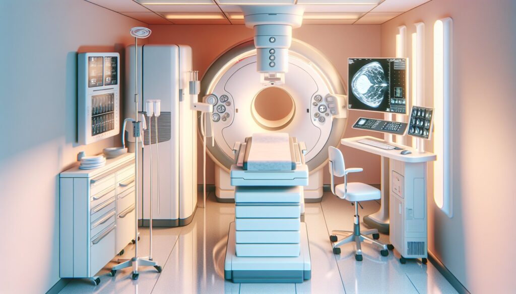
8. Mammography for Implant Evaluation Imaging Protocol — Views and Technique
The cornerstone of a successful mammographic implant evaluation is a protocol that includes both standard and specialized views. The goal is to image the implant and its immediate surroundings, as well as to visualize the maximum amount of native breast tissue possible.
The protocol consists of a total of eight views. The standard views provide an overall assessment of the breast and implant, while the Eklund views are a specialized technique critical for cancer detection. During the Eklund maneuver, the technologist carefully pushes the implant posteriorly against the chest wall while simultaneously pulling the native breast tissue forward into the compression paddle.
| View | Purpose | Key Technical Detail |
|---|---|---|
| Standard CC (Bilateral) | Overall view of implant and breast tissue. | Implant is included in the field of view. |
| Standard MLO (Bilateral) | Includes axillary tail and pectoral muscle. | Implant is included in the field of view. |
| Eklund CC (Bilateral) | Visualize anterior breast tissue without implant obscuration. | Implant is displaced posteriorly. |
| Eklund MLO (Bilateral) | Visualize anterior breast tissue without implant obscuration. | Implant is displaced posteriorly. |
A common pitfall is inadequate displacement on the Eklund views, which can leave significant amounts of breast tissue obscured. This compromises the diagnostic quality of the exam and is why communication between the radiologist and the mammography technologist is key.
9. The 3-Months-Free Offer for Radiology Residents and Fellows
Look like a rockstar on your reports. We’re offering 3+ months of free access to GigHz Precision AI for all radiology residents and fellows. You can dictate your positive findings in free form, and the AI will generate a complete, structured report using the latest ACR and SIR templates. It helps you work faster and ensures your reports are consistently comprehensive, with the appropriate Clinical Decision Support (CDS) firing automatically.
All we ask in return is your feedback so we can keep improving the product for trainees.
To apply, just send us the following three items:
- Your PGY year (e.g., PGY-2, PGY-4)
- Your training type (radiology residency or fellowship specialty)
- Your training program / hospital name
It’s that simple. No credit card, no long forms. To get started, apply for the residents free-access program.
10. Frequently Asked Questions
Is GigHz Precision AI HIPAA-compliant?
Yes. The platform is designed for de-identified workflows by default. No Protected Health Information (PHI) is required to generate a report, and we recommend users follow institutional best practices for de-identification before using any third-party tool.
Do I need my hospital’s IT department to set this up?
No. GigHz Precision AI is browser-based and requires no local installation or IT involvement. It works on any modern web browser, including the one on your call-room computer or personal iPad.
Does this replace PowerScribe or other dictation systems?
No, it works alongside them. You can dictate into the GigHz web interface, and then copy-paste the final, structured report into your PACS/RIS/PowerScribe system. It’s a workflow enhancement, not a replacement.
Can I use this on my phone or iPad?
Yes, the platform is fully responsive and works well on mobile devices and tablets, making it useful for reviewing or building reports away from a dedicated workstation.
Can I customize the templates?
Yes. While the system comes pre-loaded with ACR and other society-standard templates, you can create, save, and modify your own templates to match your personal or institutional preferences.
What happens after my residency or fellowship ends?
After your free access period as a trainee, you can choose to subscribe to a paid plan for practicing radiologists. There is no obligation, and your free access will simply expire if you choose not to continue.
Free GigHz Tools That Pair With This Article
Three free tools that complement the material above:
- ACR Appropriateness Criteria Lookup — Type an indication or clinical scenario in plain language and get the imaging studies the ACR rates for it, with adult and pediatric radiation levels. Built directly from 297 ACR topics, 1,336 clinical variants, and 15,823 procedure ratings.
- GigHz Imaging Protocol Library — A searchable library of 131 imaging protocols with the physics specs surfaced and the matching ACR Appropriateness Criteria alongside. Plain-English narratives readable in 60 seconds, organized by modality.
- GigHz Radiation Dose Calculator — Pick the imaging studies a patient has had and see total dose in millisieverts (mSv) with comparisons to natural background radiation, transatlantic flights, and chest X-rays. Useful for shared decision-making.
Reviewed by Pouyan Golshani, MD, Interventional Radiologist — May 7, 2026