Managing Mesenteric Ischemia in Low Cardiac Output: An ACR-Guided Workflow
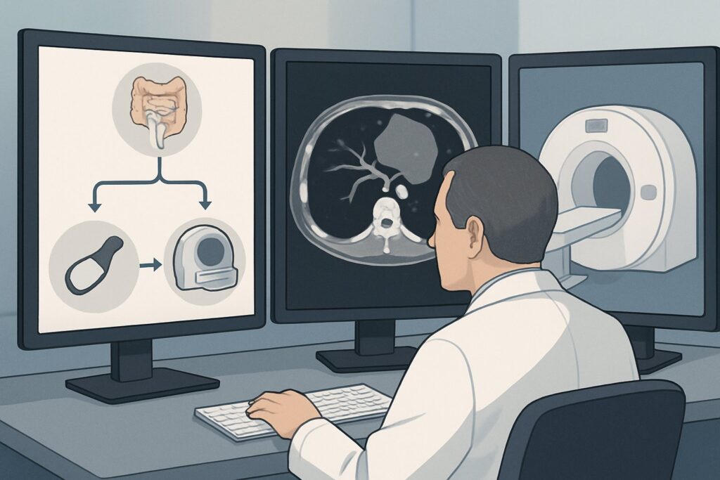
It’s 2 AM in the intensive care unit, and your patient with severe heart failure is developing new, diffuse abdominal pain. Their exam is deceptively benign, with no guarding or rebound tenderness, but their lactate is climbing. A stat Computed Tomography Angiography (CTA) of the abdomen and pelvis is performed, but the report is puzzling: the major mesenteric arteries—the celiac, Superior Mesenteric Artery (SMA), and Inferior Mesenteric Artery (IMA)—are all patent at their origins. However, the distal SMA branches show diffuse, irregular narrowing. This isn’t a typical clot or plaque. You are now faced with a critical decision for managing suspected Non-Occlusive Mesenteric Ischemia (NOMI). This article details the specific clinical workflow for this scenario, where the American College of Radiology (ACR) finds ‘Angiography with infusion of vasodilator’ to be Usually appropriate.
Who Fits This Clinical Scenario?
This workflow is specifically for patients presenting with signs of acute mesenteric ischemia in the setting of a low-flow state, where CTA has ruled out a major vessel occlusion. The key inclusion criteria are:
- Underlying Low-Flow State: The patient typically has a known condition causing systemic hypoperfusion, such as cardiogenic shock, septic shock, severe heart failure, or has recently undergone major cardiac surgery or is on potent vasopressors.
- Clinical Presentation: The hallmark is abdominal pain that seems “out of proportion” to the physical exam findings. Peritoneal signs like rigidity and rebound tenderness are notably absent, as their presence would suggest bowel infarction and necessitate a different, more aggressive surgical pathway.
- Specific CTA Findings: Imaging confirms patent origins and proximal segments of the celiac, SMA, and IMA. The crucial finding is diffuse, segmental, or irregular narrowing and spasm of the distal mesenteric branches, particularly within the SMA territory.
This scenario must be distinguished from other causes of acute mesenteric ischemia. This guidance does not apply if the CTA shows a clear filling defect in the proximal SMA (suggesting an embolic source, often from atrial fibrillation), high-grade stenosis from calcified plaque (suggesting acute-on-chronic thrombosis), or occlusion of the superior mesenteric vein.
What Diagnoses Are You Working Up in This Scenario?
In this clinical context, the differential diagnosis is narrow and centered on the consequences of systemic hypoperfusion on the gut.
Non-Occlusive Mesenteric Ischemia (NOMI) is the primary diagnosis. It accounts for a significant portion of all acute mesenteric ischemia cases and carries a high mortality rate. NOMI is a physiological response to a low cardiac output state. The body shunts blood away from the splanchnic circulation to preserve perfusion to the brain and heart. This protective mechanism leads to intense vasoconstriction of the small mesenteric arteries, causing ischemia without a physical blockage. The CTA findings of patent main arteries with distal spasm are classic for this entity.
Severe Ischemic Colitis is another consideration. While often localized to watershed areas like the splenic flexure and rectosigmoid junction (IMA territory), severe systemic hypoperfusion can cause more extensive colonic ischemia that can mimic the presentation of small bowel ischemia. The clinical management principles are similar, focusing on restoring perfusion, but the anatomic focus may differ.
Small-Vessel Vasculitis is a much less common cause but could theoretically produce diffuse narrowing of mesenteric branches. However, the acute presentation in a patient with a clear cause for a low-flow state makes NOMI vastly more probable. Vasculitis would typically present in a different clinical context, often with a more subacute course and multisystem involvement.
Why Is Angiography with Vasodilator Infusion the Recommended Therapy?
The therapeutic goal in NOMI is to reverse the intense splanchnic vasoconstriction and restore blood flow before irreversible bowel infarction occurs. Because the problem is functional vasospasm rather than a fixed mechanical obstruction, the management strategy is fundamentally different from other forms of mesenteric ischemia.
According to the ACR Appropriateness Criteria, Angiography with infusion of vasodilator is rated Usually appropriate. This procedure is both diagnostic and therapeutic. A catheter is advanced into the SMA, and a diagnostic angiogram confirms the diffuse narrowing and “string of sausages” appearance of the distal branches characteristic of NOMI. Subsequently, a vasodilator, most commonly papaverine, is infused directly through the catheter into the SMA. This targeted delivery maximizes the therapeutic effect on the splanchnic bed while minimizing systemic side effects like hypotension—a critical consideration in an already hemodynamically fragile patient.
In contrast, other interventions are less suitable for this specific scenario:
- Angiography with percutaneous transluminal angioplasty is rated Usually not appropriate. There is no focal stenosis to dilate with a balloon. Attempting angioplasty on diffusely spastic vessels is ineffective and risks vessel injury or dissection.
- Systemic infusion of prostaglandin E1 is rated May be appropriate. While it can induce vasodilation, its systemic administration can worsen hypotension, further compromising the patient’s overall perfusion. Catheter-directed therapy is more precise and hemodynamically stable.
Alongside interventional management, Systemic anticoagulation is also rated Usually appropriate. It is often used as an adjunct to prevent the formation of microthrombi in the low-flow, ischemic bowel, but it does not address the underlying vasospasm. The two therapies are complementary, not mutually exclusive.
What’s Next After Angiography? Downstream Workflow
The patient’s course after angiography and vasodilator infusion dictates the subsequent steps. Close clinical monitoring in an ICU setting is essential.
If the Study is Positive and Effective: A successful intervention is marked by both angiographic and clinical improvement. Post-infusion angiographic runs should show improved caliber of the mesenteric branches. Clinically, the patient’s abdominal pain should resolve, and metabolic markers like serum lactate should begin to normalize. The vasodilator infusion is typically continued for 24-48 hours, and a follow-up angiogram may be performed before catheter removal. The primary focus then returns to managing the underlying cause of the low-flow state.
If the Study is Ineffective or Symptoms Worsen: If the vasospasm does not respond to vasodilator infusion, or if the patient develops peritoneal signs (e.g., rigidity, rebound tenderness) during or after the procedure, it signifies progression to transmural bowel infarction. This is a surgical emergency. The interventional procedure should be terminated, and the patient requires an immediate consultation with general surgery for an exploratory laparotomy and probable bowel resection.
If the Response is Indeterminate: In some cases, there may be a partial angiographic response but persistent, albeit improved, symptoms. This situation requires a multidisciplinary discussion between the interventional radiologist, intensivist, and surgeon. Management may involve continuing the infusion, optimizing medical therapy, and performing serial abdominal exams to watch for any sign of deterioration.
Pitfalls to Avoid (and When to Get Help)
Navigating NOMI requires vigilance to avoid common and often fatal pitfalls. The key is to maintain a high index of suspicion in any patient with a low-flow state who develops abdominal pain.
- Waiting for Peritoneal Signs: The absence of peritoneal signs is a defining feature of early, reversible NOMI. Waiting for them to appear means you are likely too late, as their presence indicates transmural infarction has already occurred.
- Attributing Pain to Ileus: In critically ill patients, vague abdominal pain is often dismissed as being from a functional ileus. However, in the setting of shock, NOMI must be actively ruled out.
- Inadequate Resuscitation: Catheter-directed vasodilation is a temporizing measure that treats the regional consequence of a systemic problem. Aggressive management of the underlying cardiac dysfunction or sepsis is paramount for patient survival.
If at any point the patient develops clear peritoneal signs or shows evidence of perforation on imaging, escalate immediately to a general surgery service for emergent operative exploration.
Related ACR Topics and Tools
This article covers one specific, high-stakes scenario in mesenteric ischemia. The clinical presentations and appropriate management strategies vary widely based on the underlying cause and patient presentation. For a comprehensive overview of all related scenarios, from embolic occlusion to chronic ischemia, please consult the resources below.
- For breadth across all scenarios in Radiologic Management of Mesenteric Ischemia, see our parent guide: Radiologic Management of Mesenteric Ischemia: ACR Appropriateness Decoded.
- To look up other clinical scenarios, use the Imaging Appropriateness Selector.
- For details on imaging techniques, browse the Imaging Protocol Library.
- To discuss radiation exposure with patients, reference the Radiation Dose Calculator.
Frequently Asked Questions
Why not just use systemic IV vasodilators instead of an invasive angiogram?
Systemic vasodilators can cause profound hypotension, which is extremely dangerous in a patient who is already in a low-flow state from cardiac disease or shock. Catheter-directed infusion delivers the medication directly to the affected mesenteric arteries, maximizing the local therapeutic effect while minimizing systemic side effects.
What is the role of systemic anticoagulation in this specific scenario?
Systemic anticoagulation is also rated ‘Usually appropriate’ by the ACR for this scenario. It is used as an adjunctive therapy. The primary problem is vasospasm, but the resulting low-flow state within the bowel wall can lead to the formation of microthrombi. Anticoagulation helps prevent this secondary thrombosis but does not treat the underlying vasospasm. Therefore, it is often used in conjunction with vasodilator infusion.
How does this presentation differ from mesenteric ischemia caused by an embolism from atrial fibrillation?
The underlying cause and imaging findings are completely different. Embolic ischemia is caused by a clot (usually from the heart in atrial fibrillation) that travels and lodges in a major vessel, typically the proximal SMA, causing a complete blockage. CTA shows a clear filling defect. In contrast, Non-Occlusive Mesenteric Ischemia (NOMI) is caused by diffuse vasospasm from a systemic low-flow state, and CTA shows patent major arteries with narrowing of only the smaller, distal branches. The treatments are distinct: embolectomy for an embolus versus vasodilator infusion for NOMI.
What if the initial CTA is read as normal but my clinical suspicion for NOMI remains very high?
CTA can be falsely negative in the very early stages of NOMI before significant vasospasm is established. Conventional catheter-based angiography is the gold standard for diagnosis. If clinical suspicion is high (e.g., severe pain out of proportion to the exam, rising lactate, profound shock), proceeding directly to angiography is warranted. It can serve as both the definitive diagnostic test and the first step of therapy.
Is surgery ever the first step for this type of mesenteric ischemia?
Surgery is not the initial therapy if there are no peritoneal signs. The goal of early intervention with vasodilator infusion is to reverse the ischemia and avoid surgery. However, if the patient presents initially with peritoneal signs (suggesting bowel infarction has already occurred) or develops them during treatment, then emergent surgery for exploratory laparotomy and bowel resection becomes the primary and necessary intervention.
Reviewed by Pouyan Golshani, MD, Interventional Radiologist — May 26, 2026