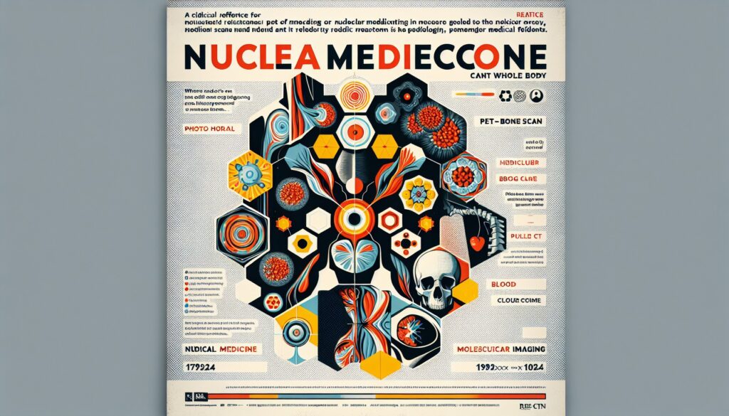
Nuclear Medicine Bone Scan (Whole Body) — Dictation, Appropriateness, and Dose for Residents
Outpatient bone scan on a prostate cancer patient with a rising PSA. The list is long, and your attending expects you to not just spot the lesions but characterize them—axial vs. appendicular, linear vs. focal, and call out any red flags like a superscan or photopenia. It’s a classic nuclear medicine study, but the nuances separate a good read from a great one. When I was a resident, I’d keep a cheat sheet of bone scan pearls just for these cases. The goal is to be efficient without missing the subtle findings that change management. This guide is built from that same playbook—giving you the structure and clinical context to dictate with confidence. For more tools like this, check out the residents and fellows resource hub we’ve put together.
What a Nuclear Medicine Bone Scan (Whole Body) Covers and What Attendings Look For
A whole-body bone scan is a functional study, not an anatomic one. It uses a radiopharmaceutical (most commonly Technetium-99m methylene diphosphonate, or Tc-99m MDP) that localizes to areas of active bone turnover, specifically osteoblastic activity. This makes it incredibly sensitive for detecting skeletal abnormalities long before they’re visible on plain films.
This study is a workhorse for answering key clinical questions:
- Are there osteoblastic metastases anywhere in the skeleton?
- Is a focal hot spot on another study a fracture or metastasis?
- Is there an occult fracture (stress, insufficiency, sacral)?
- Is there evidence of metabolic bone disease like Paget’s disease?
- Does the pattern support a diagnosis of complex regional pain syndrome (CRPS)?
Your attending is looking for a systematic interpretation. They expect you to comment on the distribution and intensity of uptake, correlate with any available prior imaging, and synthesize the findings into a clear, clinically relevant impression. Don’t just list hot spots; characterize them and provide a differential.
Radiology Report Template for Nuclear Medicine Bone Scan (Whole Body)
This template provides a solid foundation for most whole-body bone scan reports. Adapt the findings and impression based on the specific clinical scenario and imaging results.
Technique
Radiopharmaceutical: [25.0] mCi of Technetium-99m MDP administered intravenously.
Imaging Time: Whole-body planar images were obtained in the anterior and posterior projections approximately [3] hours after radiopharmaceutical administration.
Additional Imaging: [No additional spot views or SPECT/CT imaging was performed. OR: Additional spot views of the [location] were obtained. OR: SPECT/CT of the [pelvis/thoracic spine/etc.] was performed for further characterization of an equivocal finding.]
Findings
Distribution: The radiopharmaceutical is distributed physiologically throughout the skeleton. The soft tissues are clear. Both kidneys are visualized and appear symmetric in uptake and excretion. The bladder is well-visualized.
Skeleton:
There is symmetric and homogeneous uptake throughout the axial and appendicular skeleton, with no focal areas of abnormally increased or decreased radiotracer uptake to suggest metastatic disease.
(or, for positive findings)
There are multiple focal areas of intensely increased radiotracer uptake in a distribution classic for metastatic disease, including in the following locations:
- Skull: [Specify location]
- Spine: [Specify levels, e.g., T8 vertebral body, L4 pedicle]
- Ribs: [Specify right/left and number, e.g., right 5th anterior rib, left 7th posterior rib. Note if linear (fracture) vs. focal (metastasis)]
- Pelvis: [Specify location, e.g., right iliac wing, left ischium]
- Extremities: [Specify location, e.g., proximal right humerus, left femoral neck]
Degenerative Changes: Mild degenerative uptake is noted in the [glenohumeral joints, acromioclavicular joints, spine, hips, knees, feet].
Other Findings: [Note any incidental findings, such as renal asymmetry, bladder abnormalities, or soft tissue uptake.]
Impression
1. NO EVIDENCE OF SKELETAL METASTASIS. Mild degenerative changes as described above.
(or, for positive findings)
1. MULTIPLE FOCI OF ABNORMAL RADIOTRACER UPTAKE throughout the axial and appendicular skeleton, as described above, highly suspicious for widespread osseous metastatic disease.
2. MILD DEGENERATIVE CHANGES.
Free Template Sources
Building your own macro library is a rite of passage, but you don’t have to start from scratch. Two great free repositories exist that are worth bookmarking. The Radiological Society of North America (RSNA) curates a comprehensive library at RadReport.org, covering nearly every modality. For a slightly different flavor, an excellent Australian-maintained library is available at RadiologyTemplates.com.au.
The Next-Level Move: AI-Assisted Structured Reporting
A static template is a good start, but the real friction on call comes from manually slotting your findings into the right places and remembering the specific classification systems your attendings prefer. This is where AI-driven tools can streamline your workflow. Instead of clicking through a structured template, you can dictate your positive findings in free form—”multiple hot spots in the T-spine, ribs, and pelvis consistent with mets”—and the software does the rest. The GigHz Precision AI tool is designed to parse that free-form dictation, generate a clean report using pre-loaded ACR and SIR templates, and surface the appropriate Clinical Decision Support (CDS) frameworks automatically. It helps you create a high-quality, structured report without the tedious manual formatting.
When Should You Order a Nuclear Medicine Bone Scan (Whole Body)? ACR Appropriateness Criteria
The American College of Radiology (ACR) provides evidence-based guidelines on when a bone scan is the right test. For a resident, knowing these helps you recommend the right study and defend your choice on rounds.
In prostate cancer, a whole-body bone scan is Usually Appropriate for staging clinically established high-risk disease and for follow-up in patients with metastatic disease being treated with systemic therapy. It’s worth noting that PSMA PET/CT is increasingly preferred for its higher sensitivity where available.
For invasive breast cancer, a bone scan is Usually Appropriate for evaluating distant disease in patients presenting with late-stage (clinical stage IIB-III) cancer. For early-stage (I-IIA) disease or routine surveillance without symptoms, it is generally not indicated.
In the musculoskeletal setting, a bone scan is often the go-to for occult fractures. For an adult with a suspected stress or insufficiency fracture (including sacral fractures) after negative or equivocal x-rays, a bone scan with SPECT or SPECT/CT is Usually Appropriate. It’s also Usually Appropriate when a pathologic rib fracture is suspected in the context of malignancy.
When MRI is contraindicated, a 3-phase bone scan can be a key part of the workup for suspected osteomyelitis, often paired with a tagged white blood cell scan for improved specificity.
Key alternatives to consider include:
- Whole-body MRI: Superior for lytic-only metastatic disease, such as multiple myeloma, where bone scans are often falsely negative.
- FDG PET/CT: Often used for staging many cancers, as it assesses both skeletal and visceral disease simultaneously.
- PSMA PET/CT: The emerging standard for staging and restaging prostate cancer due to its high sensitivity and specificity.
- Skeletal Survey (X-rays): Still the primary modality for the initial workup of multiple myeloma.
How Much Radiation Does a Nuclear Medicine Bone Scan (Whole Body) Deliver?
Patients often ask about radiation, and having a clear, concise answer is crucial. A typical whole-body bone scan delivers an estimated effective dose of 4-6 mSv from a standard 25 mCi injection of Tc-99m MDP. This places it in a low-to-moderate exposure tier, comparable to a few years of natural background radiation. The critical organ receiving the highest dose is the bladder wall, which is why patient instructions are so important.
You can explain to patients that the single most important thing they can do to reduce their dose is to hydrate well and void frequently for 24 hours after the injection. This helps flush the unbound radiopharmaceutical from their system, significantly lowering the dose to the bladder.
| Imaging Study | Typical Effective Dose (mSv) | Equivalent Background Radiation |
|---|---|---|
| Chest X-ray (2 views) | 0.1 mSv | ~10 days |
| NM Bone Scan (Whole Body) | 4-6 mSv | ~1.5 – 2 years |
| CT Abdomen/Pelvis | 10 mSv | ~3 years |
| FDG PET/CT | 14-25 mSv | ~5-8 years |
Nuclear Medicine Bone Scan (Whole Body) Imaging Protocol — Phases, Contrast, and Technical Details
The standard protocol involves a single delayed imaging phase, but it can be expanded to a 3-phase study for specific indications like osteomyelitis. The “contrast” is the radiopharmaceutical itself, Tc-99m MDP or HDP, which are functionally equivalent. After a 2-3 hour uptake period, whole-body planar sweeps are acquired, supplemented by high-count spot views or SPECT/CT for any equivocal findings.
Below are the key technical parameters for a standard whole-body bone scan.
| Parameter | Specification |
|---|---|
| Radiopharmaceutical | 20-30 mCi Tc-99m MDP or HDP (adult dose) |
| Uptake Time | 2-3 hours post-injection |
| Camera | Dual-head gamma camera |
| Collimator | Low-energy high-resolution (LEHR) parallel-hole |
| Energy Peak & Window | 140 keV with a 20% window (126-154 keV) |
| Whole-Body Scan Speed | 10-15 cm/min |
| Spot View Target | ~500,000 counts per view |
| Additional Imaging | SPECT or SPECT/CT for equivocal findings (especially spine/pelvis) |
Common protocol pitfalls:
A common challenge is differentiating degenerative changes from metastases, especially a solitary spinal lesion. This is where SPECT/CT is invaluable; it fuses the functional uptake data with anatomic localization from a low-dose CT, dramatically reducing equivocal interpretations. Don’t hesitate to recommend it. Another pearl is to remember that purely lytic lesions (common in myeloma, renal cell, and thyroid cancers) may be “photopenic” (cold) or normal on a bone scan. If the clinical suspicion is high for these cancers, a bone scan may not be the right test.
3+ months free for radiology residents and fellows
Look like a rockstar on your reports. With the GigHz Precision AI reporting assistant, you can dictate positive findings in free form, and the AI generates a structured report using ACR and SIR templates, firing the appropriate clinical decision support automatically. We’re offering extended free access for trainees to help you master structured reporting before you graduate.
All we ask is feedback so we can keep improving the product for trainees. To apply, just send us these three items:
- Your PGY year (e.g., PGY-2, PGY-4)
- Your training type (radiology residency or fellowship specialty)
- Your training program / hospital name
There’s no credit card required and no long forms. It’s a simple process designed for busy residents. To get started, apply for the residents free-access program.
Free GigHz Tools That Pair With This Article
Three free tools that complement the material above:
- ACR Appropriateness Criteria Lookup — Type an indication or clinical scenario in plain language and get the imaging studies the ACR rates for it, with adult and pediatric radiation levels. Built directly from 297 ACR topics, 1,336 clinical variants, and 15,823 procedure ratings.
- GigHz Imaging Protocol Library — A searchable library of 131 imaging protocols with the physics specs surfaced and the matching ACR Appropriateness Criteria alongside. Plain-English narratives readable in 60 seconds, organized by modality.
- GigHz Radiation Dose Calculator — Pick the imaging studies a patient has had and see total dose in millisieverts (mSv) with comparisons to natural background radiation, transatlantic flights, and chest X-rays. Useful for shared decision-making.
Frequently Asked Questions
Is it HIPAA-compliant?
Yes. The platform is designed for de-identified workflows by default. No patient-identifying information is required to use the reporting assistant, ensuring compliance with institutional and federal privacy standards.
Do I need my hospital’s IT department to set it up?
No. GigHz Precision AI is browser-based and requires no local software installation or IT involvement. It works on any modern web browser, including on the call-room computer or your personal iPad.
Does this replace PowerScribe or other dictation systems?
No, it works alongside them. You can dictate your findings into the GigHz interface, let the AI structure the report, and then copy-paste the final, clean text into your institutional PACS/RIS or dictation system like PowerScribe.
Can I use this on my phone or iPad?
Yes, the tool is fully responsive and designed to work on desktops, tablets, and mobile devices, making it accessible whether you’re at a workstation or on the go.
Can I customize the templates?
Yes, you can create and save your own custom templates and macros within the platform. This allows you to tailor the output to match your personal preferences or the specific style of your attendings.
What happens after I finish my residency or fellowship?
The free access program is specifically for trainees. After you graduate, you would transition to a standard subscription if you choose to continue using the service in your practice. We offer straightforward plans for practicing radiologists.
Reviewed by Pouyan Golshani, MD, Interventional Radiologist — May 7, 2026