Pediatric US Appendicitis — Dictation, Appropriateness, and Dose for Residents
1. The Call Every Resident Knows
It’s a busy shift. The ED calls with a 9-year-old who has right lower quadrant pain, a fever, and a mildly elevated white count. The surgeon is already asking for your read. They want to know: is it appendicitis? Is it perforated? Or is it one of the classic mimics? This is the bread-and-butter pediatric call case, and your attending expects a clean, decisive report that directly answers the clinical question. Getting the key measurements and descriptors right—compressibility, diameter, presence of an appendicolith, secondary signs—is non-negotiable.
When I was a resident, the pressure wasn’t just finding the appendix (which can be an adventure on its own), but also dictating a report that covered every base without sounding like a rambling checklist. You need a solid framework. For more high-yield guides and tools, we’ve put together the residents and fellows resource hub with free calculators and references to help you on call.
2. What a Pediatric Appendicitis Ultrasound Covers and What Attendings Look For
A graded compression ultrasound for suspected pediatric appendicitis is the first-line imaging test for a reason: it’s accurate and uses no ionizing radiation. The goal is to directly visualize the appendix and evaluate for signs of inflammation, while also surveying the surrounding anatomy for alternative causes of pain.
Your attending (and the surgical team) will expect your report to clearly address:
- Appendix Visualization: Was the appendix seen in its entirety? If not, the study is non-diagnostic, not negative.
- Key Diagnostic Criteria: Is it a blind-ending, non-peristaltic tubular structure? Is it compressible? What is the maximal outer diameter (MOD)? A non-compressible appendix ≥6 mm is the classic finding.
- Signs of Inflammation: Note any wall thickening, hyperemia on color Doppler, or the presence of an appendicolith (a calcified deposit in the lumen).
- Evidence of Perforation: Look for peri-appendiceal complex fluid, phlegmon, a defined abscess, or loss of the normal wall layers.
- Alternative Diagnoses: Did you rule out the common mimics? This includes mesenteric adenitis, terminal ileitis, intussusception, and (in girls) ovarian pathology like torsion.
3. Radiology Report Template for Ultrasound Pediatric Appendicitis (Graded Compression)
Use this template as a starting point for your macros. It’s designed to be comprehensive, ensuring you hit all the key points your attending and the clinical team need.
Technique
Grayscale and color Doppler ultrasound images of the right lower quadrant were obtained using a high-frequency linear transducer with graded compression technique. The visualized portions of the abdomen and pelvis were also surveyed.
Findings
APPENDIX: [Not visualized, limiting the exam’s diagnostic value. / Visualized and appears normal. / Visualized and appears abnormal.]
If visualized:
The appendix arises from the cecal base. It is a [blind-ending, peristalsing/non-peristalsing] tubular structure.
Compressibility: [Compressible / Non-compressible / Incompletely compressible].
Maximum Outer Diameter: ___ mm (normal <6 mm).
Appendicolith: [Present, measuring ___ cm, with posterior acoustic shadowing / Not present].
Wall: [Normal thickness / Thickened and edematous]. Hyperemia [is/is not] demonstrated on color Doppler imaging.
Peri-appendiceal Changes: [No] surrounding inflammatory fat stranding. [No] peri-appendiceal fluid collection or abscess.
RIGHT LOWER QUADRANT:
Bowel: The terminal ileum and cecum appear [unremarkable/thickened]. No evidence of intussusception.
Lymph Nodes: [No] enlarged mesenteric lymph nodes.
Free Fluid: [No] free fluid in the right lower quadrant or pelvis.
PELVIC ORGANS (if female):
Right Ovary: Measures ___ x ___ x ___ cm. Appears normal in size and echotexture. Normal arterial and venous Doppler flow is present.
Left Ovary: Measures ___ x ___ x ___ cm. Appears normal in size and echotexture. Normal arterial and venous Doppler flow is present.
Uterus: Unremarkable.
Impression
Example 1: Positive for Appendicitis
1. Findings consistent with acute, non-perforated appendicitis, including a non-compressible, dilated appendix measuring up to [e.g., 9 mm] in maximum outer diameter with wall hyperemia.
2. No sonographic evidence of perforation, abscess, or appendicolith.
Example 2: Negative/Normal Study
1. Visualized normal appendix, compressible and measuring less than 6 mm in diameter. No sonographic evidence of acute appendicitis.
2. No alternative sonographic explanation for right lower quadrant pain.
Example 3: Non-Diagnostic Study
1. The appendix was not definitively visualized, limiting the diagnostic utility of the study. Clinical correlation is recommended. Further evaluation with MRI or low-dose CT may be considered if clinical suspicion for appendicitis remains high.
4. Free Radiology Template Sources
Building out your personal library of dictation macros is a rite of passage. If you’re looking for more templates beyond this one, two great free repositories exist. These are excellent, community-driven resources that cover a wide range of modalities and specialties.
- RadReport.org: Curated by the RSNA, this is one of the most comprehensive and standardized libraries available. https://radreport.org/
- Radiology Templates (AU): An excellent, well-maintained library from Australia with practical, clinically-focused templates. https://www.radiologytemplates.com.au/home-page/
5. The Next-Level Move: Free-Form Dictation to Structured Report
Templates are great, but clicking through fields and manually filling in measurements can break your flow, especially on a complex case. The real goal is to dictate your findings naturally, as if you were presenting to your attending, and have the report structure itself automatically. This is where AI-powered tools come in.
Instead of navigating a rigid template, you can simply dictate the positive findings: “Non-compressible appendix measuring 8 mm with surrounding inflammatory fat stranding and a 1 cm appendicolith.” GigHz Precision AI is designed to take that free-form dictation and generate a clean, structured report based on ACR-standard templates. It helps ensure all the critical elements are included in the right places, which makes your reports clearer and your attendings happier.
6. When Should You Order a Pediatric Appendicitis Ultrasound? ACR Appropriateness Criteria
The decision of which imaging study to order is critical. The American College of Radiology (ACR) provides evidence-based guidelines to help clinicians choose the most appropriate exam. For a child with suspected appendicitis, the guidance is clear.
According to the ACR Appropriateness Criteria® for Suspected Appendicitis-Child, an ultrasound of the abdomen is the first-line, “Usually Appropriate” imaging study. Its lack of radiation makes it the preferred initial test in the pediatric population. If the ultrasound is non-diagnostic because the appendix cannot be visualized (which can happen in 10-30% of cases, even with experienced sonographers), the ACR suggests alternative imaging. An MRI of the abdomen and pelvis without and with contrast is the next “Usually Appropriate” step. If MRI is unavailable or contraindicated, a low-dose CT of the abdomen and pelvis with IV contrast “May Be Appropriate.” These guidelines are designed for initial imaging, and follow-up recommendations may differ based on clinical context.
7. How Much Radiation Does a Pediatric Appendicitis Ultrasound Deliver?
This is a straightforward and important question, especially in pediatric imaging. A pediatric appendicitis ultrasound delivers an effective radiation dose of 0 mSv.
Ultrasound uses sound waves, not ionizing radiation, to create images. This is why it’s the preferred first-line modality for children and pregnant patients. The ACR’s Relative Radiation Level (RRL) designation for this exam is “O,” for none. This compares favorably to alternatives like CT, even when low-dose protocols are used.
| Imaging Study | Effective Radiation Dose | ACR RRL |
|---|---|---|
| Ultrasound Abdomen | 0 mSv | None |
| Low-Dose CT Abdomen/Pelvis | 3-10 mSv | Moderate |
| MRI Abdomen/Pelvis | 0 mSv | None |
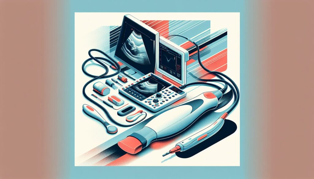
8. Pediatric Appendicitis Ultrasound Protocol — Graded Compression Technique and Key Parameters
A successful pediatric appendicitis ultrasound relies on a meticulous technique. The core principle is “graded compression,” where the sonographer applies slow, steady pressure with the transducer to displace overlying bowel gas and bring the appendix into view. The exam can take 15-20 minutes, so patience is key.
The technologist will start away from the point of maximal tenderness to gain the child’s trust. Using the psoas muscle and iliac vessels as landmarks, they identify the cecum and terminal ileum before searching for the blind-ending appendix, which typically arises from the posteromedial aspect of the cecum.
| Parameter | Specification |
|---|---|
| Primary Transducer | Linear high-frequency (9-15 MHz) for optimal resolution |
| Secondary Transducer | Curved low-frequency (4-9 MHz) for deeper penetration in larger patients |
| Key Technique | Graded compression to displace bowel gas and test for non-compressibility |
| Core Measurements | Maximum outer diameter (wall-to-wall) |
| Doppler | Color/spectral Doppler to assess for wall hyperemia (inflammation) or lack of flow (necrosis) |
Common protocol pitfalls: A common pitfall is giving up too early if the appendix isn’t immediately visible. It’s crucial to take time and use both high- and low-frequency transducers to fully survey the right lower quadrant. Another is mistaking a terminal ileum or prominent lymph node for the appendix; confirming a blind-ending structure connected to the cecum is essential.
9. 3+ months free for radiology residents and fellows
Look like a rockstar on your reports. We’re offering trainees a free, extended trial of GigHz Precision AI. You can dictate your positive findings in free form, and the AI will generate a structured report using ACR and SIR templates, with the appropriate Clinical Decision Support (CDS) firing automatically.
All we ask is your feedback so we can keep improving the product for trainees on the front lines. The signup is simple—no credit card, no long forms. Just provide the following three items:
- Your PGY year (e.g., PGY-2, PGY-4)
- Your training type (radiology residency or fellowship specialty)
- Your training program / hospital name
To get started, apply for the residents free-access program and reply to the application email with those three details.
10. Frequently Asked Questions
Is GigHz Precision AI HIPAA-compliant?
Yes. The platform is designed for de-identified workflows by default. It processes the clinical details of your dictation without requiring or storing patient health information (PHI). All data is encrypted in transit and at rest.
Do I need my hospital’s IT department to set it up?
No. GigHz Precision AI is browser-based and requires no local software installation. It works on any modern computer, including the workstations in your reading room or a personal laptop or iPad on call.
Does it work with PowerScribe or other dictation systems?
Yes. It works alongside your existing dictation system. You can dictate into the GigHz web app, let the AI structure the report, and then copy-paste the final, clean text into your PACS/RIS. It’s a workflow enhancement, not a replacement for your core dictation microphone.
Can I use it on my phone or iPad?
Yes, the web application is fully responsive and works well on tablets like the iPad, which is great for reviewing cases or drafting reports while on call away from a dedicated workstation.
Can I customize the templates?
Yes. While the system comes pre-loaded with ACR and society-standard templates, you can create, modify, and save your own templates to match your personal or institutional preferences.
What happens after my residency or fellowship ends?
The free access program is specifically for trainees. After you graduate, you’ll have the option to transition to a paid plan for practicing radiologists. We’ll reach out with details as you approach the end of your training.
Free GigHz Tools That Pair With This Article
Three free tools that complement the material above:
- ACR Appropriateness Criteria Lookup — Type an indication or clinical scenario in plain language and get the imaging studies the ACR rates for it, with adult and pediatric radiation levels. Built directly from 297 ACR topics, 1,336 clinical variants, and 15,823 procedure ratings.
- GigHz Imaging Protocol Library — A searchable library of 131 imaging protocols with the physics specs surfaced and the matching ACR Appropriateness Criteria alongside. Plain-English narratives readable in 60 seconds, organized by modality.
- GigHz Radiation Dose Calculator — Pick the imaging studies a patient has had and see total dose in millisieverts (mSv) with comparisons to natural background radiation, transatlantic flights, and chest X-rays. Useful for shared decision-making.
Reviewed by Pouyan Golshani, MD, Interventional Radiologist — May 7, 2026