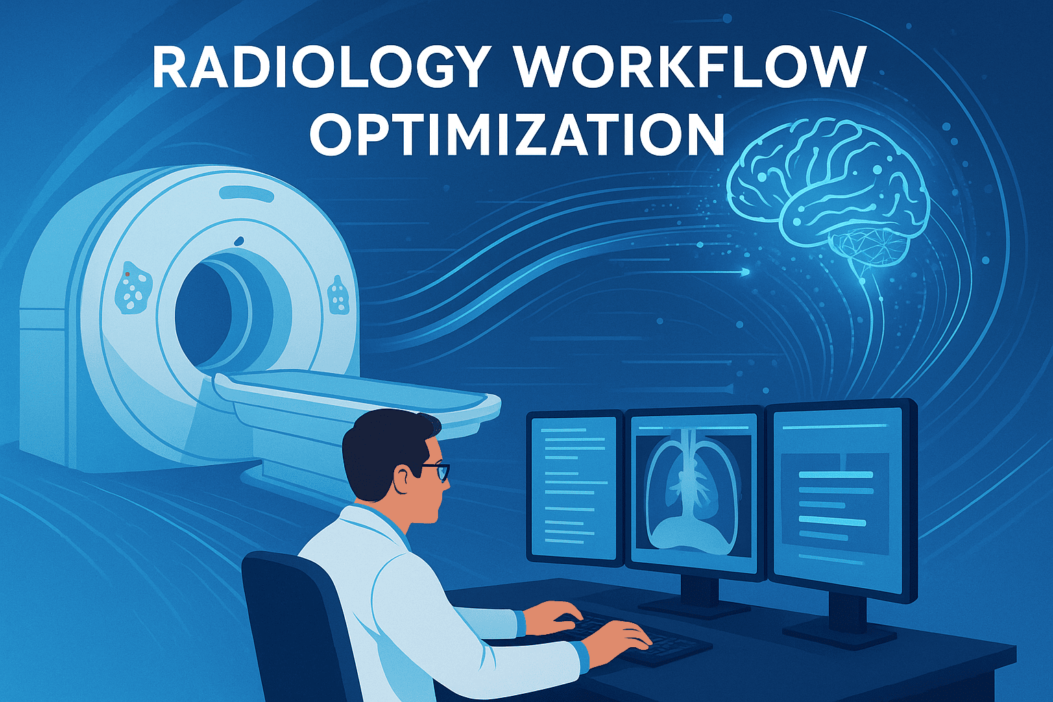
Radiology Workflow: Modern Solutions for Efficiency, Reporting, and AI Integration
Radiology is the backbone of modern diagnosis, but its workflow often feels like a complex relay race. From the moment a scan is ordered to the instant a report reaches a patient’s chart, countless steps and stakeholders are involved. An efficient radiology workflow isn’t just about speed – it’s about accuracy, collaboration, and patient care. Yet, radiologists today face mounting pressures: rising imaging volumes, staff shortages, and fragmented systems that don’t always talk to each other link.springer.com. The result? Bottlenecks that frustrate radiologists and clinicians alike, and delays that can affect patient outcomes.
For radiologists on the front lines, an optimized workflow means less time clicking and more time interpreting critical images. For hospital administrators, it means better resource utilization and throughput. And for med-tech founders aspiring to improve healthcare, understanding the nuances of radiology workflow is the key to building tools that actually help rather than hinder. This article dives into what radiology workflow really entails – from scheduling to report generation – and examines common pain points and emerging solutions. We’ll explore how modern software and AI (including the new GigHz Radiology Report Assistant – an AI-driven reporting tool at gighz.com/radiology-report-assistant) are reshaping the process, and what innovators need to know about the daily realities of radiologists.
Understanding the Radiology Workflow from Order to Report
In simple terms, radiology workflow encompasses the entire journey of an imaging study: ordering the exam, scheduling and preparing the patient, acquiring the images, interpreting those images, and generating and delivering the report. Each step is interconnected – a delay or error in one stage can ripple down the line. Here’s a high-level overview of the typical workflow:
-
Study Ordering & Scheduling: A clinician decides an imaging study (e.g. MRI, CT, X-ray) is needed and places an order. Scheduling staff coordinate the appointment, ensuring patient prep instructions are given (like fasting for an abdominal CT) and any contraindications (e.g. kidney function for contrast dye) are checked. In this planning stage, clear communication and proper protocol selection are vital to avoid repeat exams.
-
Image Acquisition (Scan Room): The patient arrives and the radiology technologist performs the scan. This involves positioning the patient, selecting the right protocol (settings on the machine), and capturing quality images. The goal is “first-time-right” imaging – getting good images without repeats. Variabilities in patient cooperation or technologist experience can impact this step. Once acquired, images are sent to the PACS (Picture Archiving and Communication System) – the digital repository of imaging.
-
Image Interpretation (Reading Room): Now the radiologist takes over. Using a PACS workstation, they review the images – often alongside prior studies and the patient’s clinical information from the RIS (Radiology Information System) or EMR (Electronic Medical Record). The radiologist’s task is to detect findings and formulate a diagnosis or report. This stage is mentally intensive: a radiologist might sift through hundreds of images per study, correlate with history, and perhaps consult colleagues or reference materials for complex cases. The worklist (queue of cases) is often long, and cases are prioritized by urgency.
-
Report Generation & Delivery: After interpreting the images, the radiologist creates a report of the findings and conclusions. Traditionally this is done via dictation – speaking into voice recognition software like Nuance PowerScribe or recording for a transcriptionist. Many radiologists use structured templates or macros for efficiency. The report is then finalized, signed, and automatically sent to the ordering clinician (and made available in the patient’s chart). In critical or unexpected findings, radiologists also directly call referring physicians to ensure timely communication. The workflow “ends” when the report is delivered and appropriately acted upon, closing the loop with the patient’s care team.
Throughout this process, there are numerous handoffs (from scheduler to tech to radiologist to clinician) and multiple software systems in play. A radiology workflow system usually involves a RIS for managing orders/appointments and a PACS for images; often these are integrated, but not always perfectly. The complexity of coordinating people and technology means inefficiencies can creep in at every stage. To appreciate why modernization is needed, let’s look at some common pain points that slow down imaging services today.
Common Inefficiencies and Pain Points in Imaging Workflow
Even the best radiology departments encounter friction in their daily process. Here are some of the most prevalent workflow pain points and bottlenecks that radiologists, administrators, and IT teams grapple with:
-
Scheduling Delays and No-Shows: Booking the right exam at the right time is harder than it looks. Outpatient imaging centers often deal with 7%+ patient no-show rates, which disrupts schedules and wastes machine time. Gaps from no-shows or last-minute cancellations mean idle staff and delayed diagnoses for other patients. On the flip side, overbooked slots or emergency add-ons can overwhelm daily operations. Poor prep (like a patient not fasting or misunderstanding instructions) can result in rescheduling an exam entirely. These scheduling inefficiencies trickle down, creating backlogs and frustrated patients.
-
Fragmented Systems and Manual Data Entry: Radiologists frequently juggle multiple platforms – viewing images in PACS, reporting in a voice dictation software, checking patient history in the EMR, and perhaps consulting prior reports in yet another system sarcmediq.com. This context-switching isn’t just annoying; it actively slows workflow and introduces error risk (e.g. copying an ID number incorrectly). In some hospitals, basic tasks like obtaining outside prior images involve chasing CDs or logging into separate portals. Lack of integration between systems means radiologists and staff act as the “human glue,” manually transferring information between siloed software. Every extra click or login drains a bit of focus and time.
-
Heavy Workload and Burnout Risks: The demand for imaging has exploded in recent decades. One experienced radiologist noted that “the workload in one day in 2018 is equal to a week in 2008 and a month in 1998”. While that might be a hyperbole, it rings true – radiologists are reading more studies, often with greater complexity (thin-slice CTs, multi-sequence MRIs) than ever before. A high case volume combined with pressure for fast turnaround can lead to fatigue. Fatigue in radiology isn’t just a wellness issue; it directly impacts accuracy and consistency. Documentation fatigue is real too. Spending late hours fixing voice recognition errors or typing long reports adds to burnout. This all circles back – burned-out radiologists may produce less thorough reports or cut corners, affecting patient care quality.
-
Imaging Quality Issues and Repeated Exams: Not every scan goes perfectly. Sometimes patients move, a contrast injection fails, or a technologist uses a suboptimal protocol – resulting in subpar images. If the radiologist can’t interpret an exam due to poor quality, that study needs to be repeated. Re-scans inconvenience the patient and double the work. Ensuring “first-time-right” image acquisition is a constant challenge, especially with varying skill levels among technologists and anxious patients who might not hold still. When imaging workflow isn’t standardized, one tech’s output might consistently require re-dos, silently bogging down the department’s efficiency.
-
Reporting Bottlenecks and Communication Gaps: After interpretation, getting the report finalized and communicated can be another choke point. Traditional dictation systems sometimes struggle with medical jargon or different accents, forcing radiologists to spend extra time correcting transcripts. Even with good voice recognition, radiologists have to structure the report in a clear, clinically useful way – which can be tedious for routine findings. And once the report is signed, ensuring it actually reaches the right people is crucial. Many institutions still rely on calls or faxes for critical findings. If the workflow for alerting a critical result (like acute bleed on a scan) is cumbersome, precious minutes can be lost. Similarly, sharing images and reports with outside physicians often relies on burning CDs or insecure email, causing delays.
These pain points underscore why radiology workflow optimization has become such a hot topic. In fact, a 2024 study highlighted 31 distinct operational pain points across the radiology workflow link.springer.com – from the moment an exam is planned to when the report is used in treatment. The stakes are high: inefficiencies don’t just cost time, they can impact patient outcomes (e.g. delayed diagnosis) and staff morale. Fortunately, recognition of these issues has spurred a wave of innovation. Modern software solutions and AI tools are now tackling each step of the workflow, with varying degrees of success. The next sections explore how technology is changing the game – and what pitfalls remain.
AI and Software Tools: Streamlining the Process (or Not)
Technology has long been intertwined with radiology – after all, this is a field that transitioned from darkrooms and film to digital images and PACS decades ago. Today, a new generation of software and AI (Artificial Intelligence) promises to further streamline radiology workflows. The idea is compelling: let machines handle the repetitive or data-heavy tasks so humans can focus on the nuanced decision-making. In practice, some tools are delivering major efficiency boosts, while others create new headaches. Let’s break down the areas where AI and modern software are making an impact in radiology workflow – and where they sometimes fall short:
-
Smarter Scheduling and Protocoling: One emerging application of AI is in the planning stage – helping to optimize how studies are ordered and protocolled. For example, AI can assist radiologists or schedulers by parsing through a patient’s electronic record and suggesting the appropriate imaging study (or even recommending against an unnecessary scan) based on clinical indications. This ensures the right test is done the first time. AI can also automate parts of protocol selection – choosing the correct scan parameters. Rather than a radiologist manually reviewing labs and prior allergies to approve a contrast MRI, an AI system could auto-check those factors and suggest the proper protocol, saving time on routine cases. Some hospitals are deploying systems that auto-generate the scanning protocol for common indications, freeing radiologists to focus only on unusual or complex planning. The payoff is fewer delays and less guesswork upfront. However, these tools must be deeply integrated with ordering systems and EHR data to work well, which can be a barrier. If an AI scheduler exists in a silo, clinicians won’t bother logging into yet another system just to book an exam.
-
Enhancing Image Acquisition and Quality Control: In the scanner room, modern tech is assisting technologists to get it right on the first try. Workflow automation tools can guide less experienced techs through complex scans or even automatically position patients and set machine parameters for certain exams philips.com. For instance, smart MRI software might auto-align and plan the next sequence based on the scout images, shaving minutes off scan time and reducing variability between techs. AI-based algorithms can also perform immediate QC (quality control) on images – checking if an X-ray is too blurry or a CT slice misses part of the anatomy – and alert the tech before the patient leaves. This kind of real-time feedback can drastically cut down repeat exams. During the COVID-19 pandemic, we even saw the rise of remote scanning assistants: an expert technologist at a central hub can virtually “drop in” to oversee or adjust settings for scanners at multiple sites. This telepresence model, like a radiology command center, ensures that even understaffed locations produce high-quality images without delays of on-site support. These innovations in the acquisition phase directly address one of the silent workflow killers: the need to redo scans or call patients back due to technical issues.
-
AI Triage and Worklist Prioritization: One of the most mature uses of AI in radiology workflow is triaging images for urgent findings. AI algorithms can analyze images in parallel with acquisition and flag suspected critical abnormalities within minutes – sometimes before the patient is even off the table. For example, AI can screen a head CT for signs of intracranial hemorrhage or a chest CT for a large pulmonary embolism. If a positive finding is detected, the system bumps that study to the top of the radiologist’s worklist and may even send an alert. This intelligent workload balancing ensures the most urgent cases get read first by an available radiologist (ideally a subspecialist suited to the case). Such triage AI is already in use in stroke workflows (e.g., flagging brain scans for clot retrieval candidates) and ER settings, effectively acting as a second set of eyes that never sleep. Radiologists report that these systems, when accurate, truly streamline the day – there’s less time lost scrolling the worklist to find critical cases. However, when AI is overly sensitive or not well-calibrated, it can “cry wolf” with false positives, disrupting workflow by prompting radiologists to double-check normal cases. The best results seem to come when AI triage is tightly integrated into the PACS or workflow software (so the radiologist doesn’t have to open a separate app) and when algorithms target findings with clear urgent implications. Triage AI isn’t replacing the radiologist’s judgment; it’s an extra safety net and prioritization tool.
-
Computer-Aided Detection and Diagnosis (CAD) in Reading: Beyond triage, AI is also assisting during image interpretation itself. Computer-aided detection (CAD) algorithms have existed for years (e.g. for mammography), but modern AI is far more powerful and versatile. Today’s FDA-cleared radiology AI applications (over 500 of them and counting) can highlight lung nodules on CT, flag possible fractures on X-rays, measure organ volumes, and more. In practice, a radiologist might see AI annotations or quantified results right on their PACS viewer – like boxes around suspected lung nodules or an automated calculation of cardiac ejection fraction. When thoughtfully deployed, these tools streamline workflow by reducing the radiologist’s burden of tedious measurements or ensuring subtle findings aren’t overlooked. One example is AI that automatically detects and measures liver lesions on serial MRI scans, then populates those measurements into the report – saving the radiologist a manual step. Another example is an algorithm that compares a current and prior chest CT to highlight any new nodule growth, so the radiologist doesn’t have to meticulously side-by-side every slice. However, if not well integrated, such tools can frustrate rather than help. Radiologists do not want to click out of their primary viewer to launch a separate AI application and wait for results. Hence, many vendors (and platforms like deepcOS) focus on embedding AI results directly into existing workflow with minimal context-switching deepc.ai. When AI results appear seamlessly in the normal reading flow, radiologists are more likely to use them. The other challenge is trust: if an AI frequently flags things that turn out to be nothing, radiologists will learn to ignore it. Therefore, developers are aiming for high specificity and providing a level of explanation (or at least an obvious visual cue) so the radiologist can quickly validate the AI’s suggestion. Used wisely, these AI assistants function like a junior colleague pointing out “hey, take a second look here,” which can be quite valuable in a busy day.
-
Voice Recognition and Reporting Aids: Generating the final report is another area ripe for optimization. Most radiologists have already transitioned from human transcription to voice recognition systems like PowerScribe or Dragon Medical, which expedite turning speech into text. Still, anyone who’s dictated a complex report knows that editing errors can be a time sink (“No, I said ileum, not ilium”). Here, AI is stepping up in two ways: better speech-to-text engines and auto-generated content. On the speech side, companies like Deepgram (with their deep learning speech recognition API) are working on more accurate medical speech recognition, boasting the ability to handle different accents and noisy backgrounds with fewer errors. On the content side, AI is now being used to draft parts of the report itself. A prominent example is Rad AI Omni, which analyzes the radiologist’s findings (the dictated observations) and then produces a draft impression section – the summary and conclusion – tailored to that radiologist’s style. Essentially, it saves the radiologist from having to manually compose the concluding paragraphs, which is both a timesaver and a fatigue reducer. Early users report significant time savings per shift radai.com, though of course the radiologist still reviews and edits the AI-generated text. Another emerging approach is using large language models (LLMs) – the same tech behind ChatGPT – to generate or improve radiology reports. The GigHz Radiology Report Assistant is one such AI tool integrating advanced language models into the workflow, helping radiologists create clear, thorough reports with less hassle. For instance, GigHz’s assistant can suggest standardized phrasing, catch inconsistencies, or even auto-insert relevant clinical information from the EMR into the report. By using AI as a co-pilot for documentation, radiologists can ensure nothing is overlooked and spend less time on keyboard work. The cautionary side: automatic report generation must be handled carefully. Radiology reports carry legal weight – an AI that overstates or misstates a finding could pose liability issues thedoctors.com. Thus, most AI report tools today focus on assisting the radiologist (e.g. providing a draft or checklist) rather than independently issuing final reports. When kept as an assistant, these tools are proving their worth by accelerating reporting while maintaining accuracy. It’s a fine balance of efficiency with human oversight.
In summary, AI and modern software are touching every part of the radiology workflow: from scheduling to image acquisition, from reading to reporting. They offer solutions to long-standing bottlenecks – but only if implemented in harmony with radiologists’ needs. The hard lessons so far have shown that technology can fail to streamline if it’s not integrated (a fancy AI tool that requires five extra clicks will likely gather dust). Moreover, tech must adapt to the real-world variability in radiology; for example, an AI might need retraining when the hospital upgrades its CT scanners or changes its protocol. Despite these challenges, the momentum is clearly toward more intelligent, connected workflows. Radiologists are gradually embracing AI as a partner – a way to amplify their expertise rather than replace it. As one radiologist put it, the goal is to “return time to the patient.” That means every minute saved by AI triaging scans or auto-filling reports is a minute the radiologist can spend thinking about the case in depth, consulting with clinicians, or simply catching their breath to avoid errors.
Designing Solutions with Radiologists in Mind: Advice for Innovators
For med-tech founders and healthcare IT developers eyeing the radiology space, building a product that truly improves workflow requires more than coding skills or clinical buzzwords. It demands an appreciation of the daily realities and constraints radiologists face. Here are some key considerations for anyone aiming to introduce a new workflow solution or AI tool into radiology:
-
“Invisible” Integration is Non-Negotiable: A recurring theme we’ve seen is that add-on tools must integrate seamlessly into existing workflows. Radiology departments have invested heavily in PACS, RIS, and EMR systems – your shiny new AI app will fail if it can’t plug into those. Radiologists will not tolerate a solution that makes them log into a separate portal or manually transfer data. The best innovations function almost like a native feature of the radiologist’s existing workstation. As an example, the deepcOS platform emphasizes embedding AI results directly into the PACS viewer or report, eliminating the need for context switching for the user. This principle is golden: reduce clicks, don’t add them. If your product alerts the radiologist to something, have it show up in the systems they already check regularly (the worklist or the PACS viewport) rather than a new dashboard. Integration also means respecting standards – DICOM for images, HL7 or FHIR for health data, IHE profiles for workflows. Hospitals are understandably wary of any tool that requires a complex IT overhaul, so designing with plug-and-play interoperability in mind is a huge plus.
-
Understand the Human Workflow (and Don’t Disrupt It): Spend time in actual reading rooms to see how radiologists work. You might discover that a seemingly logical feature is actually a nuisance in practice. Radiologists develop a rhythm using tools like the scroll wheel, hotkeys, voice commands, and dual monitors. If your solution breaks that rhythm – say, by forcing a radiologist to use a mouse when they typically use keyboard shortcuts, or by covering the images with pop-ups – it will face resistance. Remember that radiologists often work in dark environments with high concentration; a garish interface or anything that pulls their eyes off the image is unwelcome. The GigHz team, for instance, has taken care to design their Radiology Report Assistant’s interface in a way that complements radiologists’ existing reporting habits, acting like a helpful sidebar rather than an obtrusive popup. The more your tool feels like a natural extension of the radiologist’s thought process rather than an external mandate, the better adoption you’ll see.
-
Radiologist Behavior and Culture: Radiologists as a group are methodical, evidence-driven, and yes, sometimes skeptical of hype. After all, patient lives hinge on their interpretations. Med-tech founders should appreciate that any new tool needs to earn trust. This means providing transparency about how the AI works (to the extent possible) and giving radiologists control. A good approach is to allow an AI recommendation to be easily accepted or dismissed with a single click – empowering the physician to decide quickly. Furthermore, radiologists value consistency; they often use structured templates to ensure nothing is missed. A product that introduces variability (like unpredictable AI outputs) may be met with caution. It helps to allow customization: let radiologists adjust the tool to fit their reporting style or workflow preferences. Early physician champions can be your best allies if you incorporate their feedback – they’ll spread the word if the solution truly makes their day better.
-
Constraints of the Clinical Environment: Innovators should also recognize constraints like privacy, security, and regulatory compliance. Any solution handling patient data must be HIPAA-compliant and likely needs to go through an IT security review at the hospital. Integration into hospital networks can be slow (underfunded IT teams are a common bottleneck), so plan for that in your deployment timeline. On the regulatory side, if your tool provides diagnostic guidance (e.g. an AI that identifies pathologies), it may require FDA clearance or CE marking. Founders sometimes underestimate these hurdles – but being prepared (with proper validation studies, cybersecurity measures, etc.) will smooth your path to real-world use. Also, be mindful of workflow variability: a solution that works well in a large academic hospital might need tweaks for a small outpatient imaging center, and vice versa. Radiology practices differ in their case mix, staffing, and protocols, so flexibility is key.
-
Prove the ROI and Impact: Hospital administrators and department heads care about outcomes and return on investment. To win their buy-in, a new workflow solution should demonstrate measurable improvements – whether it’s faster report turnaround times, higher throughput, cost savings, or improved patient satisfaction scores. If your AI tool can cut the report dictation time by 30% or reduce missed follow-ups, gather that data and make it clear. Increasingly, decision-makers want real-world evidence that a product works in practice, not just in a controlled study. Having reference sites or pilot results that show concrete benefits will go a long way. For example, if the GigHz Radiology Report Assistant helped a pilot hospital achieve 20% faster report finalizations, that’s a compelling figure to share (and track over time). Founders should also consider workflows beyond just the radiologist – does the solution also make life easier for technologists? For referring physicians? A broader impact can strengthen the value proposition.
-
Avoiding Unintended Consequences: Finally, appreciate that changing a workflow can have side effects. If you speed up one part of the process, ensure you’re not creating a new bottleneck elsewhere. A classic example: an AI might help radiologists read faster, but if the IT infrastructure can’t handle the increased volume of studies being pulled from archives, you’ve simply moved the wait to a different spot. In one sense, med-tech innovators need to think like systems engineers – understanding the whole radiology service as an interconnected system, not just optimizing one widget in isolation. Continuous user feedback loops and the ability to update and iterate your product will help address these issues. In healthcare especially, listening to the end-users (and adjusting accordingly) is crucial; what looks good on paper might need fine-tuning in practice.
In short, successful radiology workflow solutions marry technological prowess with deep empathy for the end-user – the radiologist (and their team). Those aiming to help need to respect the complexity of the clinical environment and the limited bandwidth of the people working in it. The payoff for getting it right, however, is immense: not only is there a market need, but you’re directly contributing to better patient care by unburdening the experts who make critical diagnoses.
Emerging Solutions and Examples of Workflow Optimization
The push to streamline radiology workflow has led to a variety of innovative solutions, from startups to established healthcare IT players. Here we highlight a few notable trends and examples (without endorsements) to paint a picture of where things are headed:
-
Integrated AI Reporting Assistants: We are witnessing the rise of AI copilots tailored for radiologists. Companies like Rad AI, for example, offer tools that automatically draft report impressions or follow-up recommendations, learning a radiologist’s preferences over time. Similarly, the GigHz Radiology Report Assistant leverages advanced AI (including large language models) to help radiologists generate reports more efficiently – think of it as an AI scribe that knows radiology. These assistants don’t replace the radiologist’s voice but enhance it, ensuring consistency and saving time by handling rote documentation tasks. Early adopters say such tools can save significant minutes per case, which adds up to hours saved in a day. More importantly, by reducing mundane typing, radiologists can focus on the images and the clinical correlation. As these AI assistants improve, we can expect them to integrate images and text more closely – for instance, auto-inserting measurement data from PACS or suggesting comparison statements when prior exams are present. The key will be tight integration with existing reporting systems (as discussed, seamless workflow is king) and maintaining radiologist oversight to catch any AI mistakes.
-
Voice Tech 2.0: Voice recognition remains a cornerstone of reporting, and it’s getting better with AI. Apart from the big players in dictation (like Nuance’s Dragon/PowerScribe), newer entrants and AI platforms are tackling speech nuances. Deepgram and other AI-driven speech engines claim to handle medical vocabulary and even complex accents/dialects with greater accuracy by training on vast datasets. There’s also movement toward more naturalistic interfaces – for example, a system that could understand a radiologist’s spoken summary and automatically structure it into a well-formatted report. Imagine dictating in a free-form way (“There’s a 5mm nodule in the left upper lobe, unchanged from prior…”) and the system places each finding in the correct section, cross-references prior reports for that nodule measurement, and maybe even alerts if you forgot to address something like the appendix on an abdominal scan. We’re not fully there yet, but such intelligent voice assistants are on the horizon.
-
Workflow Orchestration Platforms: In large radiology groups or multi-site hospital networks, managing the flow of cases is a logistical challenge that some software solutions address via workflow orchestration. These are systems that automatically distribute cases to the right radiologist at the right time, often using rules or AI. For instance, if one site is overloaded, the system can route exams to a radiologist at another site who is available. Or it can ensure that a pediatric MRI goes to a pediatric radiologist first, while a high-priority ER trauma CT goes to the on-call trauma radiologist. This smart load balancing (sometimes built into modern PACS or as an overlay) helps utilize specialist skills better and avoid backlogs in one place while capacity sits idle in another. Vendors in this space focus on integrating with worklists and leveraging real-time data on radiologist availability. We can expect these systems to get more granular with AI – perhaps predicting how long a case will take to read (based on the number of images and complexity) and scheduling accordingly, or automatically escalating a study if it’s been waiting too long. The end goal is a smooth, enterprise-wide workflow that maximizes efficiency and minimizes patient wait times.
-
Cloud-Based PACS and Remote Collaboration: Traditional on-premises PACS can be limiting, especially as remote work and teleradiology expand. Newer cloud-based imaging platforms aim to let radiologists access studies anytime, anywhere with diagnostic-quality viewing. This not only aids remote readings (e.g., nighthawk radiologists covering after-hours from home), but also facilitates collaboration. Picture a subspecialist logging in from across the country to consult on a difficult case seamlessly, or a multi-disciplinary team simultaneously viewing images during a tumor board meeting via a cloud viewer. Some platforms also integrate chat or live screen sharing for radiologists to discuss cases in real time. The COVID-19 pandemic accelerated adoption of these cloud solutions out of necessity, and now many radiologists appreciate the flexibility. Cloud PACS can also incorporate AI more readily – updates to algorithms can be rolled out centrally without local installs. Companies like Google and Amazon have also dipped into medical imaging cloud services, hinting that the future radiology workflow might be as much about managing data streams and AI in the cloud as it is about local software. For hospitals, a cloud approach can reduce IT burden, though it requires robust internet connectivity and careful attention to data security.
-
Patient Engagement and Scheduling Tools: Not all innovation is on the radiologist side; some target the patient-facing part of the workflow to prevent problems at the source. As mentioned earlier, digital platforms that send automated reminders, preparation instructions, and even do online check-ins are reducing no-show rates and improving patient preparedness. For example, a system might text a patient a day before: “Remember your MRI is tomorrow at 10am. Don’t forget to fast after midnight. Reply YES to confirm or call if you need to reschedule.” This not only cuts no-shows but ensures the patient arrives properly prepped, avoiding wasted slots. Some tools go further and allow patients to fill out safety questionnaires (for metal implants, allergies, etc.) online in advance – feeding that info to the technologist and radiologist so they can plan accordingly. Especially in the outpatient setting, these conveniences go a long way. By smoothing the front end of the workflow (scheduling and intake), these solutions indirectly speed up the clinical part by reducing last-minute surprises and delays.
-
Multimedia and Structured Reporting: Recognizing that the classic paragraph-style radiology report has its limitations, some solutions are introducing richer reporting formats. Interactive multimedia reporting is one innovation – allowing key images, graphs, or hyperlinks to be embedded in the report for clinicians to click and view. For example, instead of just saying “see Figure 1,” the actual image or a link is in the digital report. This can improve communication with referring physicians by highlighting the most pertinent findings visually. Additionally, structured reporting templates (sometimes with AI assistance) ensure that important checklist items are always addressed (e.g. in a liver ultrasound report, the template ensures you comment on liver size, echotexture, any masses, common bile duct, etc.). While some radiologists feel templates can be rigid, the combination of structured elements with free-text where needed can yield more complete and comparably formatted reports. Companies are working on making these templates smarter – for instance, auto-filling normal findings and letting radiologists just modify exceptions, or auto-populating measurements and calculations. The benefit for workflow is that a well-structured report can be generated faster and is easier for downstream providers to digest.
-
AI for Quality Assurance and Follow-up: Beyond the immediate reading workflow, AI is also being applied to workflow around the radiologist. One example is tools that scan finalized reports to ensure critical results were communicated or to detect any incidental findings that need follow-up. If a radiologist mentions a lung nodule in an abdominal scan report, an AI-driven system could flag that and ensure it’s added to a follow-up tracking list (so that 6 months later, someone can confirm a follow-up chest CT was done). This addresses a workflow gap in many systems where incidental findings can fall through the cracks. Another example is using analytics on workflow data – monitoring turnaround times, protocol efficacy, discrepancies – to provide managers with insights for continuous improvement. Real-time dashboards might show how many cases are pending, average read times per modality, or whether certain shifts consistently have delays. By identifying these trends, departments can adjust staffing or processes proactively. Essentially, data-driven management tools treat the radiology workflow itself as something to be analyzed and optimized, just like any complex process in a high-tech domain.
The breadth of these solutions shows that radiology workflow optimization is a multi-faceted effort. No single tool is a silver bullet – improving workflow is about addressing numerous small inefficiencies and integrating the fixes into a coherent whole. It’s also clear that the industry is moving toward greater synergy: AI tools are being built into PACS systems, cloud platforms are enabling easier AI updates, and patient-side and doctor-side improvements are both being pursued in parallel. Radiologists often say they don’t need gimmicks; they just want tools that work reliably. The most successful modern solutions seem to be those that solve a specific problem in the chain (like reducing dictation time or eliminating repeat scans) while fitting naturally into the existing environment. A few years ago, the buzz was that “AI will replace radiologists,” but the reality in 2025 is that AI is helping radiologists be more efficient and effective, which is exactly what busy imaging departments need.
Conclusion: Streamlining Workflow for Better Care and Collaboration
Radiology workflow may never be simple, but it can certainly be smarter. By identifying common pain points – whether it’s a scheduling snafu, a slow software interface, or a reporting overload – we can implement targeted solutions that add up to significant improvements. For radiologists, a well-oiled workflow means less stress and more focus on what truly matters: interpreting images accurately and engaging in patient care decisions. For hospital administrators, it translates to higher productivity, better use of expensive imaging equipment, and possibly shorter hospital stays thanks to faster diagnostics. And for patients, even though they may never think about “radiology workflow,” they feel the benefits in quicker exam scheduling, shorter wait times for results, and more face-to-face time with physicians who are not bogged down by clerical tasks.
The road to an optimized workflow is a continuous journey. It involves not just adopting new software, but also fostering a culture of collaboration between radiologists, technologists, IT staff, and referring clinicians. Change can be daunting in healthcare, but the success stories are piling up – from AI catching a critical finding that might have been missed, to an automated assistant like GigHz Radiology Report Assistant shaving minutes off each case and preventing documentation errors. These modern tools, when thoughtfully integrated, act as force multipliers for radiology teams.
In closing, the phrase “time is brain” is often used in stroke care to emphasize speed. The same principle holds for radiology workflow: time is diagnosis. Every efficiency gained in the process can lead to an earlier answer and an earlier start to treatment. By embracing modern solutions for efficiency, reporting, and AI integration, radiology can continue to elevate its impact on patient care. The technology is ready – and with radiologists and innovators working hand in hand, the classic image of a harried radiologist drowning in paperwork and unread studies may soon become a thing of the past.
Radiologists, healthcare leaders, and tech founders all have a stake in this evolution. Streamlining radiology workflow isn’t just about doing work faster; it’s about doing it better, with more clarity and less friction. Whether it’s implementing an AI triage system for urgent cases or deploying a reporting assistant from GigHz to eliminate tedious tasks, each step moves us toward a future where radiologists can practice at the top of their expertise. The result is a win-win-win: better outcomes for patients, a more manageable workday for radiologists, and efficient operations for hospitals. In the end, an optimized radiology workflow exemplifies what healthcare technology should always strive for – more care, less hassle.
