Should You Order MRA or CT for a Recent Ischemic Stroke? An ACR-Guided Workflow

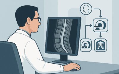
It’s 10 PM on a busy hospital service. A 71-year-old patient, admitted earlier in the day for new-onset aphasia and right-sided weakness, has already had a non-contrast head CT. The CT confirmed a small, established ischemic infarct in the left middle cerebral artery territory, ruling out hemorrhage. The acute event occurred about 18 hours ago, and the patient is outside the window for thrombolysis. Now, the primary clinical question shifts from acute intervention to secondary prevention: what caused this stroke? You need to evaluate the cervicocerebral vasculature to guide treatment, but which study is the right next step? This article details the American College of Radiology (ACR) Appropriateness Criteria workflow for this specific scenario. For an adult with a recent ischemic infarct less than 24 hours old, MRA head without IV contrast is rated Usually Appropriate as the initial imaging choice for etiologic workup.
Who Fits This Clinical Scenario for a Recent Ischemic Infarct?
This guidance applies to a specific and common clinical situation: an adult patient with an ischemic infarct confirmed on initial imaging (typically a non-contrast CT) that occurred within the last 24 hours. The focus of this workflow is not on the hyperacute diagnosis or determining eligibility for thrombolysis or thrombectomy, but rather on the subsequent, urgent workup to determine the stroke’s etiology and guide secondary prevention strategies.
Inclusion Criteria:
- Adult patient.
- Ischemic infarct confirmed on a prior imaging study.
- Symptom onset or time last known well was less than 24 hours ago.
- The clinical goal is to identify the underlying vascular cause (e.g., stenosis, occlusion, dissection).
Exclusion Criteria (These patients follow different ACR guidelines):
- Patients with active, evolving deficits being evaluated for acute intervention: A patient presenting with a focal neurologic deficit who is a potential candidate for thrombolysis or mechanical thrombectomy falls under the ACR scenario for suspected acute ischemic stroke, which prioritizes speed and often involves a CT/CTA/CTP protocol.
- Patients with resolved symptoms and negative initial imaging: If the patient’s symptoms have completely resolved and the initial head CT is negative for an infarct, the diagnosis is a transient ischemic attack (TIA). This routes to the ACR scenario for TIA.
- Patients presenting more than 24 hours after the event: The urgency and imaging considerations may differ slightly for subacute infarcts, which are covered in the ACR scenario for infarcts greater than 24 hours old.
What Diagnoses Are You Working Up in This Scenario?
After confirming an ischemic infarct, the imaging workup aims to uncover the underlying cause to prevent a recurrence. The differential diagnosis for the etiology of the stroke drives the choice of study.
Large Artery Atherosclerosis
This is a leading cause of ischemic stroke. Atherosclerotic plaque can build up in the large arteries of the head and neck, leading to stenosis (narrowing) or occlusion. A stroke can occur either from reduced blood flow past a severe stenosis (hemodynamic mechanism) or, more commonly, from a piece of plaque or a thrombus breaking off and traveling to a smaller downstream vessel (artery-to-artery embolism). Imaging must clearly visualize the intracranial arteries (e.g., middle cerebral, basilar) and extracranial arteries (e.g., internal carotid, vertebral).
Cardioembolism
An embolus originating from the heart, often due to atrial fibrillation, is another major cause of stroke. While vascular imaging cannot directly confirm a cardioembolic source, it plays a crucial role by exclusion. If a comprehensive evaluation of the cervicocerebral arteries reveals no significant stenosis or other culprit lesion, the likelihood of a cardioembolic or other embolic source increases, prompting a more focused cardiac workup (e.g., prolonged cardiac monitoring, echocardiography).
Arterial Dissection
Particularly important in younger patients but possible at any age, a tear in the wall of a carotid or vertebral artery can lead to stroke. The resulting intramural hematoma can narrow or occlude the vessel, or a thrombus can form at the site of injury and embolize distally. Imaging needs to be sensitive enough to detect the subtle vessel wall abnormalities, intimal flaps, or characteristic “flame-like” tapering associated with dissection.
Other Vasculopathies
Less common but consequential causes include vasculitis (inflammation of the vessel walls), fibromuscular dysplasia (FMD), or Moyamoya disease. These conditions often present with multifocal stenoses or characteristic vessel morphology that can be identified with high-quality vascular imaging, guiding further diagnostic tests and specific treatments.
Why Is MRA Head Without Contrast a Recommended Study for a Recent Infarct?
For evaluating the cause of a known recent infarct, several studies are rated as Usually Appropriate, but MRA head without IV contrast offers a powerful combination of diagnostic accuracy and safety, making it an excellent first choice.
The primary rationale is its ability to provide high-resolution images of the major intracranial arteries without exposing the patient to ionizing radiation (adult_rrl=O 0 mSv) or intravenous contrast agents. The standard technique, 3D Time-of-Flight (TOF), is highly sensitive for detecting significant stenosis and occlusion within the Circle of Willis and its proximal branches—the most common locations for intracranial atherosclerotic disease. This directly addresses the leading diagnosis on the differential.
Let’s compare it to other highly rated alternatives:
- CTA head with IV contrast: This study is also rated Usually Appropriate and is a valid alternative. It is significantly faster than MRA and can be superior for visualizing vessels affected by calcified plaque. However, it requires both IV iodinated contrast (a consideration in patients with renal impairment or contrast allergies) and involves ionizing radiation (adult_rrl=☢☢☢ 1-10 mSv). In a non-emergent setting focused on etiologic workup, avoiding these exposures is often preferable if MRA is available and the patient has no contraindications (e.g., incompatible implanted device).
- US duplex Doppler carotid artery: This study is rated May be appropriate. While it is excellent for evaluating the extracranial carotid arteries in the neck, it provides no information about the intracranial circulation. Given that a significant portion of strokes are caused by intracranial pathology, relying solely on carotid ultrasound is an incomplete initial workup. It is often performed as a complementary study but not as the primary, standalone investigation.
In practice, a comprehensive evaluation requires assessing both intracranial and extracranial vessels. Therefore, ordering an MRA of the head is often paired with an evaluation of the neck vessels, for which MRA neck (with or without contrast) and CTA neck are also rated Usually Appropriate. The decision between these modalities depends on institutional preference, scanner availability, and patient-specific factors. Once you’ve decided on MRA head without contrast, our protocol guide covers the technique, contrast, and reading principles: MRA Brain Without Contrast (3D TOF).
What’s Next After MRA? Downstream Workflow
The results of the initial vascular imaging study will guide the subsequent management and diagnostic pathway. The goal is to build a complete picture of the stroke’s cause to implement effective secondary prevention.
- If the study is positive for significant stenosis: If the MRA reveals a high-grade stenosis (>70%) in an artery supplying the area of the infarct (e.g., an M1 segment stenosis for an MCA territory stroke), this is identified as the likely culprit. The next steps involve optimizing medical management (antiplatelet therapy, statins, blood pressure control) and, in some cases, considering procedural intervention like stenting or endarterectomy, depending on the vessel location and clinical trial data.
- If the study is negative: A normal intracranial and extracranial MRA/CTA makes large artery atherosclerosis much less likely. The focus of the workup should pivot strongly toward an embolic source. This triggers a downstream workflow that includes an echocardiogram (transthoracic or transesophageal) to look for cardiac sources of emboli (e.g., patent foramen ovale, thrombus) and prolonged cardiac monitoring (e.g., Holter monitor or event recorder) to screen for paroxysmal atrial fibrillation.
- If the study is indeterminate or suggests dissection/vasculitis: Sometimes, findings are ambiguous. For example, there may be a subtle vessel irregularity concerning for a dissection or vasculitis. In these cases, a follow-up or alternative imaging modality may be necessary. A vessel wall imaging (VWI) MRI sequence can be particularly helpful to directly visualize inflammation or hematoma within the artery wall, confirming or refuting these diagnoses.
Pitfalls to Avoid (and When to Get Help)
Navigating the post-stroke workup requires careful consideration to avoid common diagnostic errors.
- Incomplete Vascular Evaluation: Do not stop the workup after only imaging the head or only imaging the neck. A comprehensive evaluation of the entire cervicocerebral arterial system, from the aortic arch to the distal intracranial branches, is essential.
- Over-relying on Non-Contrast CT: While a non-contrast head CT is critical initially to rule out hemorrhage, it provides no information about the blood vessels. Delaying the vascular workup (MRA or CTA) can delay the initiation of appropriate secondary prevention.
- Ignoring Patient Contraindications: Before ordering an MRI/MRA, ensure the patient is properly screened for contraindications such as pacemakers, certain aneurysm clips, or other metallic implants. If contraindications exist, CTA is the appropriate alternative.
- Misinterpreting Flow Gaps: On TOF MRA, complex or turbulent blood flow can sometimes create a “flow gap” that mimics a stenosis. Radiologist expertise is key to differentiating this artifact from true pathology.
If the imaging findings are complex, unclear, or discordant with the clinical picture, a consultation with a vascular neurologist or neuroradiologist is the appropriate next step to formulate a plan for further investigation.
Related ACR Topics and Tools
The ACR Appropriateness Criteria are a powerful resource for evidence-based imaging decisions. For breadth across all scenarios in Cerebrovascular Diseases-Stroke and Stroke-Related Conditions, see our parent guide: Cerebrovascular Diseases-Stroke and Stroke-Related Conditions: ACR Appropriateness Decoded. To explore other clinical scenarios or refine your imaging choices, use the tools below.
- ACR Appropriateness Criteria Lookup — for adjacent scenarios
- Imaging Protocol Library — for technique on the recommended study
- Radiation Dose Calculator — for cumulative dose conversations
Frequently Asked Questions
Why not just get a CTA of the head and neck on every patient?
CTA of the head and neck is also rated ‘Usually Appropriate’ and is an excellent choice, especially if MRA is contraindicated or unavailable, or if speed is critical. However, MRA without contrast is often preferred when possible because it avoids both ionizing radiation and the risks associated with iodinated IV contrast, such as allergic reaction or contrast-induced nephropathy in susceptible patients.
If the MRA head is negative, is the vascular workup complete?
No. A complete workup requires evaluation of both the intracranial and extracranial arteries. If you only ordered an MRA of the head, you still need to evaluate the neck vessels (carotid and vertebral arteries), typically with a neck MRA, CTA, or carotid ultrasound. A negative intracranial study shifts suspicion to the neck arteries or a cardioembolic source.
Does this guidance apply if the initial non-contrast CT was negative?
No. This workflow is for patients with a *confirmed* recent infarct. If the initial CT is negative but there was a transient neurologic deficit, the patient fits the ‘Transient Ischemic Attack (TIA)’ scenario, which has its own distinct imaging pathway. A negative CT does not rule out a small or early infarct, which is why further imaging like MRI with DWI is often performed in the TIA workup.
Is contrast ever needed for an MRA in this scenario?
For the initial evaluation of stenosis or occlusion, non-contrast Time-of-Flight (TOF) MRA of the head is usually sufficient. However, contrast-enhanced MRA is often used for the neck vessels to improve image quality and is essential for specific techniques like vessel wall imaging, which is used to diagnose conditions like vasculitis or dissection.
What if the patient has a pacemaker or other MRI contraindication?
If a patient has an absolute contraindication to MRI, then CTA of the head and neck with IV contrast is the best alternative. It is also rated ‘Usually Appropriate’ by the ACR and provides excellent visualization of the vasculature. You must weigh the benefits of the diagnostic information against the risks of radiation and IV contrast for that specific patient.
Reviewed by Pouyan Golshani, MD, Interventional Radiologist — August 4, 2026