Should You Order MRI or CT for Follow-Up of Treated Salivary Gland Cancer?

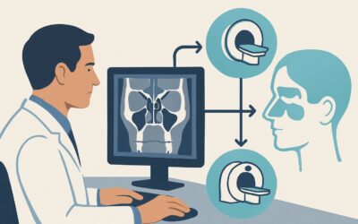
It’s a Tuesday afternoon clinic, and you’re seeing a 62-year-old patient for their two-year follow-up after a partial parotidectomy and adjuvant radiation for mucoepidermoid carcinoma. They report a new, persistent, deep ache near the surgical site and you palpate a subtle, ill-defined firmness. The clinical question is immediate: Is this post-treatment fibrosis or tumor recurrence? Deciding on the right imaging study is critical for guiding the next steps, from reassurance to biopsy and potential salvage therapy. For this specific scenario, the American College of Radiology (ACR) Appropriateness Criteria rates MRI orbits face neck without and with IV contrast as Usually Appropriate.
Who Fits This Clinical Scenario?
This guidance is specifically for patients who have completed definitive treatment (such as surgery, radiation therapy, or chemoradiation) for a malignancy of a major salivary gland—the parotid, submandibular, or sublingual glands. The clinical indication is either routine surveillance in an asymptomatic patient or, more commonly, the evaluation of new signs or symptoms concerning for recurrence. This includes new pain, a palpable mass, facial nerve weakness, or other focal neurologic deficits.
It is crucial to distinguish this scenario from others that require a different imaging approach. This workflow does not apply to:
- Initial Staging: Patients with a newly diagnosed, untreated salivary gland mass fall under a different ACR variant. The imaging goals for initial staging—defining the primary tumor extent, nodal status, and distant disease—differ from those of post-treatment surveillance.
- Other Head and Neck Cancers: Patients treated for cancers of the oral cavity, oropharynx, larynx, or nasopharynx have distinct patterns of recurrence and require tailored imaging protocols. While there is overlap, the specific focus on perineural spread in salivary gland tumors makes the choice of modality particularly important.
- Benign Salivary Gland Tumors: This guidance is for malignant histologies. The follow-up for benign tumors like pleomorphic adenomas, if performed, follows a different rationale.
What Diagnoses Are You Working Up in This Scenario?
In the post-treatment setting, the differential diagnosis for a new clinical finding is narrow but consequential. The primary goal of imaging is to differentiate between expected post-therapy changes and pathologic processes requiring intervention.
Local or Regional Recurrence
This is the most critical diagnosis to confirm or exclude. Recurrence can manifest as a discrete enhancing mass in the surgical bed, along the resection margins, or as new or enlarging cervical lymph nodes. The original tumor’s histology (e.g., adenoid cystic carcinoma, mucoepidermoid carcinoma) influences the likelihood and pattern of recurrence.
Perineural Spread (PNS)
Certain salivary gland malignancies, particularly adenoid cystic carcinoma, have a high propensity to spread along nerves. Recurrent disease can present as thickening and abnormal enhancement of cranial nerves, most commonly the facial nerve (CN VII) or trigeminal nerve (CN V). This can be clinically silent initially, and MRI is uniquely sensitive for its detection.
Post-Treatment Fibrosis and Inflammation
Surgery and radiation therapy invariably cause scarring, fibrosis, and chronic inflammation. These changes can create palpable firmness and may demonstrate enhancement on imaging, closely mimicking recurrent tumor. Differentiating between scar and recurrence is a primary challenge and a key reason for the choice of imaging modality.
Radiation-Induced Changes
Beyond simple fibrosis, radiation can cause osteoradionecrosis of the mandible, soft tissue necrosis, or second primary malignancies within the radiation field. While less common, these are important considerations in the long-term follow-up of irradiated patients.
Why Is MRI of the Orbits, Face, and Neck the Recommended Study?
The ACR rates MRI orbits face neck without and with IV contrast as Usually Appropriate because of its superior soft-tissue contrast resolution, which is essential for navigating the complex anatomy of the post-treatment head and neck.
The primary advantage of MRI is its ability to distinguish between post-surgical scar tissue and recurrent tumor. While both can enhance after contrast administration, the patterns of enhancement and signal characteristics on different MRI sequences (e.g., T2-weighted, diffusion-weighted imaging) can often differentiate benign fibrosis from viable malignancy. Furthermore, MRI is unparalleled in its ability to detect perineural spread. It can visualize subtle thickening and enhancement along the entire course of cranial nerves from the periphery to the skull base, a critical pathway for recurrence that is often missed by other modalities.
While MRI is the top-rated study, other modalities are also considered:
- CT neck with IV contrast is also rated Usually Appropriate. It is faster and more accessible than MRI and is excellent for assessing bone invasion (e.g., of the mandible or skull base) and evaluating cervical lymph nodes. However, its inferior soft-tissue contrast makes it more difficult to differentiate scar from tumor in the primary surgical bed. It also involves ionizing radiation (☢☢☢ 1-10 mSv).
- FDG-PET/CT skull base to mid-thigh is rated Usually Appropriate and is highly valuable for detecting metabolically active disease. It can be particularly useful when MRI findings are equivocal or when there is a high suspicion of distant metastases. However, it has a higher radiation dose (☢☢☢☢ 10-30 mSv) and can be falsely positive due to post-treatment inflammation, especially in the first few months after therapy.
- US neck is rated May be appropriate. It is a useful, non-invasive tool for evaluating superficial palpable abnormalities and guiding fine-needle aspiration (FNA) of suspicious lymph nodes. Its utility is limited for assessing the deep tissues of the primary site or detecting perineural spread.
When ordering the recommended study, it is critical to specify the full protocol: “MRI orbits face neck without and with IV contrast.” A limited “MRI neck” may not include the dedicated thin-section images through the face and skull base necessary to evaluate the relevant cranial nerves.
What’s Next After MRI? Downstream Workflow
The results of the surveillance MRI will dictate the subsequent clinical pathway. The workflow branches based on whether the findings are clearly positive, negative, or indeterminate.
If the study is positive for recurrence:
A definitive finding of recurrent disease, such as a new, enlarging mass or clear perineural enhancement, requires histopathologic confirmation. The next step is typically a core needle biopsy or FNA of an accessible lesion. Once recurrence is confirmed, the patient’s case should be presented at a multidisciplinary tumor board to determine the best course for salvage therapy, which may include re-operation, re-irradiation, systemic therapy, or a combination.
If the study is negative:
A negative MRI in the setting of nonspecific symptoms is highly reassuring. The patient can typically return to their standard clinical follow-up schedule. If a strong clinical suspicion for recurrence persists despite a negative scan (e.g., progressive, unexplained facial nerve palsy), a different imaging modality like FDG-PET/CT may be considered to look for metabolic evidence of disease.
If the study is indeterminate:
This is a frequent outcome, as post-treatment changes can be difficult to distinguish from early recurrence. An indeterminate report might describe subtle, non-mass-like enhancement or a stable but nonspecific finding. In this situation, the most common next step is a short-interval follow-up MRI in 2-3 months to assess for stability or progression. If the finding is highly suspicious or progressing, proceeding to FDG-PET/CT or biopsy may be warranted.
Pitfalls to Avoid (and When to Get Help)
Navigating post-treatment imaging for salivary gland cancer requires careful attention to detail to avoid common errors.
- Inadequate Clinical History: Failing to provide the radiologist with the original tumor histology, date of treatment, and specific clinical concern can lead to a non-specific or less confident interpretation.
- Ignoring Priors: Comparison with previous imaging studies is not just helpful; it is essential. The trajectory of a finding over time (new, growing, or stable) is often the most important factor in determining its significance.
- Incorrect Protocol: Ordering a generic “MRI neck” instead of the comprehensive “MRI orbits face neck” protocol can miss crucial findings at the skull base or along the facial nerve.
- Over-reliance on a Single Modality: When results are equivocal, remember that different modalities provide complementary information. A metabolically active spot on PET/CT can clarify an indeterminate finding on MRI.
If you encounter a complex case with discordant clinical and imaging findings, escalation to a multidisciplinary head and neck tumor board is always the appropriate next step.
Related ACR Topics and Tools
This article covers one specific clinical scenario. For a comprehensive overview of imaging for all head and neck cancer presentations, or to explore the tools used to develop this guidance, please refer to the resources below.
- For breadth across all scenarios in Staging and Post-Therapy Assessment of Head and Neck Cancer, see our parent guide: Staging and Post-Therapy Assessment of Head and Neck Cancer: ACR Appropriateness Decoded.
- To explore other clinical situations, use the ACR Appropriateness Criteria Lookup.
- For details on imaging techniques, consult the Imaging Protocol Library.
- To discuss radiation exposure with patients, use the Radiation Dose Calculator.
Frequently Asked Questions
Why is MRI generally preferred over CT for salivary gland cancer follow-up?
MRI is preferred due to its superior soft-tissue contrast, which allows for better differentiation between post-treatment scar tissue and recurrent tumor. It is also significantly more sensitive for detecting perineural spread—the extension of tumor along nerves—which is a common pattern of recurrence for certain salivary gland cancers like adenoid cystic carcinoma. While CT is also a useful tool, especially for assessing bone, MRI provides more detailed information about the soft tissues in the surgical bed and along cranial nerve pathways.
How often should surveillance imaging be performed after treatment?
There is no universal consensus, and surveillance schedules are often tailored to the individual patient’s risk of recurrence, based on factors like tumor stage, grade, and histology. Many institutional protocols involve more frequent imaging in the first 2-3 years post-treatment, when recurrence risk is highest, followed by less frequent scans. However, imaging is most often performed in response to new signs or symptoms rather than as routine screening in an asymptomatic patient.
What should I order if my patient has a pacemaker or other contraindication to MRI?
If a patient cannot undergo an MRI, CT neck with IV contrast is the best alternative and is also rated as ‘Usually Appropriate’ by the ACR. It provides excellent evaluation of lymph nodes and bony structures. In cases with high clinical suspicion or equivocal CT findings, an FDG-PET/CT can provide valuable metabolic information to help detect recurrent disease.
Is the ‘without and with IV contrast’ part of the MRI order essential?
Yes, both the non-contrast and post-contrast sequences are critical. The non-contrast T1-weighted images help define anatomy and identify perineural spread by looking for effacement of fat planes around nerves. The post-contrast images are essential for identifying and characterizing enhancing tissue, helping to distinguish active tumor from scar. Omitting either part of the study significantly compromises its diagnostic value.
What exactly is perineural spread and why is it so important for salivary gland cancers?
Perineural spread (PNS) is a process where cancer cells invade and migrate along nerves, often far from the original tumor site. It is a hallmark of certain malignancies, especially adenoid cystic carcinoma. Detecting PNS is critical because it indicates a more aggressive disease, carries a poorer prognosis, and significantly alters treatment planning. For example, it may require a larger surgical resection or a more extensive radiation field to cover the entire affected nerve up to the skull base.
Reviewed by Pouyan Golshani, MD, Interventional Radiologist — May 30, 2026