Should You Order Ultrasound or CT for a Child with a Large Parapneumonic Effusion?
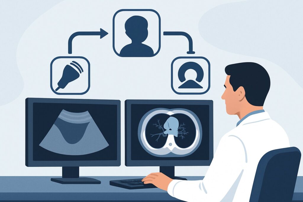
A 5-year-old with community-acquired pneumonia is not improving on appropriate antibiotics after 48 hours. She remains febrile, tachypneic, and has diminished breath sounds over the right lung base. A chest radiograph confirms a persistent right lower lobe consolidation and now shows a significant opacity blunting the costophrenic angle, concerning for a moderate-to-large parapneumonic effusion. You need to characterize this fluid collection to guide the next step in management—is it simple fluid that can be managed medically, or a complex collection requiring drainage? This clinical workflow article details the American College of Radiology (ACR) guidance for this specific scenario. For a child with pneumonia complicated by a suspected moderate or large parapneumonic effusion on radiograph, the ACR rates `US chest` as Usually Appropriate.
Who Fits This Clinical Scenario?
This guidance applies to a specific patient population: an immunocompetent child with a clinical and radiographic diagnosis of pneumonia, where the initial chest radiograph suggests the presence of a moderate or large parapneumonic effusion. The key decision point is selecting the next imaging study to better characterize this effusion and guide further management, which may include drainage.
Inclusion criteria for this workflow are straightforward:
- The patient is a child (typically older than 3 months).
- The patient is immunocompetent.
- There is a confirmed diagnosis of pneumonia.
- A chest radiograph indicates a moderate or large pleural effusion.
This workflow does not apply to several similar-appearing but distinct clinical situations. If the patient is immunocompromised, has recurrent pneumonia in different locations, or if the initial radiograph is more suspicious for a different complication, the imaging strategy changes. Specifically, this guidance is not intended for patients with suspected lung abscess or a bronchopleural fistula, as these scenarios have their own dedicated ACR Appropriateness Criteria variants and often warrant different imaging approaches from the outset.
What Diagnoses Are You Working Up in This Scenario?
When a parapneumonic effusion is identified, the primary goal of subsequent imaging is to differentiate between effusions that will likely resolve with antibiotics and those that require procedural intervention. The differential diagnosis centers on the character and complexity of the pleural fluid.
Simple Parapneumonic Effusion: This is the most common finding, representing a transudative or early exudative, free-flowing collection of sterile fluid in the pleural space. On imaging, it appears as a simple, anechoic (black) collection without internal complexity. These effusions often resolve with effective antibiotic treatment of the underlying pneumonia and may not require drainage unless they are very large and causing significant respiratory compromise.
Complicated Parapneumonic Effusion: As the inflammatory process progresses, the effusion can evolve into a fibrinopurulent stage. This fluid is thicker, and fibrin strands begin to form, creating septations that divide the pleural space into multiple pockets (loculations). This prevents the fluid from being drained with a simple needle or catheter and is a key finding that pushes management toward more invasive drainage procedures.
Empyema: This represents the final, organized stage where frank pus collects in the pleural space. The fluid is often thick and filled with echogenic debris. The visceral and parietal pleura can become thickened and inflamed, forming a “pleural rind” that can trap the lung and impair its expansion. Empyema is a clear indication for drainage, often requiring video-assisted thoracoscopic surgery (VATS).
Underlying Necrotizing Pneumonia: Less commonly, a large effusion can be the presenting sign of a more severe underlying parenchymal process, such as necrotizing pneumonia. While the effusion itself needs characterization, identifying signs of lung necrosis (e.g., lack of enhancement, abscess formation) is a critical secondary finding that alters prognosis and management.
Why Is Chest Ultrasound the Recommended Study for This Presentation?
The ACR designates `US chest` as Usually Appropriate for this scenario because it directly, safely, and effectively answers the primary clinical questions without exposing the child to ionizing radiation. Ultrasound excels at characterizing fluid, making it the ideal tool for differentiating simple from complex effusions.
The key advantages of ultrasound in this setting include:
- No Ionizing Radiation: This is a paramount consideration in pediatric imaging. Chest ultrasound carries a radiation dose of 0 mSv, making it unequivocally safer than CT.
- Excellent Fluid Characterization: Ultrasound can readily identify the internal characteristics of the effusion. It can distinguish simple, anechoic fluid from complex collections with septations, loculations, or echogenic debris suggestive of empyema. This information is crucial for determining the need for and type of drainage procedure.
- Real-Time, Bedside Capability: Ultrasound is portable and can be performed at the patient’s bedside, which is a significant advantage for a sick, potentially unstable child. It also allows for real-time guidance of thoracentesis or chest tube placement, increasing the safety and success rate of the procedure.
Alternative studies are rated lower for specific reasons. `CT chest with IV contrast` is rated May be appropriate. While it provides excellent anatomical detail of the pleura, lung parenchyma, and mediastinum, it carries a significant radiation burden for a child (pediatric RRL ☢☢☢☢ 3-10 mSv). Its use is generally reserved for cases where ultrasound is non-diagnostic, or when there is a strong suspicion for a concomitant complication not well-visualized by ultrasound, such as a lung abscess or bronchopleural fistula.
A `Radiography chest decubitus view` is also rated May be appropriate. It can confirm that an effusion is free-flowing (by showing it layering out when the child is on their side), but it provides no information about the internal complexity of the fluid. It cannot detect septations or differentiate simple fluid from pus. Therefore, it is far less informative than ultrasound and rarely adds sufficient value to be the preferred next step.
What’s Next After Chest Ultrasound? The Downstream Workflow
The results of the chest ultrasound create a clear decision tree for managing the parapneumonic effusion. The findings directly inform whether to continue medical management or proceed with a drainage procedure.
If the ultrasound shows a simple, free-flowing, anechoic effusion:
The next step is typically continued medical management with appropriate intravenous antibiotics. If the effusion is large and causing significant respiratory distress (e.g., mediastinal shift, severe tachypnea), a therapeutic thoracentesis (simple needle aspiration) may be performed for symptomatic relief. The fluid should be sent for analysis (cell count, Gram stain, culture, pH, glucose, LDH).
If the ultrasound shows a complicated effusion (loculated, septated) or empyema (echogenic debris, thick pleural rind):
This finding indicates that the effusion is unlikely to resolve with antibiotics alone and that simple needle drainage will likely fail. The patient should be evaluated by pediatric surgery or interventional radiology for definitive drainage. Options include placement of a chest tube (often with administration of fibrinolytics) or a primary video-assisted thoracoscopic surgery (VATS) procedure to break up adhesions and evacuate the purulent material.
If the ultrasound is negative or equivocal:
In the rare case that a technically adequate ultrasound does not identify a significant effusion despite high suspicion on chest radiograph, the initial interpretation of the radiograph should be reconsidered. Findings like extensive atelectasis or consolidation can mimic an effusion. If the clinical picture remains highly concerning for a complex, undiagnosed process, this is the point where escalating to a `CT chest with IV contrast` may be appropriate to evaluate for alternative diagnoses.
Pitfalls to Avoid (and When to Get Help)
Navigating this clinical scenario requires careful interpretation and timely action. Here are a few common pitfalls to avoid:
- Delaying Imaging: In a child who is not responding to antibiotics and appears toxic, delaying the characterization of a pleural effusion can lead to a longer hospital stay and increased morbidity. Prompt ultrasound is key.
- Misinterpreting Atelectasis: A complete lobar collapse can appear as a dense opacity on a chest radiograph, sometimes mimicking a large effusion. Ultrasound is excellent at differentiating consolidated lung from pleural fluid.
- Attempting Blind Thoracentesis: With the widespread availability of ultrasound, performing a blind needle aspiration is no longer the standard of care. Always use real-time ultrasound guidance to identify the largest fluid pocket and avoid injury to the lung, diaphragm, or intercostal vessels.
If the ultrasound reveals an empyema or a highly complex, loculated effusion, it is critical to escalate care promptly. This requires immediate consultation with pediatric surgery and/or interventional radiology to facilitate timely and effective drainage.
Related ACR Topics and Tools
This article focuses on one specific decision point in pediatric pneumonia. For a comprehensive overview of all related scenarios and for tools to help with ordering and patient communication, please refer to the following resources.
- For breadth across all scenarios in Pneumonia in the Immunocompetent Child, see our parent guide: Pneumonia in the Immunocompetent Child: ACR Appropriateness Decoded.
- ACR Appropriateness Criteria Lookup — For exploring adjacent scenarios or different clinical questions.
- Imaging Protocol Library — For technical details on how chest ultrasound and other studies are performed.
- Radiation Dose Calculator — For discussing cumulative radiation exposure with families when a CT scan is being considered.
Frequently Asked Questions
Why not just go straight to a CT scan to see everything at once?
While a CT scan provides comprehensive anatomical detail, it exposes the child to significant ionizing radiation (3-10 mSv). Chest ultrasound is radiation-free and is superior for the primary clinical question in this scenario: characterizing the pleural fluid. The ACR recommends a radiation-sparing approach, reserving CT for cases where ultrasound is inconclusive or other complex complications like a lung abscess are suspected.
What if the parapneumonic effusion looks small on the chest radiograph?
For small, clinically insignificant parapneumonic effusions in a well-appearing child who is responding to antibiotics, further imaging is often unnecessary. This specific ACR guidance applies when the effusion is suspected to be moderate or large, as these are more likely to be clinically significant and require characterization to guide management.
Can ultrasound tell me if the fluid is infected?
Ultrasound can strongly suggest infection by identifying features of a complicated effusion or empyema, such as septations, loculations, and echogenic debris (pus). However, it cannot definitively confirm infection. The definitive diagnosis of an infected effusion (empyema) requires sampling the fluid via thoracentesis for Gram stain, culture, and chemical analysis.
Is a chest decubitus X-ray ever useful in this situation?
A decubitus radiograph is rated as ‘May be appropriate’ by the ACR. It can confirm that an effusion is mobile and free-flowing, but it provides no information about the internal complexity of the fluid. Since ultrasound can determine both mobility and complexity without radiation, it has largely replaced the decubitus view as the preferred next step after an initial upright chest X-ray.
Does the patient need IV contrast for the chest ultrasound?
No, intravenous contrast is not used for standard diagnostic chest ultrasound. One of the benefits of ultrasound is that it does not require IV access or contrast agents, avoiding potential risks like allergic reactions or contrast-induced nephropathy.
Reviewed by Pouyan Golshani, MD, Interventional Radiologist — May 29, 2026