US Pediatric Pyloric Stenosis — Dictation, Appropriateness, and Dose for Residents
The Peds ED Call: Nailing the Pyloric Stenosis Ultrasound
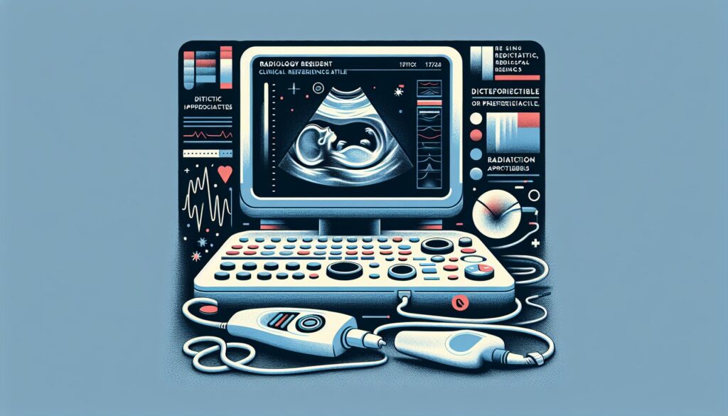
The pediatric ED calls. Six-week-old with projectile vomiting, classic story. The surgeon is waiting on your read. They want to know: is it hypertrophic pyloric stenosis (HPS), or just pylorospasm? Getting the measurements right—and more importantly, doing the dynamic observation—is the difference between a definitive diagnosis and an equivocal report that forces an upper GI series. Your attending expects you to know the exact measurement criteria and what to look for during that crucial five minutes of scanning.
When I was a junior resident, the fear was always mismeasuring the muscle or calling it positive when it was just a transient spasm. It’s a high-stakes read with a clear surgical outcome. Let’s walk through a rock-solid approach to the exam and the report. For more high-yield guides like this, check out the residents free-reference hub.
What a Pediatric Pyloric Stenosis Ultrasound Covers and What Attendings Look For
The ultrasound for hypertrophic pyloric stenosis is the gold-standard diagnostic study for infants between 2 and 12 weeks of age presenting with non-bilious projectile vomiting. It has effectively replaced the upper GI series for this indication due to its high accuracy and lack of ionizing radiation. The exam is focused, answering one primary question: is the pyloric muscle pathologically thickened and causing a fixed gastric outlet obstruction?
Your attending expects a report that is concise but complete. They will be looking for these key elements:
- Pyloric Muscle Measurements: The single-wall muscle thickness (transverse) and the pyloric channel length (longitudinal).
- Dynamic Observation: A clear statement on whether the pylorus relaxed and allowed gastric contents to pass into the duodenum over a minimum of five minutes.
- Classic Signs: Mention of the “cervix sign” or “antral nipple sign” if present.
- Exclusion of Mimics: Confirmation that the findings are fixed and not transient pylorospasm.
- Anatomic Survey: A comment on the position of the superior mesenteric vessels to screen for malrotation, especially if the clinical picture is at all atypical.
Radiology Report Template for Ultrasound for Pediatric Pyloric Stenosis
This template provides a reliable starting point for your dictations. You can adapt it for your institution’s specific requirements or use it as a base for a personal macro in your dictation system.
Technique
Real-time grayscale and color Doppler ultrasound of the upper abdomen was performed using a high-frequency linear transducer. The pylorus was evaluated in both short and long axis. Dynamic observation was performed for [e.g., 5-10] minutes after feeding.
Findings
The stomach is distended with fluid and debris. The pyloric muscle is identified. The pyloric muscle single-wall thickness measures [e.g., 5] mm (normal <3 mm). The pyloric channel length measures [e.g., 18] mm (normal <15 mm).
During dynamic real-time observation, the pyloric channel remained closed with no passage of gastric contents into the duodenum. The hypertrophied muscle did not relax. The “antral nipple sign” and “cervix sign” are present. There is no evidence of pylorospasm.
The superior mesenteric artery is to the left of the superior mesenteric vein, a normal orientation. The duodenum and proximal jejunum appear unremarkable.
Impression
Ultrasound findings are diagnostic of hypertrophic pyloric stenosis.
Key measurements:
– Pyloric muscle single-wall thickness: [e.g., 5] mm
– Pyloric channel length: [e.g., 18] mm
Free Radiology Template Sources
Building a personal library of templates is a rite of passage in residency. If you’re looking for more examples beyond this one, two great free repositories exist. The Radiological Society of North America (RSNA) curates a comprehensive library at RadReport.org, which covers a vast range of modalities and subspecialties. Another excellent resource is maintained by Australian radiologists at RadiologyTemplates.com.au.
The Next-Level Move: From Free-Form Dictation to Structured Report
Dictating the positive findings is the easy part. Structuring them perfectly, adding the right negative statements, and ensuring the impression matches the findings takes time and mental energy. This is where AI-powered tools can streamline your workflow. Instead of meticulously editing a template, you can dictate the key positive findings in free form—”pyloric muscle is 5 mm thick and 18 mm long, no passage of fluid on dynamic scan”—and let the software handle the rest.
GigHz Precision AI is designed for this exact task. It takes your free-form dictation of findings and generates a complete, structured report based on pre-loaded templates from the American College of Radiology (ACR) and Society of Interventional Radiology (SIR). It helps ensure your reports are consistent, complete, and use standardized language, which makes you look sharp and saves you time on every read.
When Should You Order a Pediatric Pyloric Stenosis Ultrasound? ACR Appropriateness Criteria
The decision to order a specific imaging study is guided by established best practices. The American College of Radiology (ACR) provides these through its Appropriateness Criteria. For the clinical scenario of vomiting in infants up to 3 months old, the guidance is clear.
According to the ACR, for an infant presenting with suspected pyloric stenosis (typically with non-bilious vomiting), an abdominal ultrasound is the first-line, “Usually Appropriate” imaging study. It is non-invasive, does not use radiation, and has excellent diagnostic accuracy. The primary alternative, an upper GI series, is now considered a historical second-line option for this specific indication, mainly used if the ultrasound is equivocal or if there is a strong suspicion of other pathology like malrotation with midgut volvulus (often suggested by bilious vomiting).
How Much Radiation Does a Pediatric Pyloric Stenosis Ultrasound Deliver?
A key advantage of ultrasound is the complete absence of ionizing radiation. The estimated effective radiation dose for a pediatric pyloric stenosis ultrasound is 0 mSv.
This is categorized by the ACR as Radiation Level “O” for “None.” For parents concerned about radiation exposure in their infant, you can confidently state that this exam is entirely radiation-free, making it the safest and most appropriate initial imaging test for this condition.
Pediatric Pyloric Stenosis Ultrasound Protocol — Technique, Measurements, and Pitfalls
A standardized protocol is essential for a diagnostic HPS ultrasound. The exam begins by positioning the infant (supine or right lateral decubitus) and using the fluid-filled stomach as an acoustic window. The key is to identify the pylorus and perform specific measurements and a dynamic evaluation.
The table below summarizes the critical parameters of the exam.
| Parameter | Specification | Purpose |
|---|---|---|
| Transducer | Linear high-frequency 9-15 MHz | Provides high-resolution images of the superficial pylorus. |
| Approach | Right epigastrium; supine or right lateral decubitus | Uses the stomach and gallbladder as landmarks to locate the pyloric channel. |
| Muscle Thickness | Measure a single hypoechoic muscle wall in transverse view | The most critical measurement. ≥3 mm is positive. Do not include mucosa or measure both walls. |
| Channel Length | Measure from antrum to duodenal bulb in longitudinal view | The second key measurement. ≥15 mm is positive. |
| Dynamic Observation | Scan continuously for at least 5 minutes | Differentiates fixed HPS from transient pylorospasm, which will relax and open. |
Common protocol pitfalls: The most common error is measuring the entire pyloric diameter instead of a single muscle wall thickness. Another pitfall is not waiting long enough during the dynamic scan; pylorospasm can be stubborn, and giving it a full five minutes or more is crucial to avoid a false positive. Finally, remember that some institutions use slightly higher thresholds (e.g., ≥4 mm thickness) for older infants to increase specificity.
The 3-Months-Free Offer for Radiology Residents and Fellows
3+ months free for radiology residents and fellows.
Look like a rockstar on your reports. With GigHz Precision AI, you can dictate positive findings in free form, and the AI generates a structured report using ACR + SIR templates with the appropriate clinical decision support firing automatically. This helps you create clear, consistent, and attending-ready reports faster.
All we ask in return is your feedback so we can keep improving the product for trainees. The signup process is simple—no credit card, no long forms. To get started, you just need to provide three items:
- Your PGY year (e.g., PGY-2, PGY-4)
- Your training type (radiology residency or specific fellowship)
- Your training program or hospital name
To get access, apply for the residents free-access program and reply to the application with the three items above. We’ll get you set up.
Free GigHz Tools That Pair With This Article
Three free tools that complement the material above:
- ACR Appropriateness Criteria Lookup — Type an indication or clinical scenario in plain language and get the imaging studies the ACR rates for it, with adult and pediatric radiation levels. Built directly from 297 ACR topics, 1,336 clinical variants, and 15,823 procedure ratings.
- GigHz Imaging Protocol Library — A searchable library of 131 imaging protocols with the physics specs surfaced and the matching ACR Appropriateness Criteria alongside. Plain-English narratives readable in 60 seconds, organized by modality.
- GigHz Radiation Dose Calculator — Pick the imaging studies a patient has had and see total dose in millisieverts (mSv) with comparisons to natural background radiation, transatlantic flights, and chest X-rays. Useful for shared decision-making.
Frequently Asked Questions
Is GigHz Precision AI HIPAA-compliant?
Yes. The platform is designed for de-identified workflows by default. No patient-identifying information is required to use the tool to structure your report findings.
Do I need my hospital’s IT department to set this up?
No. GigHz Precision AI is browser-based and requires no local software installation or IT involvement. It works on any modern computer, including the call-room PC or your personal iPad.
Does this replace or interfere with PowerScribe?
It works alongside your existing dictation system. You can use it to generate the structured report text and then simply copy and paste it into PowerScribe, Epic, or any other PACS/EMR. It complements your workflow, it doesn’t replace it.
Can I use this on my phone or iPad?
Yes, the platform is fully responsive and works well on mobile devices and tablets, making it easy to use on the go or in different reading rooms.
Can I customize the templates?
Yes, you can create and save your own custom templates or modify the existing ACR/SIR templates to match your personal preferences or your institution’s specific formatting requirements.
What happens after I finish my residency or fellowship?
The free access program is specifically for trainees. After you graduate, you can transition to a standard subscription plan for practicing radiologists to continue using the service without interruption.
Reviewed by Pouyan Golshani, MD, Interventional Radiologist — May 7, 2026