What Follow-Up Imaging Is Best for Nontraumatic Intraparenchymal Hemorrhage in Adults?
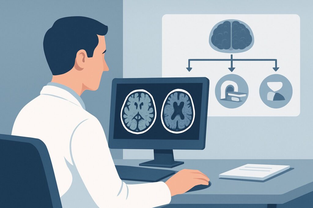
A 62-year-old patient with a history of hypertension is stabilized in the neuro-intensive care unit following a spontaneous basal ganglia hemorrhage, which was confirmed on an initial non-contrast head CT. Now, several days later, the immediate crisis has passed, and your focus shifts to the underlying cause. You need to determine the optimal follow-up imaging study to investigate the etiology of the bleed and guide long-term management to prevent recurrence. This article details the clinical workflow for this exact scenario, grounded in the American College of Radiology (ACR) Appropriateness Criteria. For this presentation, the ACR rates MRI head without and with IV contrast as Usually Appropriate, providing the most comprehensive diagnostic information.
Who Fits This Clinical Scenario for Intraparenchymal Hemorrhage Follow-Up?
This guidance applies to a specific patient population: an adult with a known, non-traumatic intraparenchymal hemorrhage (ICH) who is now clinically stable enough for a comprehensive diagnostic workup. The initial bleed has typically been confirmed on a prior study, most often an emergent non-contrast CT of the head. The primary goal of this follow-up imaging is not to re-diagnose the hemorrhage but to identify its underlying cause.
This workflow is distinct from several related but different clinical situations. You should seek other guidance if your patient presents with:
- Initial suspicion of an acute stroke: For a patient presenting with new focal neurologic deficits where the diagnosis is not yet confirmed, the initial imaging workup follows a different pathway. This is covered in the ACR variant for a clinically suspected acute ischemic stroke.
- Transient, resolved symptoms: If the patient experienced temporary neurologic symptoms that have completely resolved, the workup is for a transient ischemic attack (TIA), which has its own dedicated imaging algorithm.
- Signs of venous thrombosis: If the clinical picture suggests cerebral venous sinus thrombosis (e.g., headache, seizures, papilledema), the imaging strategy is tailored specifically to evaluating the dural venous sinuses.
The focus here is exclusively on the secondary, etiologic workup after a spontaneous ICH has already been diagnosed and the patient is no longer in the hyperacute phase.
What Underlying Causes Are You Investigating with Follow-Up Imaging?
After confirming a nontraumatic intraparenchymal hemorrhage, the crucial next step is to uncover the “why.” Follow-up imaging is a diagnostic investigation aimed at identifying the structural cause, which directly informs prognosis and treatment. The differential diagnosis is broad, but imaging helps systematically narrow the possibilities.
Hypertensive Vasculopathy: This is the most common cause of spontaneous ICH, particularly for deep hemorrhages located in the basal ganglia, thalamus, pons, and cerebellum. Chronic hypertension leads to lipohyalinosis of small perforating arteries, forming Charcot-Bouchard microaneurysms that are prone to rupture. Imaging may reveal evidence of chronic hypertensive microangiopathy, such as old lacunar infarcts or other microhemorrhages.
Cerebral Amyloid Angiopathy (CAA): A frequent cause of ICH in the elderly (typically over 65), CAA involves the deposition of beta-amyloid in the walls of small- to medium-sized cortical and leptomeningeal arteries. This makes the vessels fragile and prone to bleeding. CAA characteristically causes lobar or cortico-subcortical hemorrhages, often with multiple, recurrent bleeds over time. MRI is particularly sensitive for the associated cortical microbleeds.
Underlying Vascular Malformations: These are critical to identify as they often carry a high risk of re-bleeding and may have specific treatments. This category includes arteriovenous malformations (AVMs), cavernous malformations (cavernomas), developmental venous anomalies (DVAs), and dural arteriovenous fistulas (dAVFs). Each has a characteristic appearance on advanced imaging.
Intracranial Tumors: Both primary brain tumors (like glioblastoma) and metastatic disease (especially from melanoma, renal cell carcinoma, choriocarcinoma, and lung cancer) can present with hemorrhage. The presence of a discrete, enhancing mass associated with the hematoma is a key imaging finding.
Other Less Common Causes: The differential also includes ruptured saccular (berry) aneurysms that bleed into the parenchyma, mycotic (infectious) aneurysms, cerebral vasculitis, and coagulopathies. Imaging helps differentiate these from more common etiologies.
Why Is MRI Head Without and With IV Contrast Usually Appropriate for ICH Follow-Up?
The ACR designates MRI head without and with IV contrast as a Usually Appropriate study because it offers unparalleled soft-tissue characterization and sensitivity for the wide range of potential underlying causes of a nontraumatic ICH. It is the most comprehensive single test for this clinical question.
The power of this study lies in its combination of sequences:
- Non-contrast sequences are foundational. Susceptibility-Weighted Imaging (SWI) or Gradient-Recalled Echo (GRE) sequences are exceptionally sensitive to blood products. They can detect tiny, chronic microhemorrhages that are the hallmark of hypertensive vasculopathy or cerebral amyloid angiopathy. These sequences are also key for identifying the characteristic “popcorn” appearance with a hemosiderin rim seen in cavernous malformations. Standard sequences like T1, T2, and FLAIR help determine the age of the hematoma and visualize surrounding edema or non-hemorrhagic parenchymal abnormalities.
- Post-contrast sequences are essential for detecting pathology that enhances with intravenous gadolinium. This is critical for identifying an underlying tumor, an abscess, the nidus of an arteriovenous malformation, or signs of active inflammation from vasculitis. Without contrast, these etiologies can be easily missed.
This combination provides a complete picture that no other single modality can match. Importantly, MRI achieves this with no ionizing radiation (0 mSv).
Why Other Studies Are Rated Differently
While other studies are also rated Usually Appropriate, they serve different, often more limited, roles in this specific follow-up context.
- CT head without IV contrast: This study is excellent for the initial diagnosis of ICH and for follow-up monitoring of hematoma size, mass effect, or developing hydrocephalus. However, its sensitivity for the underlying cause is low. It cannot reliably detect cavernomas, small tumors, or the microbleeds of CAA. It is a tool for stability, not etiology.
- CTA head with IV contrast: This is a fast and effective study for identifying larger vascular lesions like aneurysms and AVMs. It is often used when MRI is contraindicated or unavailable, or as a first-line study in the acute setting if a vascular cause is strongly suspected. However, it provides less information about the brain parenchyma, is less sensitive for cavernomas, and involves both ionizing radiation (☢☢☢ 1-10 mSv) and iodinated contrast.
- Arteriography cervicocerebral: Rated May be appropriate, digital subtraction angiography (DSA) is the invasive gold standard for characterizing vascular anatomy. It is typically reserved for cases where a vascular malformation is seen or highly suspected on MRI/CTA, or when non-invasive imaging is negative but clinical suspicion remains high (e.g., in a young patient with a lobar hemorrhage).
What Are the Next Steps After the Follow-Up MRI?
The results of the contrast-enhanced MRI will guide the subsequent clinical pathway. The downstream workflow is a decision tree based on the imaging findings.
If the MRI identifies a specific structural lesion:
- Vascular Malformation (AVM, dAVF): An urgent referral to neurosurgery and/or neurointerventional radiology is warranted. The next step is often a conventional catheter cerebral angiogram (DSA) to fully characterize the lesion’s angioarchitecture and plan for treatment, which may include embolization, surgical resection, or stereotactic radiosurgery.
- Tumor: A referral to neurosurgery for biopsy or resection and to neuro-oncology for further management is the standard of care. Additional systemic imaging may be needed to search for a primary malignancy if metastases are suspected.
- Cavernous Malformation: Management depends on the location and whether it has bled previously. This typically involves a neurosurgical consultation to discuss the risks and benefits of observation versus surgical removal.
If the MRI suggests an underlying small vessel disease:
- Hypertensive Vasculopathy or CAA: The findings confirm a diagnosis that is managed medically. The focus shifts to aggressive risk factor modification, primarily strict blood pressure control for hypertensive vasculopathy and avoidance of antithrombotic agents when possible for CAA.
If the MRI is negative or non-diagnostic:
- In a younger patient with a lobar hemorrhage and a negative MRI/MRA, the index of suspicion for an occult vascular lesion remains. In this case, a conventional angiogram, rated May be appropriate, may be considered to definitively rule out a small AVM or dural fistula that was not visible on non-invasive imaging.
Pitfalls to Avoid (and When to Get Help)
Navigating the workup of a nontraumatic ICH requires careful consideration to avoid common diagnostic errors.
- Stopping at the Non-Contrast CT: While essential for initial diagnosis, a non-contrast CT is insufficient for the etiologic workup. Failing to proceed to MRI or vascular imaging means potentially missing a treatable underlying cause.
- Omitting IV Contrast: Ordering a non-contrast MRI saves time but can miss critical diagnoses. An enhancing tumor, AVM nidus, or abscess will not be characterized without gadolinium. Always specify “without and with IV contrast” unless there is a strong contraindication.
- Misinterpreting the Cause by Location: While location is a strong clue (deep for hypertension, lobar for CAA), these are not absolute rules. Atypical locations should prompt a thorough search for other causes like tumors or vascular malformations.
- Delaying the Workup: Once the patient is medically stable, the diagnostic workup should proceed in a timely manner. Identifying a high-risk lesion like an AVM is critical for preventing a potentially devastating re-hemorrhage.
If the imaging findings are complex, equivocal, or suggest a rare vascular lesion, consultation with a neuroradiologist is essential. For any identified treatable lesion, immediate escalation to the appropriate neurosurgical or neurointerventional service is the critical next step.
Related ACR Topics and Tools
This article focuses on a single, specific clinical scenario. For a comprehensive overview of imaging for all stroke and stroke-related conditions, consult the parent topic guide. For additional tools to refine your imaging orders and discuss them with patients, see the resources below.
- For breadth across all scenarios in Cerebrovascular Diseases-Stroke and Stroke-Related Conditions, see our parent guide: Cerebrovascular Diseases-Stroke and Stroke-Related Conditions: ACR Appropriateness Decoded.
- To explore adjacent scenarios and their recommended workups, use the ACR Appropriateness Criteria Lookup.
- For technical details on how specific studies are performed, visit the Imaging Protocol Library.
- To help frame conversations about radiation exposure from CT or angiography, refer to the Radiation Dose Calculator.
Frequently Asked Questions
Why is MRI preferred over CT for the follow-up of an intraparenchymal hemorrhage?
While CT is excellent for initial diagnosis and monitoring for acute changes like re-bleeding, MRI provides far superior detail about the underlying brain tissue. MRI, particularly with susceptibility-weighted sequences (SWI/GRE), can detect chronic microhemorrhages suggestive of hypertensive vasculopathy or cerebral amyloid angiopathy. Furthermore, contrast-enhanced MRI is essential for identifying underlying tumors, cavernomas, or arteriovenous malformations that are often invisible on CT.
If the patient has a contraindication to MRI (e.g., an incompatible device), what is the next best test?
If MRI is contraindicated, the ACR rates CTA head with IV contrast as ‘Usually Appropriate.’ This is a strong alternative for identifying vascular causes like aneurysms or AVMs. For a suspected tumor, a contrast-enhanced CT head would be the next step. However, these CT-based studies are less sensitive than MRI for non-vascular causes like cavernomas or evidence of small vessel disease.
Is a conventional angiogram (DSA) always necessary if an AVM is suspected on MRI?
In most cases, yes. While MRI/MRA can identify an AVM, a digital subtraction angiogram (DSA) is considered the gold standard for defining the detailed angioarchitecture. It provides critical information about the feeding arteries, the structure of the nidus, and the pattern of venous drainage, all of which are essential for planning treatment (e.g., embolization, surgery, or radiosurgery).
How soon after the initial hemorrhage should this follow-up imaging be performed?
The timing depends on clinical stability. The patient must be stable enough to tolerate transport and the duration of an MRI scan. Typically, this workup is performed within a few days to a week after the initial event, once any immediate life-threatening issues like elevated intracranial pressure have been managed. The goal is to balance patient stability with the need to identify a cause to prevent re-bleeding.
What if the follow-up MRI is completely normal, aside from the resolving hematoma?
In an older patient with a deep hemorrhage and a history of hypertension, a negative workup strongly suggests hypertensive vasculopathy as the cause, even without other imaging markers. In a younger, normotensive patient, especially with a lobar hemorrhage, a negative MRI should raise suspicion for an occult vascular lesion. In this specific subgroup, proceeding to a conventional angiogram (DSA) may be warranted to search for a small AVM or fistula not visible on non-invasive imaging.
Reviewed by Pouyan Golshani, MD, Interventional Radiologist — August 4, 2026