What Imaging Is Best for a Child with TMJ Pain and Suspected Idiopathic Arthritis?
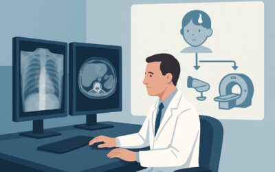
A 10-year-old patient presents to your clinic with a two-month history of progressive right-sided jaw pain and difficulty opening their mouth fully, especially in the morning. There is no history of trauma, fever, or recent dental procedures. On examination, you note tenderness over the right temporomandibular joint (TMJ) and a subtle deviation of the jaw on opening. Given the insidious onset and morning stiffness, you suspect Juvenile Idiopathic Arthritis (JIA). You need to confirm inflammation in the joint to guide therapy, but what is the correct initial imaging study? This article details the clinical workflow for this specific scenario. According to the American College of Radiology (ACR) Appropriateness Criteria, an `MRI temporomandibular joint without and with IV contrast` is Usually appropriate as the initial imaging test.
Who Fits This Clinical Scenario?
This guidance applies specifically to the initial imaging workup for a child presenting with symptoms localized to the temporomandibular joint—such as pain, swelling, limited motion, or clicking—where Juvenile Idiopathic Arthritis is a leading clinical suspicion. The patient typically has no history of significant facial trauma that would suggest a fracture or acute internal derangement. The presentation is often subacute or chronic, distinguishing it from a more acute infectious process.
It is crucial to differentiate this scenario from others that require a different diagnostic approach:
- Appendicular Joint Pain: If the child’s primary complaint is pain or swelling in a peripheral joint like the knee, wrist, or ankle, the workup follows the guidance for appendicular joint pain, which often starts with different modalities.
- Back or Sacroiliac Pain: If the symptoms point towards the axial skeleton, such as persistent back pain or buttock pain, the evaluation should follow the specific ACR variants for suspected spondyloarthropathy or sacroiliitis.
- Known Traumatic Injury: A clear history of a blow to the jaw or face shifts the primary concern to fracture, dislocation, or internal derangement, which may alter the choice or protocol of the imaging study.
This workflow is intended for the initial diagnostic phase, not for routine follow-up or surveillance of established TMJ arthritis, which may have different imaging considerations.
What Diagnoses Are You Working Up in This Scenario?
When a child presents with non-traumatic TMJ pain, your differential diagnosis guides the imaging choice. The goal is to identify active inflammation and rule out other pathologies that can mimic JIA.
Juvenile Idiopathic Arthritis (JIA) is the primary diagnosis of concern. The TMJ is one of the most commonly affected joints in JIA, yet it can be clinically silent in its early stages. Early detection is critical because untreated TMJ arthritis can lead to significant growth disturbances of the mandible, resulting in facial asymmetry, micrognathia, and long-term functional impairment. Imaging seeks to find synovitis, joint effusion, and bone marrow edema—the hallmarks of active inflammation.
Internal Derangement of the TMJ, such as anterior disc displacement, is another common cause of jaw pain and dysfunction. While it can occur with JIA, it can also be an isolated mechanical issue. MRI is excellent for visualizing the position and morphology of the articular disc.
Septic Arthritis or Osteomyelitis is a less common but critical diagnosis to exclude. This would typically present more acutely with fever, significant swelling, and severe, constant pain. Imaging is used to identify joint effusion, abscess formation, and signs of bone infection. Prompt diagnosis is essential to prevent permanent joint destruction.
Developmental or Congenital Anomalies, such as condylar hyperplasia or hypoplasia, can cause pain and altered mechanics. While often presenting as a painless facial asymmetry, pain can be a feature. Imaging helps characterize the morphology of the mandibular condyle and its relationship with the glenoid fossa.
Why Is MRI Without and With IV Contrast the Recommended Study?
For a child with suspected JIA of the temporomandibular joint, the ACR designates `MRI temporomandibular joint without and with IV contrast` as Usually appropriate. This recommendation is based on the modality’s superior ability to visualize the specific pathological changes of early inflammatory arthritis without using ionizing radiation.
The rationale for this choice over other modalities is clear:
- Detecting Early Inflammation: MRI excels at soft tissue and bone marrow characterization. It can directly visualize synovial thickening (pannus), joint effusion, and bone marrow edema. The administration of intravenous contrast is crucial, as enhancement of the synovium is the most reliable sign of active synovitis. A non-contrast MRI, rated as Usually not appropriate, can miss this key finding and falsely reassure the clinician.
- Anatomic and Functional Detail: Beyond inflammation, MRI provides detailed assessment of the articular disc position, condylar morphology, and surrounding soft tissues, helping to rule out or concurrently diagnose internal derangement.
- Radiation Safety: MRI uses no ionizing radiation (Pediatric RRL: O), a paramount consideration in children who may require future imaging and are more sensitive to the long-term effects of radiation.
Alternative imaging studies are rated lower for this specific clinical question:
- Radiography (X-ray) of the TMJ is rated Usually not appropriate. It is insensitive to the early signs of JIA, such as synovitis and marrow edema. Radiographs only show late-stage bony changes like erosions or condylar flattening, which is too late for early therapeutic intervention. It also involves a small amount of radiation (Pediatric RRL: ☢☢ 0.03-0.3 mSv).
- Computed Tomography (CT) of the maxillofacial region is also Usually not appropriate. While excellent for evaluating complex bone anatomy and fractures, it has poor soft tissue contrast and cannot reliably detect synovitis or marrow edema. Furthermore, it delivers a significant radiation dose to a child’s head and neck (Pediatric RRL: ☢☢☢ 0.3-3 mSv), making it an undesirable choice for a non-traumatic inflammatory workup.
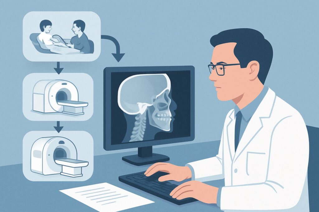
When ordering, be sure to specify “TMJ protocol without and with IV contrast” to ensure the imaging center performs the appropriate dedicated sequences, including open- and closed-mouth views, to fully assess joint mechanics and inflammation.
What’s Next After MRI? Downstream Workflow
The results of the contrast-enhanced MRI will directly guide your next steps in management and consultation. The post-imaging workflow branches based on whether active inflammation consistent with JIA is identified.
If the MRI is positive for JIA: Findings such as synovial enhancement, significant joint effusion, bone marrow edema, or erosions confirm active TMJ arthritis. The essential next step is a referral to a Pediatric Rheumatologist. They will initiate or adjust systemic medical therapy, which may include disease-modifying antirheumatic drugs (DMARDs) or biologic agents, to control inflammation and prevent long-term joint damage. A concurrent referral to an Orthodontist or Oral and Maxillofacial Surgeon with expertise in JIA may also be necessary to manage functional impairment and monitor jaw growth.
If the MRI is negative for JIA: A normal MRI effectively rules out active inflammatory arthritis. If the patient’s symptoms persist, the focus of the investigation should shift. Re-evaluate for other causes on the differential, such as myofascial pain syndrome, which is a clinical diagnosis. If the MRI shows an isolated internal derangement (e.g., disc displacement without inflammation), management may involve physical therapy, oral appliances, or referral to an oral surgeon. If the entire workup remains negative, consider less common causes or sources of referred pain from dental or otologic structures.
If the MRI is indeterminate: Occasionally, findings can be equivocal, such as a small, non-specific joint effusion without synovial enhancement. In these cases, close clinical correlation is key. A discussion with the interpreting pediatric radiologist can provide valuable context. The decision may be to proceed with a trial of conservative therapy and clinical follow-up, with a low threshold for repeat imaging in several months if symptoms worsen or fail to resolve.
Pitfalls to Avoid (and When to Get Help)
Navigating the workup for pediatric TMJ pain requires avoiding several common pitfalls to ensure timely and accurate diagnosis.
- Ordering a Non-Contrast MRI: The most critical error is omitting intravenous contrast. Active synovitis is best visualized by synovial enhancement, which is invisible on non-contrast sequences. A negative non-contrast MRI can provide false reassurance in the setting of active disease.
- Starting with X-ray or CT: Relying on modalities that use ionizing radiation and are insensitive to early inflammatory changes delays diagnosis. By the time bony erosions are visible on a radiograph or CT, irreversible joint damage may have already occurred.
- Dismissing “Mild” Symptoms: TMJ arthritis in JIA can be clinically subtle or even asymptomatic, especially early on. Do not dismiss mild jaw clicking, slight morning stiffness, or minimal deviation, as these can be the only signs of underlying inflammation that can impact jaw growth.
- Failing to Consider the Full Differential: While JIA is a primary concern, automatically attributing all TMJ pain to it without considering infection, trauma, or mechanical causes can lead to misdiagnosis.
If you encounter red flag symptoms such as fever, acute swelling, and inability to close the mouth, escalate immediately for an urgent evaluation to rule out septic arthritis.
Related ACR Topics and Tools
For a comprehensive overview of imaging recommendations across all clinical presentations of this condition, please consult the parent topic article. For specific questions about adjacent scenarios or to explore imaging techniques and safety, the following resources are available.
- For breadth across all scenarios in Joint Pain: Idiopathic Arthritis-Child, see our parent guide: Joint Pain: Idiopathic Arthritis-Child: ACR Appropriateness Decoded.
- To explore other clinical scenarios, use the ACR Appropriateness Criteria Lookup.
- For details on imaging techniques, browse the Imaging Protocol Library.
- To discuss radiation exposure with families, consult the Radiation Dose Calculator.
Frequently Asked Questions
Why is an MRI with contrast necessary if we already have a strong clinical suspicion for JIA?
An MRI with contrast is essential to confirm active inflammation within the temporomandibular joint. Clinical suspicion is a starting point, but imaging provides objective evidence of synovitis (via synovial enhancement), which confirms the diagnosis, helps determine the severity of the disease, and establishes a baseline to monitor treatment response. It also rules out other pathologies that can mimic JIA, such as internal derangement or developmental anomalies.
Can ultrasound be used as a screening tool for TMJ arthritis in a child?
According to the ACR Appropriateness Criteria, ultrasound of the head and neck is ‘Usually not appropriate’ for this specific scenario. While ultrasound can detect joint effusions and synovial thickening in more accessible joints, its utility for the TMJ is limited by the complex anatomy and bony structures that obstruct the view. It is highly operator-dependent and cannot assess for bone marrow edema, a key early sign of inflammation that is readily seen on MRI.
Does a child need to be sedated for a TMJ MRI?
The need for sedation or general anesthesia depends on the child’s age, developmental level, and ability to remain still for the duration of the scan, which is typically 30-45 minutes. Many older children and adolescents can tolerate the scan without sedation, but younger children or those with anxiety often require it to ensure high-quality, motion-free images. This should be discussed with the family and the imaging department prior to the appointment.
What if the child has braces or other dental hardware?
Orthodontic hardware can create significant artifacts on MRI scans, potentially obscuring the temporomandibular joints. It is crucial to inform the radiology department about any dental hardware when scheduling the exam. Radiologists can often use specific MRI sequences and techniques to mitigate these artifacts, but in some cases, the image quality may still be compromised. The benefit of the scan must be weighed against the potential for artifact.
If the MRI is positive for JIA in the TMJ, should we screen other joints?
Yes, a new diagnosis of JIA in the TMJ warrants a comprehensive evaluation by a pediatric rheumatologist. This will include a full physical examination of all other joints. The rheumatologist will determine if further imaging of other symptomatic or at-risk joints (like the cervical spine or sacroiliac joints) is necessary based on the clinical picture and the subtype of JIA suspected.
Reviewed by Pouyan Golshani, MD, Interventional Radiologist — May 30, 2026