What Imaging Is Best for a Sudden, Severe ‘Thunderclap’ Headache?
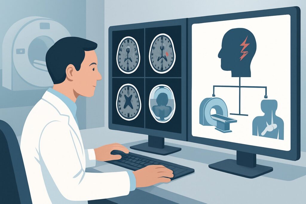
A 45-year-old patient arrives in the emergency department describing the abrupt onset of the “worst headache of my life.” It started like a switch was flipped, reaching maximal, excruciating intensity in less than a minute. Their neurologic exam is non-focal, but the story is alarming. You know that immediate imaging is required to rule out life-threatening causes, but which study provides the fastest, most accurate initial answer? This article details the American College of Radiology (ACR) evidence-based workflow for this specific, high-stakes clinical scenario. For a sudden onset severe headache reaching maximal severity within one hour, the ACR designates CT head without IV contrast as Usually appropriate, making it the clear first-line imaging test.
Who Fits This Clinical Scenario?
This guidance applies to patients presenting with a “thunderclap headache,” a term that specifically describes a severe headache that develops abruptly and reaches its peak intensity within one hour, often in less than five minutes. The key diagnostic feature is the hyperacute time course, which patients may describe as being “hit by a lightning bolt” or a “thunderclap” in their head. The patient’s neurologic examination may be normal or may show deficits such as nuchal rigidity or altered mental status.
It is critical to distinguish this presentation from other headache patterns that follow different diagnostic pathways:
- Gradual Onset Headaches: This workflow does not apply to headaches that build in intensity over several hours or days, such as typical primary migraine or tension-type headaches.
- Positional Headaches: If the headache is significantly worse when the patient is upright and improves when they lie down, this suggests intracranial hypotension, a distinct clinical scenario with a different imaging workup.
- Headaches with Papilledema: If the headache is accompanied by signs of increased intracranial pressure like papilledema or pulsatile tinnitus, the workup shifts to evaluating for intracranial hypertension.
This article is exclusively for the patient whose defining feature is the sudden, explosive onset of a severe headache, a classic red flag for a serious underlying pathology.
What Diagnoses Are You Working Up in This Scenario?
The imaging choice for a thunderclap headache is driven by a differential diagnosis that is heavily weighted toward time-sensitive, potentially catastrophic conditions. The primary goal of the initial scan is to identify or exclude these possibilities quickly.
Subarachnoid Hemorrhage (SAH): This is the single most important diagnosis to exclude. Most commonly caused by a ruptured cerebral aneurysm, SAH presents with a classic thunderclap headache in over 70% of cases. Blood collecting in the subarachnoid space is acutely life-threatening and requires immediate neurosurgical or neurointerventional consultation. The entire initial workup is designed around maximizing sensitivity for this diagnosis.
Reversible Cerebral Vasoconstriction Syndrome (RCVS): A less common but important mimic of aneurysmal SAH, RCVS involves multifocal constriction of cerebral arteries. It can also present with a thunderclap headache and may sometimes be associated with small, convexal subarachnoid hemorrhages. While the initial imaging may be negative, the clinical suspicion can trigger further vascular imaging.
Cervical Artery Dissection: A tear in the wall of a carotid or vertebral artery can lead to a thunderclap headache, often accompanied by neck pain. While the primary finding is vascular, a dissection can lead to ischemic stroke or, rarely, subarachnoid hemorrhage, which may be visible on initial imaging.
Cerebral Venous Sinus Thrombosis (CVST): Though the headache of CVST is often more subacute, it can present acutely. This condition involves a blood clot in the brain’s venous drainage system and can lead to venous infarction or hemorrhage. While non-contrast CT can show signs of CVST, it is often subtle, and dedicated venous imaging is typically required for definitive diagnosis.
Why Is CT Head without IV Contrast the Recommended Initial Study?
For a patient with a sudden, severe headache, the ACR panel rates CT head without IV contrast as Usually appropriate. This recommendation is based on its high diagnostic accuracy for the most critical diagnosis, its speed, and its widespread availability.
The primary rationale is its excellent sensitivity for detecting acute subarachnoid hemorrhage. Freshly extravasated blood is hyperdense (appears bright) on a non-contrast CT scan, making it readily visible in the basal cisterns, sulci, and ventricles. When performed within the first 6-12 hours of symptom onset, its sensitivity for SAH is very high. This makes it the ideal screening tool to rapidly rule in or rule out the most immediate life-threat.
In contrast, other powerful imaging modalities are rated lower for this initial workup for specific reasons:
- MRI head without IV contrast is rated Usually not appropriate. While MRI is excellent for many neurologic conditions, it is less sensitive than CT for hyperacute hemorrhage. Furthermore, it is slower to acquire and less accessible in most emergency settings, making it impractical for a time-sensitive workup where minutes matter.
- CTA head with IV contrast is rated May be appropriate. CTA is a crucial test, but not as the first step. Its role is to identify the source of a bleed (like an aneurysm) after hemorrhage has been confirmed on the non-contrast CT. Using it as the initial test exposes the patient to IV contrast and a higher radiation dose (☢☢☢ 1-10 mSv) unnecessarily if the non-contrast CT is negative and SAH is ultimately ruled out by other means.
The non-contrast CT provides the essential first data point—is there blood?—with a moderate radiation dose (adult relative radiation level ☢☢☢ 1-10 mSv) and no risk from IV contrast agents. It is the fastest way to triage the patient toward the correct downstream pathway. Once you’ve decided on this study, our protocol guide covers the technique and key reading principles. For a detailed review, see our guide: CT Brain Without Contrast.
What’s Next After CT Head without IV Contrast? Downstream Workflow
The result of the initial non-contrast head CT dictates the subsequent management steps in a clear, branching pathway.
If the CT is positive for subarachnoid hemorrhage: This is a medical emergency. The immediate next step is to obtain a CTA head with IV contrast to identify the source of the bleeding, most commonly a ruptured aneurysm. An urgent consultation with neurosurgery and/or neurointerventional radiology is mandatory for definitive management, which may include surgical clipping or endovascular coiling of the aneurysm.
If the CT is negative: A negative CT, especially if performed within six hours of headache onset, significantly lowers the probability of SAH. However, it does not completely exclude it, as the sensitivity of CT decreases over time. If clinical suspicion for SAH remains high despite a negative CT, the classic next step is a lumbar puncture (LP). The cerebrospinal fluid is analyzed for red blood cells and xanthochromia (the yellowish breakdown product of hemoglobin), which confirms an older bleed. If the LP is also negative, the diagnosis of aneurysmal SAH is effectively ruled out, and the workup can pivot to other causes like RCVS or migraine.
If the CT is indeterminate or shows other findings: Occasionally, the CT may be equivocal or reveal an unexpected finding like a mass, an intraparenchymal hemorrhage, or signs suggestive of venous sinus thrombosis. In these cases, the next imaging study is tailored to the suspected diagnosis. This often involves MRI/MRA or CT venography (CTV) to better characterize the abnormality and guide further management.
Pitfalls to Avoid (and When to Get Help)
In the high-stakes workup of a thunderclap headache, several common pitfalls can lead to diagnostic delays or errors. Be mindful of the following:
- Delaying the Initial CT: The sensitivity of non-contrast CT for SAH is highest in the first 12-24 hours. As time passes, the blood becomes isodense with brain parenchyma and can be missed. Do not delay the initial scan.
- Stopping the Workup Prematurely: A negative head CT does not end the workup if your clinical suspicion for SAH is high. Failing to proceed to a lumbar puncture in this setting is a frequent cause of missed SAH.
- Misinterpreting Subtle Signs: Small amounts of blood can be subtle, appearing as faint hyperdensity in the sulci or interpeduncular cistern. If the reading is equivocal, a formal radiology review is essential.
- Ordering the Wrong Initial Test: Requesting an MRI or CTA as the first-line study in the emergency setting for this presentation can waste critical time and resources. Stick to the non-contrast CT first.
If the patient has any focal neurologic deficit, a rapidly declining level of consciousness, or hemodynamic instability, escalate immediately to a senior clinician and involve critical care and neurosurgical specialists early.
Related ACR Topics and Tools
This article focuses on a single, critical decision point. For a comprehensive overview of imaging for all headache types and clinical scenarios, please refer to our parent guide. It provides the breadth that complements the depth of this workflow.
- For breadth across all scenarios in Headache, see our parent guide: Headache: ACR Appropriateness Decoded.
- To explore other evidence-based clinical pathways, use the ACR Appropriateness Criteria Lookup.
- To review technical details for other imaging studies, see the Imaging Protocol Library.
- To discuss radiation exposure with patients, our Radiation Dose Calculator can help frame the conversation.
Frequently Asked Questions
What if the non-contrast head CT is negative but I am still highly suspicious of a subarachnoid hemorrhage?
If your clinical suspicion for subarachnoid hemorrhage (SAH) remains high despite a negative CT, the standard of care is to proceed with a lumbar puncture (LP). The cerebrospinal fluid should be analyzed for an elevated red blood cell count and for xanthochromia. A negative CT followed by a negative LP effectively rules out aneurysmal SAH.
Why not order a CTA head on every patient with a thunderclap headache from the start?
While CTA is essential for finding the source of a bleed, it is not the best initial screening test. A non-contrast CT is faster, avoids IV contrast exposure, and has a lower radiation dose. If the non-contrast CT and subsequent LP (if needed) are negative for hemorrhage, the CTA is unnecessary. It is reserved for cases where a bleed has been confirmed, to guide urgent intervention.
How quickly does a non-contrast CT need to be done after headache onset?
As soon as possible. The sensitivity of non-contrast CT for detecting subarachnoid hemorrhage is over 98% within the first 6 hours of symptom onset. This sensitivity decreases over time, dropping to around 90% by 24 hours and approximately 50% after one week, as the blood is resorbed and becomes less dense on the scan.
Is MRI ever the right choice for a thunderclap headache?
MRI is generally not the appropriate *initial* imaging test for a thunderclap headache because it is slower and less sensitive for hyperacute blood than CT. However, it can be very valuable as a second-line test if the initial CT/LP workup is negative but a diagnosis is still needed. MRI is superior for detecting conditions like reversible cerebral vasoconstriction syndrome (RCVS), posterior reversible encephalopathy syndrome (PRES), or small strokes that can also present with a thunderclap headache.
Does this imaging algorithm change for pediatric patients?
The fundamental algorithm of using a non-contrast head CT as the first-line test for suspected SAH generally holds for pediatric patients. However, the differential diagnosis for thunderclap headache in children is broader and includes arteriovenous malformations (AVMs) as a more common cause of hemorrhage than in adults. Radiation dose is a more significant concern, and CT protocols must be specifically tailored to children to minimize exposure (pediatric RRL ☢☢☢ 0.3-3 mSv).
Reviewed by Pouyan Golshani, MD, Interventional Radiologist — June 27, 2026