What Imaging Is Best for Acute Respiratory Illness with Concerning Clinical Signs?
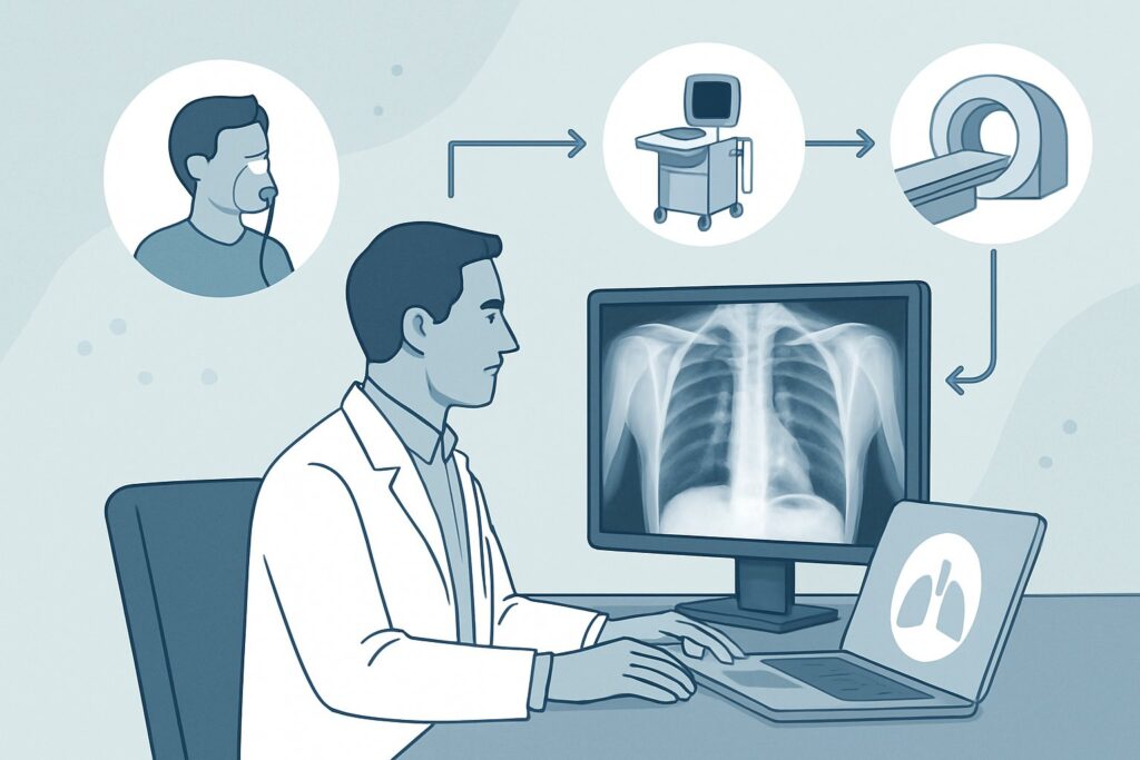
A 68-year-old man with a history of diabetes presents to the urgent care clinic with a four-day history of productive cough, fever, and worsening shortness of breath. On examination, he is febrile to 38.6°C, tachypneic with a respiratory rate of 26, and has diminished breath sounds with crackles in the left lower lung field. His oxygen saturation is 93% on room air. You are confident this patient requires imaging to confirm your suspicion of pneumonia and guide treatment, but which study is the most appropriate first step? This article provides a detailed clinical workflow for this specific scenario, guided by the American College of Radiology (ACR) Appropriateness Criteria. For this patient with risk factors for a poor outcome, the ACR rates a chest radiograph as ‘Usually Appropriate’ for initial imaging.
Who Fits This Clinical Scenario?
This guidance applies to a specific subset of immunocompetent adult patients presenting with an acute respiratory illness. The key inclusion criteria are the presence of respiratory symptoms (such as cough, dyspnea, or sputum production) combined with one or more risk factors that suggest a more severe illness or a higher likelihood of pneumonia. These risk factors include:
- Positive physical examination findings: Such as focal crackles, egophony, bronchial breath sounds, or dullness to percussion.
- Abnormal vital signs: Including fever (temperature >38°C), tachycardia (heart rate >100 bpm), tachypnea (respiratory rate >24 breaths/min), or hypotension.
- Organic brain disease: New or worsening confusion or altered mental status, which can be a sign of severe systemic illness or hypoxia.
- Other risk factors for poor outcome: This category includes advanced age (typically >65 years) or the presence of significant comorbidities like chronic obstructive pulmonary disease (COPD), diabetes mellitus, chronic heart failure, or chronic kidney disease.
This workflow is distinct from other clinical situations. It does not apply to patients with a completely normal physical exam and stable vital signs, a scenario where imaging may not be indicated at all. It also differs from cases where a specific complication, such as a parapneumonic effusion or empyema, is already strongly suspected, which may alter the choice of initial imaging.
What Diagnoses Are You Working Up in This Scenario?
When a patient presents with acute respiratory symptoms and concerning clinical signs, the primary goal of initial imaging is to confirm or exclude a few key diagnoses that require specific management.
Community-Acquired Pneumonia (CAP): This is the most common and critical diagnosis to identify. The presence of fever, cough, and focal findings on a lung exam makes CAP the leading consideration. Imaging is essential to confirm the presence of a parenchymal infiltrate, which establishes the diagnosis and provides a baseline for assessing treatment response. Early and accurate diagnosis is crucial for initiating appropriate antibiotic therapy and reducing morbidity.
Acute Exacerbation of Underlying Lung Disease: In patients with known or undiagnosed chronic lung conditions like COPD or bronchiectasis, an acute respiratory illness can represent an infectious exacerbation. While often viral, a superimposed bacterial pneumonia is a common trigger. A chest radiograph helps differentiate a simple exacerbation from one complicated by a new infiltrate, which would necessitate antibiotics.
Acute Decompensated Heart Failure: Particularly in older adults or those with known cardiac disease, symptoms of dyspnea and cough can mimic pneumonia. A chest radiograph is invaluable for identifying signs of pulmonary edema, cardiomegaly, or pleural effusions that would point toward a cardiac etiology rather than a primary infection, thereby redirecting management toward diuretics and afterload reduction.
Other Less Common Causes: While less frequent, initial imaging can sometimes reveal alternative diagnoses. These might include a large pleural effusion from a non-infectious cause, evidence of aspiration pneumonitis, or, rarely, findings suggestive of a pulmonary embolism or malignancy presenting with acute symptoms. The initial radiograph serves as a crucial screening tool to rule out these possibilities.
Why Is a Chest Radiograph the Recommended Initial Study for This Presentation?
The ACR designates a standard chest radiograph (Radiography chest) as ‘Usually Appropriate’ for this clinical scenario, making it the clear first-choice imaging study. The rationale is grounded in its diagnostic utility, safety, and efficiency for answering the primary clinical question: Is pneumonia present?
A chest radiograph is highly effective for detecting the alveolar opacities and infiltrates characteristic of community-acquired pneumonia. For the vast majority of patients in this category, it provides sufficient information to confirm the diagnosis, assess the extent of the disease, and initiate empiric antibiotic therapy. Furthermore, it is a fast, cost-effective, and universally available examination. From a safety perspective, it involves a very low radiation dose (adult relative radiation level ☢ <0.1 mSv), which is a key consideration in applying the As Low As Reasonably Achievable (ALARA) principle.
In contrast, more advanced imaging modalities are rated ‘Usually not appropriate’ for the initial evaluation of these patients:
- CT chest without IV contrast: While more sensitive than radiography for subtle or early pneumonia, a CT scan is not recommended as the first step. It delivers a significantly higher radiation dose (adult RRL ☢☢☢ 1-10 mSv) without typically altering the initial management of uncomplicated CAP. Its use is reserved for cases where the radiograph is negative but clinical suspicion remains high, or when complications are suspected.
- US chest: Point-of-care ultrasound (POCUS) can be useful for detecting pleural effusions and subpleural consolidations. However, the ACR rates it as ‘Usually not appropriate’ as a comprehensive initial study because it is highly operator-dependent, has a limited field of view for deep parenchymal disease, and is less standardized than radiography for a complete lung assessment.
The initial choice of a chest radiograph aligns with a stepwise diagnostic approach. It effectively triages patients, confirming the diagnosis in most cases while reserving higher-radiation or more complex studies for those with atypical presentations, non-resolving symptoms, or suspected complications.
What’s Next After a Chest Radiograph? Downstream Workflow
The results of the initial chest radiograph directly guide the next steps in patient management. The clinical pathway diverges based on whether the findings are positive, negative, or indeterminate.
If the radiograph is positive for pneumonia (e.g., shows a lobar consolidation or infiltrate): The diagnosis of CAP is confirmed. The immediate next step is to initiate empiric antibiotic therapy based on local resistance patterns and patient-specific factors (e.g., comorbidities, recent hospitalizations). Risk stratification scores like the CURB-65 or Pneumonia Severity Index (PSI) can help determine the appropriate site of care—outpatient, inpatient ward, or intensive care unit. Follow-up imaging is generally not needed for patients who respond well to treatment.
If the radiograph is negative: A negative chest X-ray in a patient with concerning signs presents a clinical challenge. If clinical suspicion for pneumonia remains high (e.g., persistent hypoxia, high fever), consider that the infiltrate may be in an early stage and not yet visible. Management may involve treating empirically for pneumonia and observing closely, or considering a CT chest if an alternative diagnosis (like pulmonary embolism) is suspected or if the patient is severely ill. If clinical suspicion is lower, the diagnosis is more likely acute bronchitis or another viral respiratory illness, which typically requires only supportive care.
If the radiograph is indeterminate or shows other findings: Sometimes, findings are ambiguous (e.g., atelectasis vs. infiltrate) or suggest an alternative diagnosis (e.g., signs of heart failure). In these cases, clinical correlation is paramount. If heart failure is suspected, obtaining a B-type natriuretic peptide (BNP) level and initiating diuretic therapy may be appropriate. If a large pleural effusion is seen, this may trigger a different workflow, such as diagnostic thoracentesis, which is covered in a separate ACR variant for complicated pneumonia.
Pitfalls to Avoid (and When to Get Help)
In managing patients with acute respiratory illness and risk factors, several common pitfalls can impact outcomes. First, avoid delaying initial imaging in a patient with abnormal vital signs or hypoxia; a timely chest radiograph is crucial for diagnosis and disposition. Second, do not reflexively order a CT scan as the initial test for suspected uncomplicated pneumonia; this leads to unnecessary radiation exposure and cost. Third, be cautious about over-interpreting a negative chest radiograph in a patient who appears clinically ill, as early or subtle pneumonias can be missed. Finally, remember to correlate imaging findings with the full clinical picture—a small infiltrate in an otherwise well-appearing patient is managed differently than the same finding in a septic, elderly individual. If a patient fails to improve on appropriate antibiotics or deteriorates clinically, it is time to escalate care and consider repeat or advanced imaging (like CT) to look for complications such as empyema, abscess, or an alternative diagnosis.
Related ACR Topics and Tools
The ACR Appropriateness Criteria are a comprehensive resource for evidence-based imaging decisions. This article focuses on one specific scenario, but many related situations exist. For a broader overview of all variants within this topic, please consult our parent guide. For additional tools to support your clinical workflow, see the resources below.
- For breadth across all scenarios in Acute Respiratory Illness in Immunocompetent Patients, see our parent guide: Acute Respiratory Illness in Immunocompetent Patients: ACR Appropriateness Decoded.
- Imaging Appropriateness Selector — for adjacent scenarios
- Imaging Protocol Library — for technique on the recommended study
- Radiation Dose Calculator — for cumulative dose conversations
Frequently Asked Questions
Why not order a CT scan first if it’s more sensitive for pneumonia?
While a CT scan is more sensitive, a chest radiograph is sufficient to diagnose most cases of community-acquired pneumonia and involves a substantially lower radiation dose. The ACR recommends a stepwise approach, reserving CT for cases where the radiograph is inconclusive, the patient is not responding to treatment, or complications are suspected. For initial diagnosis in this scenario, the added sensitivity of CT does not typically change immediate management enough to justify the increased radiation and cost.
What if the patient has a normal chest X-ray but is still hypoxic?
Hypoxia with a normal chest radiograph is a significant red flag. The differential diagnosis is broad and includes early or subtle pneumonia not yet visible on X-ray, pulmonary embolism, acute respiratory distress syndrome (ARDS) from a non-pulmonary source, or shunting from severe atelectasis. This situation often warrants further investigation, which may include a CT angiogram of the chest to rule out pulmonary embolism, especially if risk factors are present.
Does this guidance apply to patients who are immunocompromised?
No, this specific guidance is for immunocompetent patients. The differential diagnosis and imaging approach for immunocompromised individuals (e.g., those with HIV/AIDS, on chemotherapy, or post-transplant) are significantly different. These patients are at risk for opportunistic infections that may have atypical radiographic appearances, often necessitating earlier and more frequent use of CT scans.
Is a two-view (PA and lateral) chest radiograph necessary?
Yes, a standard two-view chest radiograph, including both a posteroanterior (PA) and a lateral view, is the recommended examination. The lateral view is critical for localizing infiltrates, detecting disease in areas obscured on the PA view (like behind the heart or diaphragm), and assessing for pleural effusions in the posterior costophrenic sulci.
When is a follow-up chest radiograph indicated after diagnosing pneumonia?
Routine follow-up chest radiographs are not recommended for patients with community-acquired pneumonia who demonstrate satisfactory clinical improvement. Follow-up imaging is typically reserved for patients who fail to improve after 48-72 hours of appropriate antibiotic therapy, or for older patients and smokers to ensure resolution and rule out an underlying obstructing lesion, usually performed 6-8 weeks after treatment.
Reviewed by Pouyan Golshani, MD, Interventional Radiologist — June 27, 2026