What Imaging Is Best for Focal, Noncyclical Breast Pain in a Woman Under 30?

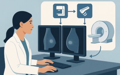
A 28-year-old woman presents to your clinic with a persistent, sharp pain in her right breast, localized to the upper outer quadrant. The pain has been present for three weeks, is unrelated to her menstrual cycle, and is significant enough to disrupt her sleep. On examination, you confirm focal tenderness without a palpable mass, skin changes, or nipple discharge. You need to determine the appropriate initial imaging study to evaluate this clinically significant, focal, noncyclical breast pain. This article provides a detailed clinical workflow for this specific scenario, grounded in the American College of Radiology (ACR) Appropriateness Criteria. For this young patient, the ACR designates breast ultrasound as a ‘Usually Appropriate’ initial imaging study.
Who Fits This Clinical Scenario for Focal Breast Pain?
This guidance is specifically for the initial imaging workup of a female patient under the age of 30 who presents with clinically significant breast pain. The key inclusion criteria are:
- Age: Less than 30 years old.
- Pain Character: Clinically significant (i.e., of sufficient severity to prompt a clinical visit or disrupt daily activities).
- Pain Location: Focal, meaning it is localized to an area of one quadrant or less.
- Pain Timing: Noncyclical, indicating the pain is not associated with the menstrual cycle.
It is crucial to distinguish this presentation from similar but distinct clinical scenarios that follow different diagnostic pathways. This workflow does not apply if:
- The pain is clinically insignificant: This includes pain that is diffuse (involving more than one quadrant), cyclical, or mild and non-focal. These presentations often do not require imaging.
- The patient is 30 or older: The pre-test probability of malignancy increases with age, altering the imaging algorithm. For women aged 30-39 or 40 and older, the recommendations change, often incorporating mammography.
- A palpable mass is present: The finding of a distinct, palpable breast lump shifts the workup from a “breast pain” scenario to a “palpable breast mass” scenario, which has its own specific ACR guidelines and a higher urgency.
- Other suspicious signs are present: Nipple discharge, skin changes (dimpling, erythema, peau d’orange), or axillary adenopathy warrant a different, more aggressive workup.
What Diagnoses Are You Working Up in This Scenario?
In a woman under 30, the differential diagnosis for focal, noncyclical breast pain is broad, but the vast majority of causes are benign. The primary goal of imaging is to identify a structural cause for the pain and, most importantly, to confidently exclude the rare possibility of malignancy.
The most common causes are benign fibrocystic changes. These can include the development of simple or complex cysts, which can become tender, or areas of glandular or stromal fibrosis. These conditions are extremely prevalent in young women and are a frequent source of focal pain.
Less common, but still important to consider, are inflammatory or infectious processes. A developing abscess or localized mastitis can present with focal pain, often accompanied by erythema and warmth, though these signs may be subtle initially. Trauma, even if minor and not well-recalled by the patient, can lead to a hematoma or fat necrosis that presents as a painful focal abnormality.
A rare but critical consideration is breast cancer. While malignancy is very uncommon in this age group, it is not impossible. Inflammatory breast cancer or a small underlying tumor can occasionally present with pain as the primary symptom, even without a palpable mass. Therefore, any persistent, focal, noncyclical pain warrants a thorough evaluation to rule out this possibility. Other rare causes include Mondor disease, a superficial thrombophlebitis of a breast vein.
Why Is Breast Ultrasound the Recommended Study for This Presentation?
The American College of Radiology (ACR) designates US breast as Usually Appropriate for the initial evaluation of focal, noncyclical breast pain in a woman under 30. This recommendation is based on the modality’s high diagnostic yield, safety profile, and suitability for the typical breast composition in this age group.
The primary rationale for choosing ultrasound is its excellent performance in dense breast tissue. Women under 30 typically have dense breasts, which appear white on a mammogram. This density can obscure underlying masses, significantly reducing the sensitivity of mammography. Ultrasound is not limited by breast density and provides superb soft-tissue resolution, making it highly effective at identifying and characterizing common benign findings like cysts and fibroadenomas. It can reliably differentiate a simple, benign fluid-filled cyst from a solid mass that might require further investigation.
Conversely, other imaging modalities are rated as Usually Not Appropriate for this specific clinical scenario:
- Mammography diagnostic and Digital breast tomosynthesis diagnostic: As mentioned, the high breast density in this age group limits the utility of these studies. Furthermore, both modalities use ionizing radiation (ACR Relative Radiation Level ☢☢). While the dose is low, the principle of ALARA (As Low As Reasonably Achievable) advises against unnecessary radiation exposure in a young population, especially when a non-radiation alternative like ultrasound is more effective.
- MRI breast without and with IV contrast: While extremely sensitive, breast MRI has lower specificity and can lead to a higher rate of false-positive findings. In a low-risk population, this can trigger unnecessary anxiety and biopsies for benign conditions. MRI is also more costly, less accessible, and requires the administration of IV contrast. It is reserved for problem-solving or for screening in very high-risk young women, not for the initial workup of focal pain.
Ultrasound’s key advantages are its lack of ionizing radiation (ACR Relative Radiation Level O) and its ability to provide real-time, dynamic evaluation of the precise area of pain, a technique known as sonographic palpation. When ordering, be specific: “Targeted ultrasound of the right breast, upper outer quadrant, to evaluate focal pain.”
What’s Next After a Breast Ultrasound? Downstream Workflow
The results of the breast ultrasound will guide the subsequent steps in the patient’s management. The workflow branches based on whether the findings are negative, benign, or suspicious.
- If the ultrasound is negative (BI-RADS 1): A negative or normal ultrasound, where no structural correlate for the pain is found, is a highly reassuring result. In this case, the pain is likely musculoskeletal or physiologic. The appropriate next step is clinical follow-up and reassurance. Management can focus on symptomatic relief with analgesics, warm compresses, and a well-fitting support bra.
- If the ultrasound shows a clearly benign finding (BI-RADS 2): The sonogram may identify a simple cyst, a classic fibroadenoma, or other definitively benign lesion. If the finding corresponds to the area of pain, it provides a clear diagnosis. For a symptomatic simple cyst, ultrasound-guided aspiration can be both diagnostic and therapeutic, often providing immediate pain relief. For other benign findings, no further imaging or intervention is typically needed beyond routine clinical follow-up.
- If the ultrasound is indeterminate or suspicious (BI-RADS 3, 4, or 5): If the ultrasound reveals a solid mass that is not clearly benign, a complex cystic lesion, or other suspicious features, further action is required.
- A BI-RADS 3 (Probably Benign) finding may warrant short-interval imaging follow-up (typically in 6 months) to ensure stability.
- A BI-RADS 4 (Suspicious) or BI-RADS 5 (Highly Suggestive of Malignancy) finding requires tissue sampling. The next step is an ultrasound-guided core needle biopsy of the lesion to obtain a definitive pathologic diagnosis.
Pitfalls to Avoid (and When to Get Help)
When managing focal breast pain in a young woman, several potential pitfalls can compromise an accurate and timely diagnosis.
- Dismissing the pain: Do not automatically attribute focal, noncyclical pain to hormones or fibrocystic change without a proper workup. While benign causes are most likely, a thorough evaluation is necessary to exclude pathology.
- Ordering the wrong initial study: Defaulting to mammography in a woman under 30 is a common error. It exposes the patient to unnecessary radiation and is less sensitive than ultrasound in dense breasts.
- Inadequate clinical information on the order: Failing to specify the exact location and character of the pain on the imaging requisition can lead to a non-targeted, and potentially non-diagnostic, study.
- Ignoring persistent symptoms after a negative ultrasound: If a patient’s focal pain persists or worsens despite a negative ultrasound, clinical re-evaluation is warranted. A referral to a breast specialist for a second opinion or further assessment may be appropriate.
If the clinical picture is concerning or the ultrasound findings are equivocal, do not hesitate to escalate. A consultation with a breast radiologist or a breast surgeon is the appropriate next step.
Related ACR Topics and Tools
For a comprehensive overview of imaging for all types of breast pain across different age groups, please see our parent guide. Additional GigHz resources can help you navigate adjacent scenarios and understand imaging techniques and safety.
- For breadth across all scenarios in Breast Pain, see our parent guide: Breast Pain: ACR Appropriateness Decoded.
- To look up other clinical scenarios, use the Imaging Appropriateness Selector.
- For details on imaging techniques, explore the Imaging Protocol Library.
- To discuss radiation exposure with patients, consult the Radiation Dose Calculator.
Frequently Asked Questions
Why isn’t mammography recommended as the first step for a 27-year-old with focal breast pain?
Mammography is not recommended as the initial study in this age group for two main reasons. First, women under 30 typically have dense breast tissue, which can obscure potential abnormalities on a mammogram, making it less sensitive than ultrasound. Second, mammography involves ionizing radiation, which should be avoided in young patients when a more effective, radiation-free alternative like ultrasound is available.
What if the patient’s focal pain persists after a completely normal breast ultrasound?
A normal ultrasound is very reassuring and effectively rules out a significant underlying structural cause, such as a large cyst or suspicious mass. If severe, focal pain persists, the cause is likely non-glandular (e.g., musculoskeletal, costochondritis). Management should focus on reassurance and symptomatic relief. However, if the pain worsens or new symptoms develop, clinical re-evaluation and potential referral to a breast specialist are appropriate.
Does a finding of ‘fibrocystic changes’ on ultrasound mean that’s the cause of the pain?
Not necessarily. Fibrocystic changes are extremely common findings on ultrasound in women under 30 and are often incidental. However, a tense or enlarging cyst within an area of fibrocystic change can certainly be a direct cause of focal pain. The radiologist will correlate the location of any specific findings with the patient’s reported area of tenderness to determine if it is the likely cause.
If the patient is 31 years old instead of 29, does the recommendation change?
Yes, age is a critical factor. The ACR guidelines for ‘Female with clinically significant breast pain (focal and noncyclical)’ have a different variant for ages 30 to 39. In that age group, both diagnostic mammography (or tomosynthesis) and breast ultrasound are considered ‘Usually Appropriate’. The rationale is the slightly increased risk of malignancy and changes in breast density with age.
Should I order a bilateral or a targeted unilateral ultrasound for focal pain?
For truly focal pain, a targeted unilateral ultrasound of the area of concern is sufficient and appropriate. However, many radiology departments have a standard protocol to perform a complete bilateral examination to provide a comprehensive baseline, which is also acceptable. The most important part of the order is to clearly specify the location of the pain so the sonographer and radiologist can perform a detailed, targeted evaluation of that specific area.
Reviewed by Pouyan Golshani, MD, Interventional Radiologist — May 26, 2026