What Imaging Is Best for Jaundice When Biliary Obstruction Is Not Suspected?
A 45-year-old male with a history of heavy alcohol use presents to your clinic with new-onset scleral icterus, fatigue, and vague abdominal discomfort. His laboratory results are notable for a conjugated hyperbilirubinemia with markedly elevated aspartate aminotransferase (AST) and alanine aminotransferase (ALT), far out of proportion to a mildly elevated alkaline phosphatase. This pattern strongly suggests hepatocellular injury rather than a mechanical blockage of the bile ducts. You have ruled out an obstructing gallstone or pancreatic head mass based on the initial clinical picture. The immediate question is which imaging study will best evaluate the liver parenchyma to confirm your suspicion and assess for underlying chronic disease. This article details the ACR-guided workflow for this specific scenario, where an abdominal ultrasound (US) is rated Usually appropriate as the initial imaging test.
Who Fits This Clinical Scenario for Non-Obstructive Jaundice?
This imaging workflow is designed for patients presenting with jaundice where the clinical evidence points toward a medical, metabolic, or functional cause. The key feature is the low suspicion for a physical blockage of the biliary tree. This patient cohort typically includes individuals with:
- Laboratory patterns suggesting hepatocellular injury (e.g., transaminase levels significantly higher than alkaline phosphatase).
- A clinical history suggestive of parenchymal liver disease, such as known viral hepatitis, significant alcohol use, recent initiation of a hepatotoxic medication (drug-induced liver injury or DILI), or signs of autoimmune disease.
- Symptoms of an acute viral prodrome (fever, malaise, myalgias) preceding the onset of jaundice.
- Known inherited disorders of bilirubin metabolism, where imaging is used to exclude coexisting structural pathology.
Conversely, this guidance does not apply to patients who fall into different clinical scenarios. For instance, a patient presenting with painless jaundice and a palpable gallbladder (Courvoisier’s sign) fits the suspected mechanical obstruction pathway. Similarly, a patient with jaundice and no clear predisposing conditions who is undergoing their very first evaluation would start with the initial imaging for undifferentiated jaundice scenario. This distinction is critical for ensuring the most efficient and appropriate diagnostic workup.
What Diagnoses Are You Working Up in Suspected Medical Jaundice?
When mechanical obstruction is unlikely, the diagnostic focus shifts to the liver parenchyma and its function. The differential is broad, but imaging helps narrow the possibilities and assess the severity of the underlying condition.
Hepatocellular Injury or Inflammation: This is the most common category and includes a wide range of etiologies. Acute viral hepatitis (e.g., Hepatitis A, B, or E), alcoholic hepatitis, and drug-induced liver injury are primary considerations. Imaging, particularly ultrasound, can reveal changes in liver size and echotexture, gallbladder wall thickening, or periportal edema, which are nonspecific but supportive signs of hepatic inflammation.
Chronic Liver Disease and Cirrhosis: Jaundice can be the first sign of decompensation in a patient with previously undiagnosed cirrhosis. Imaging is crucial for identifying the morphologic signs of cirrhosis, such as a nodular liver surface, caudate lobe hypertrophy, and splenomegaly. It also screens for complications like ascites and, most importantly, for a superimposed hepatocellular carcinoma (HCC) that could be causing the decline.
Infiltrative Diseases: Less common but important to consider, these conditions involve the diffuse deposition of substances within the liver. Examples include hemochromatosis (iron), Wilson’s disease (copper), amyloidosis, or diffuse metastatic disease. While ultrasound findings can be subtle, more advanced imaging like Magnetic Resonance Imaging (MRI) is highly sensitive for detecting abnormal iron or copper deposition.
Inherited or Functional Disorders: Conditions like Gilbert’s syndrome (unconjugated hyperbilirubinemia) or the rarer conjugated hyperbilirubinemias like Dubin-Johnson and Rotor syndromes are diagnoses of exclusion. In these cases, imaging serves a vital role: to confirm the absence of any structural or parenchymal abnormality, thereby strengthening the diagnosis of a functional disorder.
Why Is Abdominal Ultrasound the Recommended Study for This Presentation?
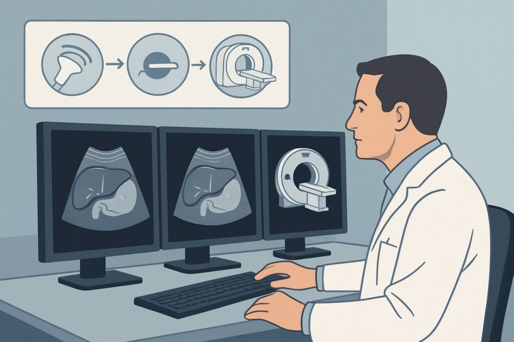
For a patient with suspected medical jaundice, the American College of Radiology designates US abdomen as Usually appropriate. This recommendation is based on its excellent safety profile, accessibility, and diagnostic capability for the key questions in this scenario.
The primary role of ultrasound here is twofold. First, it serves as a definitive, non-invasive confirmation that the biliary tree is not dilated, solidifying the conclusion that the jaundice is non-obstructive. Second, it provides a detailed assessment of the liver parenchyma. Ultrasound can effectively characterize liver echotexture to identify steatosis (fatty liver), assess for the coarse, nodular appearance of cirrhosis, and detect signs of portal hypertension such as splenomegaly, ascites, or reversed flow in the portal vein. It is a powerful tool for distinguishing between an acutely inflamed liver and one with chronic, established disease.
The lack of ionizing radiation (0 mSv) and the typical avoidance of IV contrast make it the safest initial option. While MRI abdomen without and with IV contrast with MRCP and CT abdomen with IV contrast are also rated Usually appropriate, they are generally reserved for problem-solving or when US findings are inconclusive. They offer superior tissue characterization but come with higher cost, longer acquisition times (for MRI), or radiation exposure (for CT, with an effective dose of 1-10 mSv).
Conversely, other studies are rated lower for this specific clinical question:
- CT abdomen without IV contrast is rated Usually not appropriate. Without intravenous contrast, a CT scan provides very limited information about the liver parenchyma, failing to adequately characterize inflammation, fibrosis, or small lesions. It exposes the patient to radiation (1-10 mSv) for minimal diagnostic yield in this context.
- ERCP (Endoscopic Retrograde Cholangiopancreatography) is also Usually not appropriate. ERCP is an invasive, therapeutic procedure designed to diagnose and treat biliary obstruction. In a patient where obstruction is not suspected, performing an ERCP carries substantial risks, including pancreatitis, bleeding, and perforation, with no diagnostic benefit.
What’s Next After US abdomen? Downstream Workflow
The results of the abdominal ultrasound guide the subsequent clinical and diagnostic pathway. The workflow branches based on whether the findings are definitive, negative, or indeterminate.
If the US confirms cirrhosis and/or portal hypertension: The diagnosis of chronic liver disease is established. The next steps are primarily clinical, focusing on identifying the underlying etiology (e.g., viral serologies, autoimmune markers) and managing complications. This includes initiating a screening program for hepatocellular carcinoma, typically with alternating ultrasound and cross-sectional imaging (CT or MRI), and potentially an upper endoscopy to screen for esophageal varices.
If the US is negative or shows non-specific findings (e.g., steatosis): A normal ultrasound in the face of significant jaundice and abnormal liver enzymes strongly supports a medical etiology like viral hepatitis or a drug-induced injury. The workup proceeds with comprehensive laboratory testing. If the clinical picture remains confusing or liver enzymes do not improve as expected, a follow-up MRI abdomen without and with IV contrast may be warranted to look for more subtle infiltrative processes or other parenchymal abnormalities not visible on ultrasound.
If the US is indeterminate or reveals an unexpected focal lesion: This finding fundamentally changes the clinical question. The priority becomes characterizing the newly discovered lesion. In this situation, a multiphase contrast-enhanced MRI or CT of the abdomen is the definitive next step. MRI is often preferred for its superior soft-tissue contrast in characterizing liver lesions without using ionizing radiation.
If the US unexpectedly shows biliary ductal dilation: The patient’s clinical scenario has been re-categorized. They now fit the “suspected mechanical obstruction” pathway. The next appropriate study is typically an MRI with MRCP to precisely identify the level and cause of the obstruction.
Pitfalls to Avoid (and When to Get Help)
Navigating the workup for medical jaundice requires careful integration of clinical, laboratory, and imaging data. Several common pitfalls can hinder an accurate and timely diagnosis.
- Fixating on a “normal” biliary tree: While confirming the absence of ductal dilation is a key purpose of the ultrasound, don’t stop there. The report must be scrutinized for subtle but critical details about the liver parenchyma, such as its echotexture, surface nodularity, and size, as well as signs of portal hypertension.
- Prematurely attributing jaundice to steatosis: Hepatic steatosis is extremely common and may be an incidental finding. If a patient has acute, severe jaundice, the presence of fat in the liver does not exclude a more urgent superimposed process like acute viral hepatitis or DILI.
- Delaying advanced imaging for inconclusive results: If the ultrasound is equivocal or the patient’s condition worsens despite a seemingly non-specific scan, do not hesitate to proceed to a more advanced modality like MRI. Delay can be detrimental, especially if a small tumor or an aggressive inflammatory process is the cause.
If a patient develops signs of acute liver failure, such as encephalopathy or a severe coagulopathy (elevated INR), this constitutes a medical emergency. The patient requires immediate escalation to a specialist, typically a hepatologist, and transfer to a center with transplant capabilities, as the underlying diagnosis must be determined urgently.
Related ACR Topics and Tools
This article focuses on a single, specific clinical scenario. For a comprehensive overview of all jaundice-related variants and their recommended imaging pathways, see our parent guide. For further exploration of imaging criteria, techniques, and safety, the following resources are available:
- Parent Topic Hub: For breadth across all scenarios in Jaundice, see our parent guide: Jaundice: ACR Appropriateness Decoded.
- ACR Criteria Lookup: To review guidelines for other clinical presentations, visit the Imaging Appropriateness Selector tool.
- Imaging Protocols: For detailed technical parameters of the recommended studies, consult the Imaging Protocol Library.
- Dose Calculation: To discuss radiation exposure with patients, use the Radiation Dose Calculator.
Frequently Asked Questions
My patient’s labs strongly suggest viral hepatitis. Is an ultrasound still necessary?
Yes. While the diagnosis may seem clear from lab work, an ultrasound is still considered ‘Usually appropriate’. It serves to confirm the absence of an unexpected coexisting condition, such as an underlying obstruction or a focal mass, and provides a valuable baseline assessment of the liver parenchyma for signs of chronic disease like cirrhosis, which would significantly alter long-term management.
If the initial ultrasound is negative, when should I consider a liver biopsy versus another imaging study?
If the initial ultrasound is negative but liver enzymes remain persistently elevated without a clear diagnosis from serologic and clinical workup, the next step is often a more advanced imaging study like an MRI. MRI can detect subtle infiltrative diseases (e.g., hemochromatosis) that ultrasound may miss. A liver biopsy is typically reserved for cases where even advanced imaging is non-diagnostic or when histologic confirmation of a specific condition (like autoimmune hepatitis or DILI) is required to guide therapy.
Can ultrasound with Doppler distinguish between different causes of medical jaundice?
While standard grayscale ultrasound assesses liver structure, adding Doppler imaging evaluates blood flow. Doppler is essential for assessing the portal vein for patency and direction of flow, which helps diagnose portal hypertension. It can also detect hepatic vein or inferior vena cava thrombosis (Budd-Chiari syndrome), a rare but important cause of acute liver injury and jaundice. However, it cannot reliably distinguish between different hepatocellular causes like viral hepatitis versus drug-induced injury, which look similar.
Why is MRI with MRCP also rated ‘Usually appropriate’ if ultrasound is the first choice?
MRI with MRCP is rated ‘Usually appropriate’ because it is an excellent, comprehensive test for the liver and biliary system. However, it is typically used as a second-line or problem-solving tool in this scenario due to its higher cost, lower availability, and longer scan time compared to ultrasound. It becomes the primary choice when ultrasound is inconclusive, a focal lesion needs characterization, or there is a high suspicion for a specific infiltrative disease better visualized with MRI.
The patient has a contraindication to MRI (e.g., a pacemaker). What is the best alternative if the ultrasound is not definitive?
If a patient cannot undergo an MRI and the ultrasound is inconclusive, a contrast-enhanced CT abdomen is the next best option. It is also rated ‘Usually appropriate’ by the ACR for this scenario. A multiphase CT (with arterial, portal venous, and delayed phases) provides excellent characterization of liver parenchyma and can identify vascular abnormalities or focal lesions, though with less soft-tissue detail for diffuse disease compared to MRI and with the use of ionizing radiation.
Reviewed by Pouyan Golshani, MD, Interventional Radiologist — June 27, 2026