What Imaging Is Best for Jaundice with Suspected Mechanical Obstruction?

A 68-year-old male presents to your clinic with two weeks of progressive, painless jaundice. His sclerae are icteric, his urine is dark, and his stools have become pale. Initial labs confirm a conjugated hyperbilirubinemia with a markedly elevated alkaline phosphatase, strongly suggesting a mechanical obstruction of the biliary tree. You need to identify the level and cause of the blockage, but which imaging study should you order first? This article details the American College of Radiology (ACR) guided workflow for this specific scenario, starting with the initial study rated Usually Appropriate: abdominal ultrasound.
Who Fits This Clinical Scenario for Obstructive Jaundice?
This diagnostic workflow applies to patients presenting with jaundice where the clinical suspicion points toward a physical blockage of the bile ducts, rather than a primary liver (hepatocellular) or blood (hemolytic) disorder.
Inclusion criteria for this scenario include jaundice accompanied by one or more of the following:
- Suggestive Laboratory Pattern: A predominantly conjugated (direct) hyperbilirubinemia with alkaline phosphatase (ALP) and gamma-glutamyl transferase (GGT) levels elevated out of proportion to aminotransferases (AST and ALT).
- Classic Clinical Signs: Painless jaundice, which raises concern for malignancy; pale (acholic) stools; dark (bilirubinuric) urine; or a palpable, non-tender gallbladder (Courvoisier’s sign).
- Prior Imaging Findings: An incidental finding of biliary ductal dilation on an imaging study performed for another reason.
This guidance is distinct from the workup for other presentations of jaundice. For example, a patient with undifferentiated jaundice and no clear localizing signs or labs would fall under the initial imaging for jaundice scenario. Likewise, a patient with a history of viral hepatitis, toxin exposure, and massively elevated AST/ALT levels would be evaluated under the pathway for suspected medical or metabolic etiologies.
What Diagnoses Are You Working Up with Suspected Mechanical Obstruction?
When mechanical obstruction is suspected, the primary goal of imaging is to confirm the presence of biliary ductal dilation and then to identify the level and cause of the blockage. The differential diagnosis is focused and carries significant clinical weight.
Choledocholithiasis is the most common cause of biliary obstruction. This occurs when a gallstone passes out of the gallbladder and becomes lodged in the common bile duct. While it can present with pain (biliary colic), it can also be less symptomatic, causing a progressive, obstructive jaundice.
A pancreatic head mass is a critical, can’t-miss diagnosis. Pancreatic ductal adenocarcinoma is a classic cause of painless, progressive obstructive jaundice because the tumor compresses the distal common bile duct as it passes through the head of the pancreas. Early detection is paramount.
Cholangiocarcinoma, a malignancy arising from the bile duct epithelium, is another important consideration. These tumors can occur anywhere along the biliary tree, from the small intrahepatic ducts to the distal common bile duct. Hilar cholangiocarcinomas, or Klatskin tumors, occur at the confluence of the right and left hepatic ducts and present a major surgical challenge.
Less commonly, benign biliary strictures can cause obstruction. These are often iatrogenic, developing as a complication of prior surgery (like cholecystectomy), or can be related to chronic inflammatory conditions such as chronic pancreatitis or primary sclerosing cholangitis.
Why Is Abdominal Ultrasound the Recommended First Study for Suspected Obstruction?
For a patient with suspected mechanical obstruction, the ACR designates US abdomen as Usually appropriate. It is the ideal first-line imaging test for several key reasons.
First, its primary strength is detecting biliary ductal dilation—the cardinal sign of a downstream obstruction. Ultrasound is highly sensitive for identifying a dilated common bile duct (typically >6-7 mm in diameter, though this increases slightly with age and post-cholecystectomy status) and intrahepatic ductal dilation. Confirming dilation solidifies the diagnosis of obstruction and guides the next steps.
Second, ultrasound can often identify the cause. It is excellent for detecting gallstones within the gallbladder (cholelithiasis) and can frequently visualize an obstructing stone in the common bile duct (choledocholithiasis). It can also identify a hypoechoic mass in the head of the pancreas, raising immediate concern for malignancy.
Finally, ultrasound is non-invasive, widely available, cost-effective, and, crucially, involves no ionizing radiation (adult_rrl=O 0 mSv). This makes it a safe and efficient starting point.
Why Other Studies Are Not First-Line
- CT abdomen without IV contrast is rated Usually not appropriate. While CT is a powerful tool, non-contrast scans are poor at visualizing non-calcified gallstones and offer limited soft-tissue detail of the pancreas and bile ducts compared to other modalities. It exposes the patient to radiation (adult_rrl=☢☢☢ 1-10 mSv) for inferior diagnostic information in this initial workup.
- ERCP (Endoscopic Retrograde Cholangiopancreatography) is rated May be appropriate but is not a first-line diagnostic test. ERCP is an invasive procedure that combines endoscopy and fluoroscopy. While it provides excellent visualization of the biliary tree and allows for therapeutic intervention (e.g., stone removal, stent placement), it carries significant risks, including pancreatitis, perforation, and bleeding. It is typically reserved for cases where an obstruction has been confirmed by non-invasive imaging and an intervention is planned.
Other modalities like MRI abdomen with MRCP and CT abdomen with IV contrast are also rated Usually appropriate, but they are typically used as second-line or problem-solving tools after an initial ultrasound, as described in the downstream workflow below.
What’s the Next Step After an Abdominal Ultrasound?
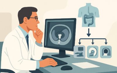
The results of the initial abdominal ultrasound dictate the subsequent diagnostic and therapeutic pathway. The workflow branches based on whether the ultrasound confirms an obstruction and if it identifies a clear cause.
If the ultrasound is positive (shows biliary dilation):
- Cause Identified: If the ultrasound clearly shows a stone in the common bile duct, the next step is often therapeutic, such as ERCP for stone extraction. If a suspicious pancreatic mass is seen, the next step is typically a high-quality, multiphase contrast-enhanced CT or MRI for staging the potential malignancy.
- Cause Unclear: If the ultrasound confirms ductal dilation but cannot definitively identify the cause of the obstruction, the best next step is MRI abdomen with MRCP (Magnetic Resonance Cholangiopancreatography). This non-invasive study provides detailed, high-resolution images of the entire biliary tree and pancreas without radiation, making it exceptional for identifying small stones, strictures, or tumors missed on ultrasound.
If the ultrasound is negative (no biliary dilation):
A normal-caliber biliary tree makes a significant mechanical obstruction much less likely. In this case, the diagnostic focus should pivot away from a blockage and toward a medical, metabolic, or hepatocellular cause of jaundice. This effectively moves the patient into a different clinical scenario, and the workup should proceed with further laboratory testing for viral hepatitis, autoimmune conditions, or genetic disorders.
Pitfalls to Avoid (and When to Get Help)
In the workup of suspected obstructive jaundice, several common pitfalls can delay diagnosis or lead to unnecessary testing.
First, avoid anchoring on a “negative” ultrasound if clinical suspicion remains high. Ultrasound is operator-dependent, and its sensitivity for distal common bile duct stones or small pancreatic tumors can be limited, especially by overlying bowel gas or patient body habitus.
Second, do not order a non-contrast CT to evaluate the biliary tree. As noted, it provides limited value and exposes the patient to radiation unnecessarily. If cross-sectional imaging is needed, a contrast-enhanced, pancreas-protocol CT or an MRI/MRCP is far superior.
Third, recognize the urgency of biliary obstruction accompanied by signs of infection (fever, chills, leukocytosis), which suggests acute cholangitis. This is a medical emergency requiring immediate biliary decompression, often via ERCP. If you suspect cholangitis, escalate immediately for gastroenterology or interventional radiology consultation.
Related ACR Topics and Tools
This article focuses on a single, specific clinical scenario. For a comprehensive overview of all imaging pathways for jaundice, refer to our parent guide. For further exploration of imaging techniques and safety, the following resources are available.
- For breadth across all scenarios in Jaundice, see our parent guide: Jaundice: ACR Appropriateness Decoded.
- To find the right imaging study for other clinical presentations, use the ACR Appropriateness Criteria Lookup.
- For detailed procedural techniques on recommended studies, consult the Imaging Protocol Library.
- To discuss cumulative radiation exposure with patients, the Radiation Dose Calculator can be a helpful tool.
Frequently Asked Questions
Why not start with CT with IV contrast if I’m worried about a pancreatic mass?
While a contrast-enhanced CT is excellent for staging a known pancreatic mass, abdominal ultrasound is the recommended initial test because it can confirm the presence of biliary obstruction without using ionizing radiation. If the ultrasound shows ductal dilation, a follow-up CT or MRI is then highly justified. If it shows no dilation, the patient is spared the radiation and contrast dose of a CT, and the workup can pivot to non-obstructive causes.
What if the ultrasound is negative but my clinical suspicion for obstruction is very high?
If there is a strong clinical or laboratory suggestion of mechanical obstruction (e.g., classic painless jaundice, Courvoisier’s sign, markedly elevated ALP) despite a negative or equivocal ultrasound, the next appropriate step is MRI with MRCP. MRCP is more sensitive than ultrasound for detecting stones in the common bile duct and for identifying strictures or small tumors that may not be visible on ultrasound.
Is endoscopic ultrasound (EUS) a good first choice?
Endoscopic ultrasound (EUS) is rated as ‘May be appropriate’ by the ACR for this scenario. It is highly sensitive for detecting small stones and pancreatic lesions. However, it is more invasive than a standard transabdominal ultrasound and is not as widely available. It is typically used as a problem-solving tool, often after non-invasive imaging is inconclusive, or to obtain tissue samples (fine-needle aspiration) from a suspected mass.
How does a patient’s prior cholecystectomy change the interpretation of the ultrasound?
After a cholecystectomy, the common bile duct often dilates slightly and can measure up to 10 mm in diameter in an asymptomatic patient. This is considered a normal post-surgical change. Therefore, in a patient without a gallbladder, a common bile duct diameter greater than 10 mm is generally used as the threshold to suspect obstruction.
When is MRCP preferred over a contrast-enhanced CT as the second-line study?
MRCP is generally preferred over CT when the primary question is to visualize the biliary tree itself to look for a non-calcified stone, a subtle stricture, or to map biliary anatomy. Contrast-enhanced CT is often preferred when the primary suspicion is a solid pancreatic mass, as it provides excellent detail for tumor staging, including evaluation of vascular involvement.
Reviewed by Pouyan Golshani, MD, Interventional Radiologist — June 27, 2026