What Imaging Is Best for Seizures in a Patient with a Known Brain Tumor?

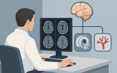
A 62-year-old patient with a history of a resected low-grade glioma and a well-controlled seizure disorder calls your office. Over the past month, he has experienced two breakthrough focal seizures, a significant change from his baseline of being seizure-free for two years on medication. As the treating clinician, you must determine if this change in seizure semiology is related to tumor progression, post-treatment effects, or another cause. The immediate clinical question is which imaging study will most effectively and safely provide the answer. This article details the American College of Radiology (ACR) Appropriateness Criteria workflow for this specific scenario, where the primary concern is evaluating a change in a known seizure disorder in a patient with a history of a tumor. For this presentation, the ACR rates MRI head without and with IV contrast as Usually Appropriate.
Who Fits This Clinical Scenario?
This guidance applies specifically to patients who meet two key criteria: a pre-existing, diagnosed seizure disorder and a known history of a primary or metastatic intracranial tumor. The clinical trigger for imaging is a change in the patient’s condition, such as increased seizure frequency, a new seizure type (semiology), a new focal neurologic deficit, or a failure to return to their prior neurologic baseline after a seizure. The core question is whether this change is driven by an alteration in the underlying structural lesion.
This workflow is distinct from several related but different clinical situations:
- New-onset seizure, no known tumor: A patient presenting with a first-time seizure requires a different diagnostic approach to identify the initial cause. This is covered in the new-onset seizure guidelines.
- Known seizure disorder, unchanged semiology: For a patient with stable epilepsy and no history of a tumor, routine surveillance imaging is often not indicated if their seizures are unchanged.
- Known seizure disorder, surgical planning: A patient being evaluated for epilepsy surgery requires specialized imaging protocols (like fMRI or MEG) to map eloquent cortex, which are not the primary goal in this scenario.
Correctly identifying your patient’s scenario is crucial for ordering the most diagnostically useful and resource-appropriate study.
What Diagnoses Are You Working Up in This Scenario?
When a patient with a history of a brain tumor presents with worsening seizures, the differential diagnosis is focused but critical. The imaging study is primarily intended to differentiate between several possibilities, each with vastly different management implications.
Tumor Recurrence or Progression
This is the most pressing concern. For both primary brain tumors (like gliomas or meningiomas) and metastatic disease, a change in seizure control can be the first sign that the tumor is growing or has recurred after treatment. Imaging must be sensitive enough to detect subtle changes in size, enhancement, or surrounding edema that indicate active tumor.
Treatment-Related Changes
Paradoxically, the effects of cancer treatment can mimic tumor progression and also be epileptogenic. Radiation necrosis, for instance, is a delayed, non-cancerous tissue injury that can cause inflammation, edema, and contrast enhancement on imaging, often appearing months to years after therapy. Differentiating this from true tumor recurrence is a classic diagnostic challenge where advanced imaging techniques can be vital.
Pseudoprogression
This phenomenon, particularly common after chemoradiation for gliomas, involves a temporary increase in contrast enhancement and swelling on imaging that looks like tumor progression but subsequently stabilizes or improves without a change in treatment. While transient, it can trigger seizures and requires careful follow-up.
Non-Tumor-Related Etiologies
While less likely to be the primary cause, it’s important to remember that these patients can develop other neurologic conditions. A new ischemic stroke, hemorrhage, or central nervous system infection could also present with new or worsening seizures and must be considered, especially if the imaging findings are atypical for tumor recurrence.
Why Is MRI Head Without and With IV Contrast the Recommended Study?
The ACR designates MRI head without and with IV contrast as Usually Appropriate because it provides the most detailed structural information needed to evaluate the differential diagnoses in this scenario. Magnetic Resonance Imaging (MRI) offers superior soft-tissue contrast compared to other modalities, allowing for precise visualization of the brain parenchyma, tumor margins, and surrounding structures.
The rationale for this specific protocol includes:
- Pre-contrast sequences (T1, T2, FLAIR): These are essential for assessing structural changes, vasogenic edema (seen on T2/FLAIR), and hemorrhage. Comparing these images to prior studies is fundamental to detecting subtle interval growth or new lesions.
- Post-contrast sequences (T1 with gadolinium): The administration of intravenous contrast is critical. Active tumors and areas of inflammation (like radiation necrosis) disrupt the blood-brain barrier, causing gadolinium-based contrast agents to leak into the brain tissue and “enhance.” The pattern and degree of enhancement are key features used to distinguish recurrence from scar tissue or stable post-treatment change.
Why are other studies rated lower for this specific presentation?
- CT head without IV contrast: Rated as May be appropriate, a non-contrast Computed Tomography (CT) scan is fast and can detect large-scale changes like significant mass effect or hemorrhage. However, its poor soft-tissue resolution makes it insensitive for detecting subtle tumor progression or differentiating tumor from post-treatment edema or gliosis. It is often inadequate for definitive assessment. (RRL: ☢☢☢ 1-10 mSv)
- FDG-PET/CT brain: Rated as May be appropriate (Disagreement), this functional study measures metabolic activity. It can be a powerful problem-solving tool when MRI is equivocal, as recurrent high-grade tumors are typically hypermetabolic, while radiation necrosis is hypometabolic. However, it is not the recommended first-line study due to higher radiation dose, lower spatial resolution than MRI, and the potential for false positives (e.g., inflammation can also be hypermetabolic). (RRL: ☢☢☢ 1-10 mSv)
The recommended MRI study involves no ionizing radiation (RRL: O 0 mSv), making it the safest option for patients who may require multiple follow-up scans over their lifetime.
What’s Next After MRI Head Without and With IV Contrast? Downstream Workflow
The results of the contrast-enhanced MRI will guide the subsequent clinical pathway. The workflow branches based on whether the findings are positive, negative, or indeterminate for tumor progression.
If the study is positive for tumor recurrence:
Clear evidence of new or progressive enhancing disease requires immediate action. The next step is a referral back to the patient’s neuro-oncology and/or neurosurgery team. This may lead to a discussion of further treatment options, including re-operation, repeat radiation, or a change in systemic therapy. Seizure management will also be re-evaluated, often requiring an adjustment or addition of anti-epileptic drugs (AEDs) to regain control.
If the study is negative or stable:
If the MRI shows no change compared to previous scans, the focus shifts away from tumor progression as the cause of worsening seizures. The next step is typically a clinical re-evaluation of AED management. This may involve checking drug levels, adjusting dosages, or considering a different medication. A consultation with an epileptologist and potentially an outpatient or inpatient video-Electroencephalogram (EEG) monitoring session may be warranted to better characterize the seizures and optimize therapy.
If the study is indeterminate:
This is a common and challenging outcome, especially when trying to distinguish tumor recurrence from radiation necrosis. If the initial MRI is equivocal, the downstream workflow may involve more advanced imaging. This could include an MRI with perfusion or spectroscopy, or proceeding to the May be appropriate FDG-PET/CT brain scan to assess the lesion’s metabolic activity. In some cases, only a surgical biopsy can provide a definitive diagnosis.
Pitfalls to Avoid (and When to Get Help)
Navigating this clinical scenario requires careful attention to detail to avoid common diagnostic and management errors.
- Pitfall 1: Ordering a non-contrast MRI. Forgetting to order the study “without and with IV contrast” is a frequent mistake. The post-contrast images are essential for assessing blood-brain barrier integrity and are non-negotiable in this workup.
- Pitfall 2: Not providing prior imaging. The radiologist’s ability to detect subtle interval change is critically dependent on comparison with previous scans. Always ensure that prior studies are available for review.
- Pitfall 3: Over-reliance on imaging alone. Remember that treatment-related effects like radiation necrosis can perfectly mimic tumor progression on imaging. Clinical correlation and sometimes advanced imaging or biopsy are necessary.
If the clinical picture and imaging findings are discordant, or if the diagnosis remains uncertain after initial advanced imaging, escalate by consulting a multidisciplinary tumor board that includes neuroradiologists, neuro-oncologists, and neurosurgeons.
Related ACR Topics and Tools
This article covers one specific variant within the broader topic of Seizures and Epilepsy. For a comprehensive overview of imaging recommendations across all related scenarios, from new-onset seizures to surgical planning, please consult our parent guide. For additional tools to help with ordering and interpreting imaging, see the resources below.
- For breadth across all scenarios in Seizures and Epilepsy, see our parent guide: Seizures and Epilepsy: ACR Appropriateness Decoded.
- ACR Appropriateness Criteria Lookup — for adjacent scenarios
- Imaging Protocol Library — for technique on the recommended study
- Radiation Dose Calculator — for cumulative dose conversations
Frequently Asked Questions
Why is contrast-enhanced MRI so critical in a patient with a known tumor history?
In patients with a history of a brain tumor, the primary concern is differentiating active tumor recurrence from stable post-treatment scar tissue or radiation-induced changes. Active tumors disrupt the blood-brain barrier, which allows the gadolinium-based contrast agent to leak into the tumor tissue, causing it to ‘enhance’ or light up on the scan. This enhancement is a key sign of active disease that would be missed on a non-contrast study.
What if my patient has a severe gadolinium allergy or renal failure?
If a patient cannot receive gadolinium-based contrast, an MRI head without IV contrast is still rated as ‘Usually Appropriate’ and is the next best test. While it lacks the information from enhancement, it can still show changes in tumor size, edema, or mass effect. In complex cases, a non-contrast CT might be used for a basic assessment, or a consultation with neuroradiology may be needed to discuss alternatives like an FDG-PET/CT scan, weighing its risks (radiation) and benefits.
How does this imaging workup differ from a patient with new-onset seizures but no tumor history?
For a new-onset seizure, the goal of imaging is broad: to find any structural cause, which could be a tumor, stroke, infection, or developmental abnormality. For a patient with a known tumor, the workup is highly focused on evaluating that specific lesion for recurrence or treatment-related changes. The choice of imaging (MRI with contrast) is often the same, but the clinical question and the interpretation of the findings are much more specific.
Is a CT scan ever the right first choice in this scenario?
A non-contrast CT head is generally reserved for emergent situations where MRI is unavailable or contraindicated, primarily to rule out acute hemorrhage or hydrocephalus. While the ACR rates it as ‘May be appropriate,’ it is not sensitive enough for the primary question of tumor recurrence. It should be considered a preliminary or screening tool, with the understanding that a follow-up MRI will almost certainly be needed for definitive evaluation.
How often should surveillance imaging be performed in an asymptomatic patient with a history of a brain tumor and seizures?
Surveillance imaging schedules are determined by neuro-oncology guidelines and depend on the tumor type, grade, and treatment history. This article’s recommendations apply specifically to when a patient becomes symptomatic with new or worsening seizures. Routine surveillance is a separate clinical question and should follow the protocol established by the patient’s oncology team.
Reviewed by Pouyan Golshani, MD, Interventional Radiologist — May 29, 2026