What Imaging Is Best for Stage I Pure Seminoma Surveillance After Orchiectomy?
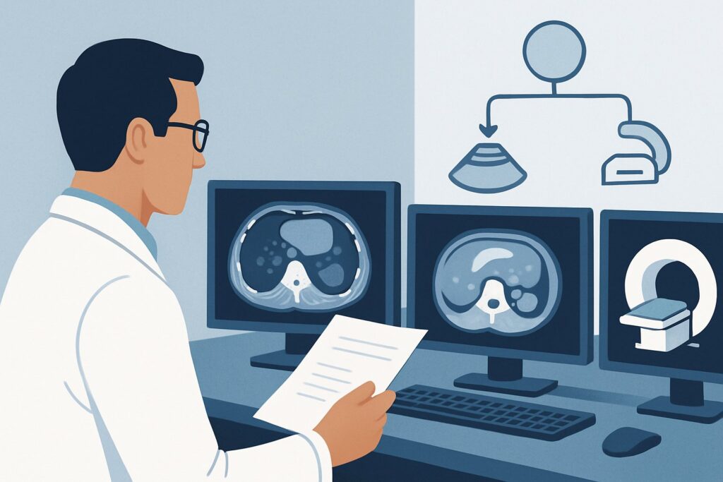
A 28-year-old man, six months post-radical orchiectomy for a stage IA pure seminoma, arrives for his scheduled follow-up. He feels well, has no complaints, and his tumor markers are normal. You are managing his active surveillance protocol, a strategy chosen to avoid the toxicity of adjuvant therapy. Now, the key clinical question arises: what is the most appropriate imaging to order for this routine, asymptomatic visit? The goal is to detect potential recurrence early while minimizing long-term risks, particularly from cumulative radiation exposure in a young patient. According to the American College of Radiology (ACR) Appropriateness Criteria, for this specific scenario, Radiography chest, MRI abdomen and pelvis without and with IV contrast, and CT abdomen and pelvis with IV contrast are all considered Usually Appropriate. This article will navigate the rationale behind these choices and help you build a safe and effective surveillance workflow.
Who Fits This Clinical Scenario for Seminoma Surveillance?
This guidance is specifically for the active surveillance of patients with early-stage pure seminoma of the testicle. The precise inclusion criteria are:
- Diagnosis: Pathologically confirmed pure seminoma following radical orchiectomy.
- Stage: Pathologic stage IA (pT1N0M0) or IB (pT2N0M0). This means the tumor was confined to the testicle (with or without lymphovascular invasion) and initial staging, including imaging and tumor markers, showed no evidence of spread.
- Clinical Status: The patient is asymptomatic and there is no clinical suspicion of recurrence. This includes normal physical examination and normal serum tumor markers (beta-hCG, AFP, LDH).
It is critical to distinguish this scenario from similar but distinct clinical situations that require different imaging strategies. This workflow does not apply to:
- Initial Staging: The imaging workup performed immediately after diagnosis but before a surveillance plan is established is a separate clinical question.
- Nonseminoma Germ Cell Tumors (NSGCT): Nonseminomas have different patterns of spread and relapse kinetics, necessitating a distinct surveillance protocol.
- Suspected Recurrence: If a patient presents with symptoms (e.g., new back pain, a palpable mass) or has rising tumor markers, this is no longer routine surveillance. This patient requires a diagnostic workup, not a screening study, which falls under a different ACR variant.
What Diagnoses Are You Working Up in This Scenario?
In the context of active surveillance for stage I seminoma, imaging is not used to work up a new differential diagnosis but to screen for specific, known patterns of disease recurrence. The primary goal is to detect metastatic disease at its earliest, most treatable stage.
Retroperitoneal Lymph Node Metastasis
This is the most common site of relapse for testicular seminoma. The lymphatic drainage of the testes flows directly to the retroperitoneal lymph nodes, specifically the para-aortic and paracaval nodal basins. The landing zone for a right-sided tumor is typically the interaortocaval region, while a left-sided tumor drains to the left para-aortic nodes. Surveillance imaging of the abdomen and pelvis is focused almost entirely on identifying new or enlarging nodes in these locations.
Pulmonary Metastasis
The lungs are the most common site of distant, hematogenous metastasis from testicular cancer. While less frequent than retroperitoneal relapse for stage I disease, it is a critical site to monitor. Small, asymptomatic pulmonary nodules are the typical target of chest imaging during surveillance.
Mediastinal or Supraclavicular Lymph Node Metastasis
Metastasis can also occur in other nodal basins, such as the mediastinum or the supraclavicular fossa, though this is less common as a first site of relapse. Chest imaging can help detect disease in these areas as well.
Contralateral Testicular Tumor
Patients with a history of a germ cell tumor have a small but increased risk of developing a new primary tumor in the remaining testicle. While imaging is not the primary screening tool for this, a scrotal examination is a key part of every follow-up visit. An ultrasound of the scrotum, rated May be appropriate, would be the correct study if any abnormality is palpated.
Why Are CT, MRI, and Chest X-Ray All ‘Usually Appropriate’ for This Presentation?
The ACR guidelines designate three different imaging studies as Usually Appropriate for stage I seminoma surveillance, reflecting a clinical consensus that the optimal strategy involves a trade-off between diagnostic accuracy and the long-term risks of radiation. The choice and frequency depend on creating a protocol that minimizes cumulative dose over a patient’s lifetime.
CT abdomen and pelvis with IV contrast is rated Usually Appropriate because it is highly sensitive and specific for detecting the most common site of relapse: retroperitoneal lymphadenopathy. It is fast, widely available, and provides excellent anatomic detail of the nodal basins. However, its significant drawback is the use of ionizing radiation (ACR Relative Radiation Level ☢☢☢ 1-10 mSv). For a young patient undergoing many scans over a 5- to 10-year surveillance period, the cumulative dose is a major concern, carrying a small but real risk of secondary malignancy.
MRI abdomen and pelvis without and with IV contrast is also Usually Appropriate and is an increasingly favored alternative to CT. Its primary advantage is the complete lack of ionizing radiation (ACR RRL O 0 mSv). Modern MRI techniques provide diagnostic accuracy for detecting retroperitoneal nodal disease that is comparable to CT. The main limitations are longer scan times, higher cost, and potential contraindications (e.g., certain implants). Many leading cancer centers have shifted to MRI-based surveillance protocols to mitigate radiation risk.
Radiography chest is the third Usually Appropriate study. It serves a different purpose: screening for pulmonary metastases. Its key benefit is an extremely low radiation dose (ACR RRL ☢ <0.1 mSv), making it ideal for frequent screening of the chest. It is not, however, capable of evaluating the retroperitoneum.
A typical surveillance protocol, guided by NCCN or EAU guidelines, often combines these modalities—for example, alternating abdominal/pelvic CT or MRI with chest radiography at specified intervals.
Why Alternatives Are Rated Lower
- US abdomen and retroperitoneum is rated Usually not appropriate. While it uses no radiation, it is highly operator-dependent, and the retroperitoneal lymph nodes are often poorly visualized due to overlying bowel gas. It lacks the sensitivity needed for reliable surveillance.
- FDG-PET/CT whole body is also Usually not appropriate for routine surveillance. It carries a very high radiation dose (ACR RRL ☢☢☢☢ 10-30 mSv) and has a higher rate of false-positive findings in an asymptomatic screening population. Its use is reserved for evaluating a suspected recurrence found on other imaging or assessing treatment response.
What’s Next After Surveillance Imaging? Downstream Workflow
The results of surveillance imaging will direct the subsequent clinical pathway. The decision tree is generally straightforward.
- If all imaging is negative: This is the most common and desired outcome. The patient continues on the established surveillance schedule. The next imaging study and clinical visit are scheduled according to the protocol (e.g., in 6 to 12 months). This provides reassurance and reinforces the success of the surveillance strategy.
- If chest radiography is positive or equivocal: An indeterminate or suspicious finding on a chest X-ray, such as a new nodule, should prompt a follow-up diagnostic study. The next step is typically a CT chest with IV contrast to better characterize the finding. While CT chest is not appropriate for routine screening in this scenario, it is the standard for working up an abnormality found on a radiograph.
- If CT or MRI abdomen/pelvis shows new or growing retroperitoneal nodes: This finding is highly suspicious for relapse. The patient should be promptly referred to a medical oncologist or urologic oncologist. Further workup may include an FDG-PET/CT to confirm the findings are metabolically active and to screen for other sites of disease. This will guide salvage therapy, which is highly effective and usually consists of either radiation therapy or platinum-based chemotherapy.
- If an indeterminate finding is noted: A common dilemma is the presence of a small, non-specific, or borderline-enlarged lymph node. Instead of proceeding directly to treatment, the most common approach is a short-interval follow-up scan (e.g., CT or MRI in 3 months) to assess for stability or growth, which helps differentiate a reactive node from metastatic disease.
Pitfalls to Avoid (and When to Get Help)
Navigating stage I seminoma surveillance requires careful attention to protocol and long-term risk management. Common pitfalls include:
- Inconsistent Imaging Protocols: Failing to adhere to a consistent, evidence-based schedule can lead to missed recurrences or unnecessary radiation exposure. Use institutional or national guidelines (e.g., NCCN) as a framework.
- Ignoring Cumulative Radiation Dose: Repeatedly ordering CT scans when a radiation-free alternative like MRI is available and appropriate can needlessly increase a young patient’s lifetime cancer risk.
- Misinterpreting Post-Orchiectomy Changes: Normal post-surgical changes in the inguinal region or small, stable retroperitoneal nodes can be mistaken for recurrence. Comparing with prior scans is essential.
- Dismissing Patient Symptoms: Never assume a new symptom like back pain is benign. Even on a surveillance protocol, new, persistent symptoms warrant a diagnostic workup, which may deviate from the standard surveillance imaging plan.
If imaging reveals unequivocal evidence of recurrence, or if you are uncertain about the significance of an indeterminate finding, consultation with a multidisciplinary tumor board including urologic oncology, medical oncology, and radiology is the standard of care.
Related ACR Topics and Tools
This article covers a single, specific clinical scenario. For a broader view of imaging across all presentations of testicular cancer, or to explore the technical details of the recommended studies, the following resources are essential.
- For breadth across all scenarios in Staging and Surveillance of Testicular Cancer, see our parent guide: Staging and Surveillance of Testicular Cancer: ACR Appropriateness Decoded.
- To explore adjacent scenarios or search for different clinical presentations, use the ACR Appropriateness Criteria Lookup.
- For detailed technical specifications on how to perform the recommended studies, consult the Imaging Protocol Library.
- To facilitate conversations with patients about radiation exposure from different imaging plans, use the Radiation Dose Calculator.
Frequently Asked Questions
Why is MRI considered ‘Usually Appropriate’ if CT has been the historical standard for seminoma surveillance?
MRI is rated ‘Usually Appropriate’ because it offers comparable diagnostic accuracy to CT for detecting retroperitoneal lymph node recurrence without using any ionizing radiation. Given that surveillance protocols for stage I seminoma involve multiple scans over many years in young men, minimizing cumulative radiation dose to reduce the risk of secondary cancers is a primary concern. Many major cancer centers now prefer MRI for this reason.
If a patient’s surveillance CT shows a new 9 mm retroperitoneal lymph node, what is the next step?
A new, sub-centimeter retroperitoneal lymph node is suspicious for recurrence but can also be a reactive (benign) node. The next step is typically a discussion with oncology and often involves either a short-interval follow-up scan in 2-3 months to assess for growth or proceeding to an FDG-PET/CT to determine if the node is metabolically active. Immediate treatment is usually deferred until recurrence is confirmed.
Is a chest CT needed for routine surveillance instead of a chest radiograph?
No, for routine, asymptomatic surveillance, a chest CT is rated ‘Usually not appropriate’ by the ACR. A standard chest radiograph is ‘Usually Appropriate’ and is sufficient for screening for pulmonary metastases in this low-risk setting. A chest radiograph provides adequate screening with a fraction of the radiation dose of a CT scan. A CT is reserved for working up an abnormality seen on the radiograph or if the patient develops pulmonary symptoms.
Do tumor markers need to be checked at every surveillance imaging appointment?
Yes. Clinical follow-up with serum tumor markers (AFP, beta-hCG, and LDH) is a cornerstone of surveillance for all testicular germ cell tumors, including pure seminoma. While pure seminomas are less likely to produce AFP or beta-hCG, a small percentage do, and LDH can be an indicator of tumor burden. Markers should be checked at each follow-up visit, in conjunction with imaging and a physical exam.
How long does a patient with stage I seminoma need to be on an active surveillance protocol?
Surveillance protocols typically last for 5 to 10 years, with the frequency of visits and imaging being highest in the first 2-3 years, when the risk of relapse is greatest. The exact schedule can vary by institution but generally follows guidelines from organizations like the NCCN or EAU. After 5 years, the risk of relapse is very low, and surveillance becomes less intensive or may be stopped.
Reviewed by Pouyan Golshani, MD, Interventional Radiologist — May 29, 2026