What Imaging Is Best for Suspected Breast Cancer Recurrence After Mastectomy?
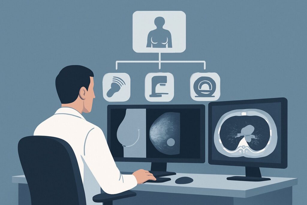
A 62-year-old woman, five years post-mastectomy for invasive ductal carcinoma, presents to your clinic for follow-up. During her self-exam, she noticed a new, firm, pea-sized nodule just medial to her mastectomy scar. It’s non-tender but fixed to the underlying tissue. Her anxiety is palpable as she asks, “Is it back?” You now face the critical decision of which imaging study to order first to evaluate this new finding. This article provides a focused, evidence-based workflow for this exact clinical scenario, guiding you to the most appropriate initial step. For a patient with a history of mastectomy presenting with symptoms or signs of local recurrence, the American College of Radiology (ACR) Appropriateness Criteria rates US breast as Usually Appropriate.
Who Fits This Clinical Scenario for Suspected Recurrence?
This guidance applies specifically to patients with a prior history of mastectomy (unilateral or bilateral) for breast cancer who now present with new signs or symptoms suggestive of a local recurrence. This includes:
- A new palpable lump or nodule on the chest wall, in the mastectomy scar, in the axilla, or in the supraclavicular region.
- New skin changes such as erythema, thickening, dimpling, or ulceration over the chest wall.
- Persistent, focal chest wall pain without another clear cause.
- Nipple changes or discharge in a patient who underwent a nipple-sparing mastectomy.
This workflow is distinct from other common clinical situations. It does not apply to:
- Asymptomatic Surveillance: Patients with a history of mastectomy who are asymptomatic and undergoing routine imaging follow-up. That situation follows a different surveillance protocol.
- Patients with Prior Lumpectomy: Individuals who underwent breast-conserving therapy (lumpectomy) have remaining breast tissue, and their imaging workup for a new finding typically begins with diagnostic mammography and tomosynthesis.
- Initial Cancer Diagnosis: This guidance is for evaluating recurrence, not for the initial workup of a newly diagnosed primary breast cancer before any surgical treatment.
What Diagnoses Are You Working Up in a Post-Mastectomy Patient?
When a patient presents with a new palpable finding after mastectomy, several diagnoses must be considered. While recurrence is the primary concern, benign post-surgical changes are common and can mimic malignancy.
Local Recurrence of Breast Cancer
This is the most consequential diagnosis to exclude. Recurrence can manifest as a solid nodule in the skin, subcutaneous fat, or underlying pectoralis muscle. It can also present as involvement of regional lymph nodes, such as the axillary, internal mammary, or supraclavicular nodes, which may become palpable.
Fat Necrosis or Scar Tissue
Perhaps the most common benign cause of a palpable lump post-surgery. Trauma to fatty tissue from the mastectomy or reconstruction can lead to fat necrosis, which evolves over time and can form a firm, irregular mass that is clinically indistinguishable from a recurrence. Similarly, scar tissue can become thickened and nodular.
Suture Granuloma
This is a benign inflammatory reaction to retained suture material. It typically presents as a firm, palpable nodule along the surgical scar line and can appear months or even years after the initial procedure.
Seroma or Hematoma
Fluid collections are common in the early post-operative period but can sometimes persist, become encapsulated, and present as a firm mass. While usually benign, the wall of a chronic seroma can, in rare instances, harbor recurrent disease.
Why Is Ultrasound the Recommended First Study for Suspected Recurrence After Mastectomy?
The ACR designates breast ultrasound (US) as Usually Appropriate for this scenario because it is the most effective, direct, and safest initial imaging modality for evaluating the superficial tissues of the chest wall.
The primary rationale is that after a mastectomy, there is no longer a breast to be imaged with mammography. The clinical question shifts to evaluating the skin, subcutaneous tissues, and chest wall muscles—anatomy for which ultrasound provides excellent high-resolution detail. Ultrasound can readily characterize a palpable finding, distinguishing a simple fluid collection (seroma) from a complex one, and a benign-appearing solid nodule (like a suture granuloma) from a solid mass with suspicious features (e.g., irregular margins, spiculation, internal vascularity).
Crucially, ultrasound is performed in real-time, allowing the radiologist to directly correlate the imaging findings with the palpable area of concern. If a suspicious lesion is identified, ultrasound provides a safe and accurate method to guide a core needle biopsy during the same visit, accelerating the diagnostic process.
Alternative studies are rated lower for this initial workup:
- Mammography or Digital Breast Tomosynthesis is rated Usually not appropriate. Without breast tissue to compress, these modalities offer very limited utility for evaluating the chest wall and can expose the patient to unnecessary radiation (☢☢ 0.1-1 mSv).
- MRI breast without and with IV contrast is also rated Usually not appropriate as the first test. While MRI is very sensitive for detecting recurrence, it is less specific and more resource-intensive. It is better reserved as a problem-solving tool if ultrasound is equivocal or if there is a strong clinical suspicion for recurrence despite a negative ultrasound.
Ultrasound involves no ionizing radiation (O 0 mSv) and does not require IV contrast, making it an exceptionally safe first step. When ordering, be specific: “Ultrasound of the right chest wall and axilla, with attention to the palpable nodule at the 3 o’clock position of the mastectomy scar.” This detail ensures the sonographer and radiologist focus on the precise area of clinical concern.
What Is the Downstream Workflow After a Chest Wall Ultrasound?
The results of the chest wall ultrasound will directly guide your next steps. The workflow typically branches into one of three paths based on the BI-RADS (Breast Imaging Reporting and Data System) assessment.
Positive for a Suspicious Mass (BI-RADS 4 or 5)
If the ultrasound identifies a solid mass with suspicious features, the definitive next step is an ultrasound-guided core needle biopsy. This procedure provides a tissue diagnosis to confirm or exclude malignancy. If the biopsy confirms recurrence, the patient will then require a full staging workup (which may include CT and/or FDG-PET/CT) and will be referred back to their oncology team to discuss treatment options, such as surgery, radiation, or systemic therapy.
Negative or Clearly Benign Finding (BI-RADS 1 or 2)
If the ultrasound shows no abnormality or a finding with classic benign features (e.g., a simple cyst, typical post-surgical scarring, or fat necrosis), the patient can be reassured. The standard management is a return to routine clinical follow-up. No further imaging is typically needed unless the clinical picture changes.
Indeterminate or Probably Benign Finding (BI-RADS 3)
This is a less common outcome in this setting. If a finding is deemed probably benign, the recommendation is typically for short-term imaging follow-up with ultrasound in 6 months to ensure stability. However, given the high-risk context of a prior cancer diagnosis, many clinicians and radiologists may opt for biopsy even for a BI-RADS 3 finding to achieve diagnostic certainty sooner.
Pitfalls to Avoid (and When to Get Help)
Navigating the workup of a suspected recurrence requires careful attention to detail. Here are a few common pitfalls to avoid:
- Ordering Mammography: Do not order a mammogram for a patient with a prior mastectomy. It provides no useful information for the chest wall and results in unnecessary radiation and delays.
- Ignoring Clinical Suspicion: If your clinical suspicion for recurrence remains high despite a negative or benign ultrasound report, do not dismiss the patient’s symptoms. Escalate by speaking directly with the breast radiologist to discuss the discrepancy and consider further imaging, such as an MRI.
- Not Biopsying Axillary Nodes: A new, palpable axillary lymph node is as concerning as a chest wall nodule. Ensure that any suspicious-appearing nodes on ultrasound are also targeted for biopsy.
- Vague Imaging Orders: An order for “breast ultrasound” is insufficient. Specify the side, that the patient is post-mastectomy, and the exact location of the palpable finding to ensure a targeted, high-yield exam.
If you encounter a complex or discordant case, a multidisciplinary discussion involving the surgeon, radiologist, and oncologist is always the best path forward.
Related ACR Topics and Tools
This article covers one specific scenario in depth. For a comprehensive overview of all clinical variants, or for tools to help with adjacent imaging decisions, the following resources are valuable.
- For breadth across all scenarios in Imaging of Invasive Breast Cancer, see our parent guide: Imaging of Invasive Breast Cancer: ACR Appropriateness Decoded.
- ACR Appropriateness Criteria Lookup — for exploring other clinical scenarios and their corresponding imaging recommendations.
- Imaging Protocol Library — for detailed technical parameters of various imaging studies.
- Radiation Dose Calculator — for discussing cumulative radiation exposure with patients.
Frequently Asked Questions
Why isn’t MRI the first choice for a palpable lump after mastectomy if it’s so sensitive?
While breast MRI is highly sensitive for detecting cancer recurrence, it is not the recommended first-line study for a focal, palpable finding. Ultrasound is faster, more cost-effective, and can directly guide a biopsy in the same session. MRI is better suited as a problem-solving tool if ultrasound is negative but clinical suspicion remains high, or for evaluating the full extent of disease once recurrence is proven by biopsy.
What if the patient had breast reconstruction with an implant?
The initial imaging recommendation remains the same: targeted ultrasound. Ultrasound is excellent for evaluating the tissues superficial to the implant and the chest wall deep to it. It can also assess the integrity of the implant itself. Mammography is contraindicated, and MRI would only be used in a problem-solving capacity.
If the ultrasound is negative, is there any role for follow-up imaging?
If the ultrasound identifies a clear benign cause for the palpable finding (e.g., a suture granuloma) or is completely negative (BI-RADS 1 or 2), routine clinical follow-up is typically all that is needed. Short-term imaging follow-up is generally reserved for BI-RADS 3 (probably benign) findings, though biopsy is often preferred in this high-risk population for diagnostic certainty.
Should I order a PET/CT scan for a new palpable nodule after mastectomy?
No, an FDG-PET/CT is not an appropriate initial diagnostic tool for a focal palpable finding. It is a systemic staging tool, not a high-resolution diagnostic imaging study for a localized concern. The ACR rates FDG-PET/CT as *Usually not appropriate* for this initial workup. Its role is in staging for distant metastatic disease *after* a local recurrence has been confirmed by biopsy.
Can a mammogram be done on the contralateral, intact breast at the same time?
Yes. While the side with the mastectomy should be evaluated with ultrasound, the contralateral (remaining) breast should continue to undergo its standard screening or diagnostic mammography as per routine surveillance guidelines. The two evaluations are separate clinical questions.
Reviewed by Pouyan Golshani, MD, Interventional Radiologist — May 30, 2026