What Imaging Should You Order for a Child Hospitalized with Community-Acquired Pneumonia?
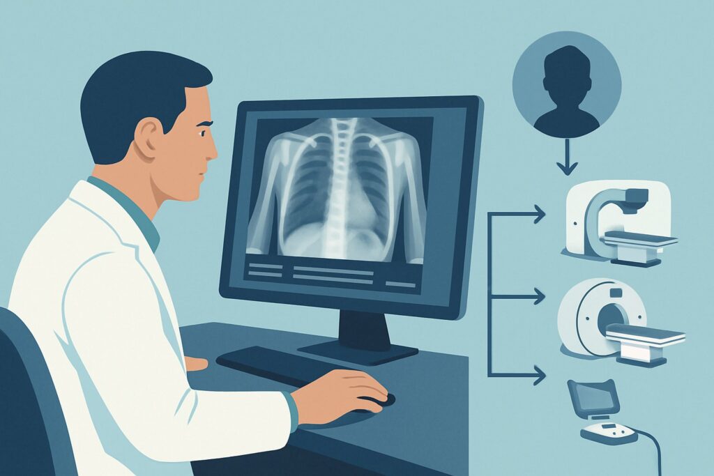
It’s 9 PM in the emergency department. A 4-year-old boy, diagnosed with community-acquired pneumonia three days ago and started on amoxicillin, is back. His fever has not resolved, he is tachypneic with subcostal retractions, and his oxygen saturation is 93% on room air. You’ve made the decision to admit him for intravenous antibiotics and respiratory support. Now, the clinical question is what initial imaging to order to guide management. This article provides a detailed workflow for this specific scenario: an immunocompetent child, 3 months or older, with community-acquired pneumonia (CAP) that is not responding to outpatient treatment or is severe enough to require hospital admission. For this presentation, the American College of Radiology (ACR) Appropriateness Criteria rate a chest radiograph as Usually Appropriate.
Who Fits This Clinical Scenario?
This guidance is specifically for an immunocompetent child, aged three months or older, with a clinical diagnosis of community-acquired pneumonia who presents with worsening symptoms or severity that necessitates a higher level of care. The key triggers for this scenario are either a failure to improve on appropriate outpatient antibiotic therapy or a presentation that is severe enough from the outset to warrant hospital admission. This includes signs like hypoxemia, significant respiratory distress, inability to maintain hydration, or concern for a developing complication.
It is critical to distinguish this situation from other related clinical presentations. This workflow does not apply to:
- The well-appearing child with suspected uncomplicated CAP: For an otherwise healthy child who does not require admission, routine chest radiography is often unnecessary as it typically does not change management.
- A child with suspected hospital-acquired pneumonia: This condition involves different pathogens and risk factors, which alters the diagnostic approach and falls under a separate ACR variant.
- A child with a known, large parapneumonic effusion already identified on a radiograph: That patient has moved to the next stage of the workup, which involves characterizing the complication, not initial diagnosis. This article focuses on the initial imaging upon admission.
What Diagnoses Are You Working Up in This Scenario?
When a child with CAP fails to improve or requires hospitalization, the differential diagnosis broadens beyond simple, uncomplicated pneumonia. The primary goal of imaging is to confirm the diagnosis, assess the severity and extent of disease, and, most importantly, identify potential complications that require a change in management.
Complicated Bacterial Pneumonia: This is the leading concern. The lack of response to first-line antibiotics suggests a more virulent pathogen (e.g., Staphylococcus aureus), a resistant organism (e.g., drug-resistant Streptococcus pneumoniae), or the development of a structural complication. Imaging helps define the extent of the parenchymal consolidation.
Parapneumonic Effusion and Empyema: A collection of fluid in the pleural space is one of the most common reasons for clinical worsening in pediatric pneumonia. A small, simple (uninfected) effusion may resolve with antibiotics alone, but a large or complex effusion (empyema) often requires drainage. Imaging is essential for detection.
Necrotizing Pneumonia or Lung Abscess: Though less common, these are severe complications where lung tissue is destroyed, potentially forming cavities or abscesses. A standard radiograph may show suggestive findings like pneumatoceles or a thick-walled cavity, signaling a more aggressive disease process that requires prolonged antibiotic therapy and specialist consultation.
Significant Atelectasis: Collapse of lung tissue due to mucous plugging or external compression can mimic or coexist with pneumonia. Differentiating large-volume atelectasis from consolidation is important, as it may prompt interventions like chest physiotherapy or bronchoscopy.
Why Is a Chest Radiograph the Recommended Study for This Presentation?
For a child being admitted with worsening community-acquired pneumonia, a chest radiograph is the cornerstone of initial imaging. The ACR designates this study as Usually Appropriate, reflecting its high diagnostic utility, wide availability, and favorable safety profile in this context.
The primary rationale is that a two-view (posteroanterior/anteroposterior and lateral) chest radiograph provides a rapid and effective assessment of the key clinical questions. It can reliably confirm the presence and location of a pulmonary infiltrate, evaluate the extent of disease, and identify most clinically significant complications like a moderate-to-large pleural effusion, pneumothorax, or developing abscess. This information is often sufficient to guide initial inpatient management, including antibiotic choice and the need for respiratory support.
From a safety perspective, the radiation dose is very low. The ACR notes a pediatric relative radiation level (RRL) of ☢ (<0.03 mSv), which is a fraction of the annual natural background radiation. This makes it an appropriate first-line study in a pediatric population, adhering to the ALARA (As Low As Reasonably Achievable) principle.
Alternative imaging modalities are rated lower for this specific initial workup for clear reasons:
- CT Chest (with or without IV contrast): This is rated Usually not appropriate as a first step. While CT provides exquisite anatomical detail, it delivers a substantially higher radiation dose (pediatric RRL ☢☢☢☢, 3-10 mSv). It is reserved for cases where a chest radiograph is inconclusive or there is high suspicion of a complex complication (like an abscess or bronchopleural fistula) that requires more precise characterization to plan an intervention.
- US Chest: Ultrasound is rated May be appropriate. It is an excellent radiation-free tool for evaluating pleural fluid, capable of distinguishing simple from complex effusions and guiding thoracentesis. However, it is less effective for assessing the lung parenchyma itself, particularly in deeper, air-filled regions. It is often used as a follow-up or adjunct to chest radiography, especially when an effusion is suspected, rather than as the primary initial study for the entire chest.
What’s Next After a Chest Radiograph? Downstream Workflow
The results of the initial chest radiograph will direct the subsequent clinical pathway. The findings create clear decision points for management and potential further imaging.
If the radiograph shows uncomplicated consolidation: If the image confirms a lobar or segmental infiltrate without evidence of a large effusion, abscess, or other complication, the next step is typically medical management. This involves IV antibiotics, hydration, and respiratory support. Repeat imaging is generally not needed unless the child fails to improve clinically after 48-72 hours of appropriate inpatient therapy.
If the radiograph is negative or shows only minor atelectasis: In a child with significant respiratory symptoms but a non-diagnostic chest radiograph, consider alternative diagnoses. This could include viral bronchiolitis with mucous plugging, asthma, or a foreign body aspiration. The clinical picture should guide the next steps, which may involve observation or further specialized testing rather than more advanced imaging.
If the radiograph shows a moderate or large pleural effusion: This finding significantly changes the workflow and often triggers a new ACR scenario. The next step is frequently a chest ultrasound to characterize the fluid collection (simple vs. complex/loculated) and to guide a potential drainage procedure (thoracentesis or chest tube placement). This moves the patient into the workup for a complicated pneumonia.
If the radiograph suggests an abscess or necrotizing pneumonia: Findings like a thick-walled cavity or multiple pneumatoceles are concerning. While the radiograph can suggest these diagnoses, a contrast-enhanced chest CT is often the next step to confirm the finding, define its extent, and evaluate for associated vascular complications. This escalation is reserved for children who are severely ill or not responding to standard therapy.
Pitfalls to Avoid (and When to Get Help)
In managing a hospitalized child with pneumonia, several common pitfalls can delay recovery or lead to unnecessary testing. Be mindful of ordering repeat “daily” chest radiographs without a specific clinical question; imaging should be guided by a change in clinical status. Avoid the premature use of CT when a chest radiograph and ultrasound can provide sufficient information for initial management. Another pitfall is failing to obtain a lateral view, which is crucial for evaluating the retrocardiac space and posterior costophrenic sulci for subtle infiltrates or effusions. If the radiograph shows a large effusion, necrotizing features, or any other sign of severe complication, it is time to escalate care by consulting with pediatric infectious disease, pulmonology, or surgery specialists.
Related ACR Topics and Tools
This article covers one specific scenario in pediatric pneumonia. For a comprehensive overview of all related variants, from uncomplicated outpatient cases to rare complications, please consult the parent topic hub. The following GigHz tools can also support your clinical decision-making:
- For breadth across all scenarios in Pneumonia in the Immunocompetent Child, see our parent guide: Pneumonia in the Immunocompetent Child: ACR Appropriateness Decoded.
- ACR Appropriateness Criteria Lookup — for adjacent scenarios
- Imaging Protocol Library — for technique on the recommended study
- Radiation Dose Calculator — for cumulative dose conversations
Frequently Asked Questions
Is a single AP view chest radiograph sufficient for a hospitalized child with pneumonia?
While a single anteroposterior (AP) or posteroanterior (PA) view may be performed in unstable patients, a two-view study including a lateral view is strongly preferred. The lateral view is critical for assessing the retrocardiac and retrosternal clear spaces and posterior costophrenic sulci, where infiltrates or effusions can be hidden on a single frontal view.
If the chest radiograph shows a small pleural effusion, is further imaging always needed?
Not necessarily. Small, free-flowing parapneumonic effusions are common and often resolve with effective antibiotic treatment for the underlying pneumonia. Further imaging, typically with ultrasound, is usually reserved for moderate to large effusions or when the patient is not improving clinically, to rule out the development of a complex effusion or empyema that may require drainage.
Why is chest CT considered ‘Usually Not Appropriate’ for the initial imaging of a hospitalized child with CAP?
Chest CT is rated ‘Usually Not Appropriate’ for initial evaluation primarily due to its significantly higher radiation dose compared to a chest radiograph. For the initial questions—confirming pneumonia and identifying common complications like effusions—a radiograph provides sufficient information. CT is reserved as a second-line study for specific indications, such as suspected lung abscess, necrotizing pneumonia, or other complex features not clearly defined by the initial radiograph.
Can chest ultrasound replace chest radiography as the initial imaging study in this scenario?
The ACR rates chest ultrasound as ‘May be appropriate’. While it is excellent for evaluating the pleura and detecting effusions without radiation, it is less reliable for assessing the lung parenchyma, especially central portions. Therefore, chest radiography remains the recommended initial study for a comprehensive evaluation. Ultrasound is best used as an adjunct to investigate a suspected effusion seen on a radiograph or in settings where radiography is not readily available.
How soon should I expect to see improvement on a chest radiograph after starting IV antibiotics?
Radiographic findings lag behind clinical improvement. It is common for a chest radiograph to look the same or even slightly worse in the first 24-48 hours of effective therapy. Clinical improvement—such as decreased fever, improved respiratory rate, and lower oxygen requirement—is the most important indicator of response. Repeat imaging should not be performed routinely and should only be considered if the child fails to show clinical improvement after 48-72 hours.
Reviewed by Pouyan Golshani, MD, Interventional Radiologist — May 29, 2026