What Imaging Should You Order for a Child’s Back Pain Without Red Flags?
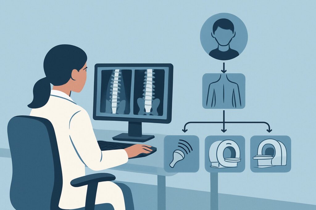
A 10-year-old presents to your clinic with several weeks of intermittent, low-level mid-back pain. The pain is worse after soccer practice but resolves with rest. The physical exam is entirely normal: no point tenderness, full range of motion, no neurologic deficits, and no skin changes. There are no constitutional symptoms like fever, weight loss, or night sweats. You suspect a simple musculoligamentous strain, but the parents are anxious and requesting imaging. This article addresses the specific American College of Radiology (ACR) guidelines for this common scenario: a child with back pain, no clinical red flags, undergoing initial imaging. For this presentation, if imaging is pursued, the ACR designates `Radiography complete spine` as Usually Appropriate.
Who Fits This Clinical Scenario?
This guidance applies specifically to pediatric patients presenting with back pain where a thorough history and physical examination have failed to identify any “red flag” findings. The key inclusion criteria are:
- Patient is a child or adolescent.
- The primary complaint is back pain (thoracic, lumbar, or sacral).
- There are no clinical red flags. This means the absence of fever, night pain that awakens the child, unexplained weight loss, constant or progressively worsening pain, neurologic symptoms (weakness, numbness, bowel/bladder dysfunction), a history of malignancy, or significant trauma.
This workflow is distinct from other pediatric back pain scenarios. If your patient presents differently, this guidance may not apply. For example, this article does not cover:
- Children with red flags: A patient with fever, night sweats, or a known malignancy falls into a separate category where more advanced imaging is often warranted upfront.
- Patients with known or suspected inflammation/infection: If there is clinical suspicion for conditions like diskitis or osteomyelitis (e.g., elevated inflammatory markers), the imaging algorithm changes significantly.
- Patients with overlying skin changes: A palpable lump, hairy patch, or draining sinus over the spine suggests a congenital anomaly and requires a different diagnostic pathway, often starting with ultrasound or Magnetic Resonance Imaging (MRI).
What Diagnoses Are You Working Up in This Scenario?
In a child with non-specific back pain and a normal exam, the differential diagnosis is heavily weighted toward benign and mechanical causes. The goal of initial imaging is to rule out common structural abnormalities while avoiding unnecessary radiation or costly advanced studies.
Musculoligamentous Strain: This is by far the most common cause of back pain in children and adolescents. It’s a diagnosis of exclusion, as imaging will be normal. The pain is typically activity-related and self-limited. While radiographs won’t confirm a strain, their primary role is to confidently exclude other structural causes.
Spondylolysis and Spondylolisthesis: These conditions, involving a stress fracture in the pars interarticularis (spondylolysis) which can lead to vertebral slippage (spondylolisthesis), are particularly common in young athletes involved in sports with repetitive hyperextension, like gymnastics or football. Oblique radiographs can often visualize the characteristic “Scotty dog” fracture, though AP and lateral views are the standard first step.
Scheuermann’s Disease: This structural kyphosis, or forward rounding of the back, is a common cause of thoracic back pain in adolescents. It is defined by specific radiographic criteria, including anterior wedging of at least three consecutive vertebrae. A standing lateral radiograph is the key diagnostic study.
While less likely in the absence of red flags, initial radiographs also serve as a baseline screen for more serious but rare conditions like benign bone tumors (e.g., osteoid osteoma), significant scoliosis, or vertebral body abnormalities that might warrant further investigation.
Why Is Complete Spine Radiography the Recommended Initial Study?
When a clinical decision is made to proceed with imaging for a child with back pain and no red flags, the ACR finds `Radiography complete spine` to be Usually Appropriate. This recommendation is based on a careful balance of diagnostic yield, radiation safety, and resource utilization for this specific low-risk population.
Anteroposterior (AP) and lateral radiographs of the spine are an excellent first-line tool for assessing bony alignment, vertebral body morphology, and potential fractures or defects. They can readily diagnose or exclude the most common structural causes in this differential, including Scheuermann’s kyphosis, significant scoliosis, and spondylolisthesis. The study is widely available, quick to perform, and does not require sedation.
In contrast, more advanced imaging modalities are designated Usually not appropriate for this initial workup:
- MRI spine area of interest without IV contrast: While MRI provides superior soft tissue detail and does not use ionizing radiation, it is not the appropriate first step in a low-risk patient. It is more expensive, has longer acquisition times, and may require sedation in younger children. Its use is reserved for cases where red flags are present, radiographs are abnormal, or pain persists despite conservative management.
- CT spine area of interest without IV contrast: Computed Tomography (CT) offers excellent bony detail, but its utility over radiography in this initial setting is minimal and comes at the cost of a significantly higher radiation dose. For a child, the radiation from spine radiographs (pediatric relative radiation level ☢☢☢, 0.3-3 mSv) is substantially lower than that from a spine CT (pediatric RRL ☢☢☢☢, 3-10 mSv). Minimizing radiation exposure is a critical principle in pediatric imaging (ALARA – As Low As Reasonably Achievable).
The choice of complete spine radiography provides a cost-effective, low-radiation screening examination that effectively evaluates for the most probable structural etiologies in this specific clinical scenario, reserving more advanced imaging for patients with concerning findings or a change in clinical status.
What’s Next After Radiography? Downstream Workflow
The results of the spine radiographs will guide your next steps, which often involve clinical management rather than more imaging.
- If the radiographs are negative: A normal radiographic study is the most common outcome in this scenario. This result strongly supports a diagnosis of musculoligamentous strain. The appropriate next step is reassurance and conservative management, including activity modification, physical therapy, and analgesics as needed. Further imaging is not indicated unless the patient develops new red flags or the pain becomes chronic and unresponsive to treatment.
- If the radiographs show spondylolysis or spondylolisthesis: A positive finding for a pars defect or vertebral slip warrants a referral to a pediatric orthopedic surgeon or sports medicine specialist for further management, which may include bracing, activity restriction, and specialized physical therapy. The specialist may consider further imaging, such as MRI or bone scan with SPECT, but this is a downstream decision outside the scope of initial imaging.
- If the radiographs are positive for Scheuermann’s Disease: Confirmation of vertebral wedging consistent with this diagnosis should also prompt a referral to pediatric orthopedics to discuss observation or bracing, depending on the severity of the kyphosis and the child’s skeletal maturity.
- If the radiographs are indeterminate or suspicious: In the rare event that radiographs reveal a finding suspicious for infection, inflammation, or neoplasm (e.g., vertebral body destruction, aggressive-appearing lesion), the clinical situation changes immediately. The patient now fits a different ACR scenario, such as “Child. Back pain. With at least one clinical red flag. Suspected infection, inflammation, or malignancy on radiography.” The next step in this pathway is almost always an MRI of the spine, often with and without IV contrast, to better characterize the abnormality.
–
Pitfalls to Avoid (and When to Get Help)
The primary pitfall in this scenario is over-imaging. Most pediatric back pain without red flags is benign and self-resolving, and a period of watchful waiting and conservative care is often the most appropriate first step before any imaging is ordered. Another common error is ordering advanced imaging like MRI or CT as the initial study, which exposes the child to unnecessary cost, potential sedation, and (in the case of CT) significant radiation without clear clinical justification. Finally, be vigilant for evolving symptoms; pain that begins as mechanical can develop red flags over time. If the pain becomes constant, awakens the child from sleep, or is accompanied by systemic symptoms or neurologic changes, escalate care and imaging immediately.
Related ACR Topics and Tools
For a comprehensive overview of all pediatric back pain scenarios and their corresponding imaging recommendations, please consult our parent guide. For additional resources on imaging selection, technique, and safety, the following tools may be helpful.
- For breadth across all scenarios in Back Pain-Child, see our parent guide: Back Pain-Child: ACR Appropriateness Decoded.
- ACR Appropriateness Criteria Lookup — for adjacent scenarios
- Imaging Protocol Library — for technique on the recommended study
- Radiation Dose Calculator — for cumulative dose conversations
Frequently Asked Questions
Is it ever appropriate to skip radiographs and go directly to MRI for a child’s back pain?
Yes, but only if clinical red flags are present. For a child presenting with back pain accompanied by fever, night sweats, neurologic deficits, or a history of cancer, MRI is often the appropriate first imaging study to evaluate for infection, inflammation, or malignancy. In the absence of these findings, radiographs are the recommended initial step.
What if the back pain is localized to one specific spot but there are no other red flags?
Focal, persistent point tenderness can be a subtle red flag, particularly if it’s over a bony prominence. While the overall scenario might still fit a low-risk profile, this finding increases the suspicion for a stress injury like spondylolysis or a benign bone lesion like an osteoid osteoma. Radiographs remain the appropriate first step, but your threshold to proceed with advanced imaging like MRI might be lower if the radiographs are negative and the focal pain persists.
Do the guidelines change if the child is very young, for example, under 5 years old?
Yes, back pain in a very young child (under 5) is considered a red flag in itself and should be investigated more thoroughly. The differential diagnosis in this age group is more concerning for serious pathology like diskitis, osteomyelitis, or tumors (e.g., neuroblastoma). This scenario would not fit the ‘no clinical red flags’ pathway, and a more aggressive imaging workup, often starting with MRI, is typically warranted.
Should I order standing or supine radiographs for a child with back pain?
For assessing alignment issues like scoliosis or kyphosis (Scheuermann’s disease), standing (weight-bearing) AP and lateral radiographs are preferred as they provide a functional assessment of the spine. For trauma or suspected fracture, supine views may be necessary. For general, non-specific pain, standing views are generally more informative.
If radiographs are normal, when should I consider a follow-up study?
If initial radiographs are normal and a diagnosis of musculoskeletal strain is made, no follow-up imaging is needed if the child’s symptoms improve with conservative care. You should only consider repeat or advanced imaging if the pain persists for an extended period (e.g., more than 4-6 weeks) despite treatment, worsens significantly, or if new red flags develop.
Reviewed by Pouyan Golshani, MD, Interventional Radiologist — June 27, 2026