What Imaging Should You Order for Suspected Esophageal Cancer Recurrence?
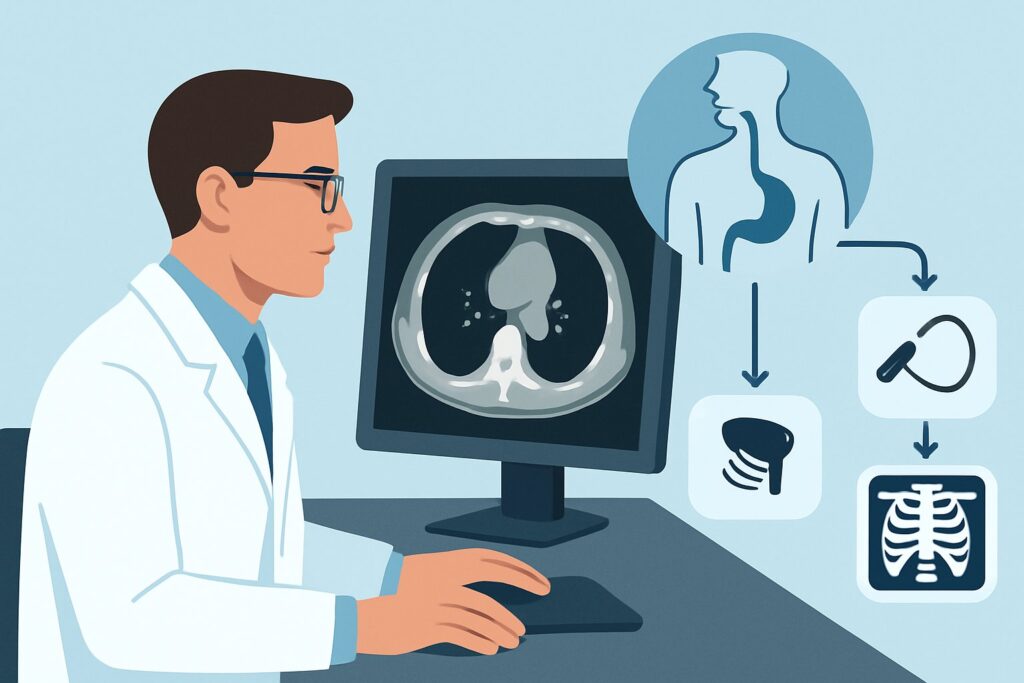
A 68-year-old man, eighteen months post-esophagectomy and adjuvant chemoradiation for esophageal adenocarcinoma, presents to your clinic with a new, persistent cough and a vague sensation of food “sticking.” His weight is down five pounds from his last visit three months ago. You suspect a potential recurrence, but the clinical signs are non-specific. The immediate question is how to best evaluate for locoregional disease versus distant metastases without delay. This scenario requires an imaging modality that can comprehensively assess the chest and abdomen, the most common sites of failure. For a patient with suspected or known recurrence of esophageal cancer after treatment, the American College of Radiology (ACR) Appropriateness Criteria rate `CT chest and abdomen with IV contrast` as “Usually Appropriate,” providing a crucial first step in the diagnostic pathway.
Who Fits This Clinical Scenario for Suspected Esophageal Cancer Recurrence?
This guidance is specifically for patients who have already completed a full course of definitive therapy for esophageal cancer—whether surgery, chemotherapy, radiation, or a combination—and now present with signs, symptoms, or laboratory findings concerning for disease recurrence. This includes new or worsening dysphagia, odynophagia, significant unintentional weight loss, persistent cough, chest or abdominal pain, or rising tumor markers.
It is critical to distinguish this situation from other, distinct clinical scenarios that require different imaging approaches:
- Initial Pretreatment Staging: This workflow is not for newly diagnosed patients who have not yet started therapy. Their initial workup involves a different set of imaging priorities focused on defining the primary tumor (T-stage), nodal involvement (N-stage), and distant metastases (M-stage) to guide the primary treatment plan.
- Routine Post-Treatment Surveillance: This guidance does not apply to asymptomatic patients undergoing routine, scheduled follow-up imaging. The choice and frequency of imaging in the absence of symptoms is a separate clinical question.
- Imaging During Treatment: This scenario is distinct from assessing treatment response while a patient is actively undergoing chemotherapy or radiation. Mid-treatment imaging has its own specific goals, such as determining if the current regimen is effective.
What Diagnoses Are You Working Up with Post-Treatment Imaging?
When ordering imaging for suspected recurrence, you are primarily investigating a differential that spans local, regional, and distant disease, while also considering non-malignant post-treatment effects.
Locoregional Recurrence: This is a primary concern and often the most challenging to diagnose. Recurrence can occur at the surgical anastomosis, in the tumor bed if the esophagus is in situ, or in nearby lymph node basins (e.g., mediastinal, supraclavicular, celiac axis). Imaging must be able to detect new or enlarging soft tissue masses and abnormal lymph nodes in these areas.
Distant Metastatic Disease: Esophageal cancer most commonly spreads to the lungs, liver, adrenal glands, and distant lymph nodes. A comprehensive imaging study must thoroughly evaluate these organs. The presence of distant disease fundamentally alters the patient’s prognosis and treatment goals, often shifting the focus from curative-intent local therapy to systemic palliation.
Post-Treatment Changes vs. Recurrence: A significant diagnostic challenge is differentiating benign post-therapeutic changes from malignant tissue. Radiation-induced fibrosis, inflammation, and granulation tissue at a surgical anastomosis can all mimic the appearance of a recurrent tumor on imaging. This is where the morphologic detail from CT becomes critical, and where functional imaging may later play a problem-solving role.
Benign Stricture: Not all post-treatment dysphagia is cancer. Benign fibrotic strictures can develop at the anastomosis, causing mechanical obstruction. While imaging can show the narrowing, endoscopy is often required for definitive diagnosis and potential therapeutic intervention.
Why Is CT of the Chest and Abdomen the Recommended First Study?
For a patient with suspected esophageal cancer recurrence, both `CT chest and abdomen with IV contrast` and `FDG-PET/CT skull base to mid-thigh` are rated “Usually Appropriate” by the ACR. CT is frequently the initial modality due to its wide availability, speed, and excellent anatomic resolution, which is essential for evaluating the common sites of recurrence.
The rationale for starting with contrast-enhanced CT is strong. It provides a detailed anatomical map of the chest and upper abdomen. IV contrast is essential for highlighting abnormal enhancement in potential liver or adrenal metastases and for delineating lymph nodes and vascular structures in the mediastinum. CT excels at detecting pulmonary nodules, which are a common site of distant spread. It can identify new soft tissue thickening or mass at the primary site or anastomosis, though differentiating it from scar tissue can be difficult.
While CT provides the anatomic detail, FDG-PET/CT provides functional information, identifying areas of high metabolic activity characteristic of cancer. It is highly sensitive for detecting viable tumor and can be superior for distinguishing post-treatment fibrosis from active recurrence. Many centers use CT as the first-line test and reserve PET/CT for confirming suspected findings, resolving equivocal CT results, or as a baseline before starting systemic therapy for confirmed metastatic disease.
Alternative studies are rated lower for specific reasons in this context:
- Fluoroscopy upper GI series: Rated “Usually Not Appropriate.” While it can visualize the esophageal lumen and identify a stricture, it provides no information about extraluminal disease, lymph nodes, or distant organs, making it insufficient for restaging.
- MRI chest and abdomen: Rated “Usually Not Appropriate.” MRI is prone to respiratory motion artifact in the chest and generally offers no significant advantage over CT for detecting the lung, liver, or nodal metastases typical of esophageal cancer.
The radiation dose for a contrast-enhanced CT of the chest and abdomen is significant (ACR Relative Radiation Level ☢☢☢☢, 10-30 mSv), but this exposure is justified by the critical need to accurately diagnose or exclude a life-threatening recurrence. Once you’ve decided on CT, our protocol guide covers the technique, contrast, and reading principles. For detailed specifications, see our guide: CT Chest/Abdomen/Pelvis with IV Contrast.
What’s Next After CT? Downstream Workflow
The results of the CT scan will guide the subsequent clinical pathway. The next steps are contingent on whether the findings are positive, negative, or indeterminate for recurrence.
- If the CT is positive for recurrence: The location and extent of disease dictate the next steps. For isolated, resectable locoregional recurrence, further workup with FDG-PET/CT is often performed to rule out occult distant disease before considering aggressive local therapies like surgery or re-irradiation. If distant metastases are confirmed, the patient is typically referred to medical oncology for systemic therapy. Biopsy of a suspicious lesion is almost always required to confirm the diagnosis histologically before initiating treatment.
- If the CT is negative: If clinical suspicion remains high despite a negative CT (e.g., worsening dysphagia), further evaluation is warranted. This often involves endoscopy with biopsy to directly visualize the mucosa and rule out a luminal recurrence that was inconspicuous on CT. An FDG-PET/CT may also be considered to look for metabolically active disease not visible on anatomic imaging.
- If the CT is indeterminate: Equivocal findings, such as mild thickening at the anastomosis or borderline-sized lymph nodes, are common. In this situation, FDG-PET/CT is an excellent problem-solving tool to determine if these areas are metabolically active. Alternatively, short-interval follow-up CT in 6-12 weeks can assess for stability or progression. The decision often depends on the degree of clinical suspicion and the patient’s fitness for further intervention.
Pitfalls to Avoid (and When to Get Help)
Navigating the workup for suspected esophageal cancer recurrence requires careful attention to detail to avoid common errors.
- Omitting IV Contrast: Ordering a non-contrast CT severely limits its diagnostic utility. It compromises the evaluation of the liver, adrenal glands, and lymph nodes. Always specify “with IV contrast” unless there is a strong contraindication.
- Not Comparing to Priors: The most critical step in interpreting follow-up scans is direct comparison with previous imaging. New or growing findings are highly suspicious, whereas stable post-treatment changes are reassuring. Ensure the radiologist has access to all relevant prior studies.
- Over-reliance on Imaging Alone: Imaging findings must be correlated with the clinical picture and endoscopic findings. A “negative” scan in a patient with progressive dysphagia should prompt an endoscopic evaluation, not a cessation of the workup.
If CT findings are complex or equivocal, or if the results will lead to a major therapeutic decision like surgery or radiation, presentation at a multidisciplinary tumor board is the standard of care.
Related ACR Topics and Tools
This article covers one specific scenario in depth. For a comprehensive overview of imaging at all stages of disease, from initial diagnosis to routine surveillance, please consult the parent topic article. You can also use the tools below to explore other scenarios, protocols, and radiation dose considerations.
- For breadth across all scenarios in Staging and Follow-up of Esophageal Cancer, see our parent guide: Staging and Follow-up of Esophageal Cancer: ACR Appropriateness Decoded.
- ACR Appropriateness Criteria Lookup — for adjacent scenarios
- Imaging Protocol Library — for technique on the recommended study
- Radiation Dose Calculator — for cumulative dose conversations
Frequently Asked Questions
Why is FDG-PET/CT also rated ‘Usually Appropriate’ for suspected esophageal cancer recurrence?
FDG-PET/CT is also rated ‘Usually Appropriate’ because it provides crucial functional information that complements the anatomical detail of CT. It is highly sensitive for detecting metabolically active cancer cells and can be particularly effective at differentiating benign post-treatment scar tissue from active tumor recurrence. It is also excellent for detecting unexpected distant metastases, which can change the entire treatment plan. Many institutions use CT first and employ PET/CT to confirm suspicious findings or when CT is inconclusive.
Should I order a CT of the chest, abdomen, and pelvis, or just chest and abdomen?
The ACR lists ‘CT chest and abdomen with IV contrast’ as ‘Usually Appropriate.’ ‘CT chest abdomen pelvis with IV contrast’ is rated ‘May be appropriate (Disagreement).’ The primary sites of distant metastasis for esophageal cancer are the lungs, liver, and adrenal glands, which are covered by a chest and abdomen CT. The pelvis is a lower-yield area. However, including the pelvis may be reasonable if the patient has specific symptoms like bone pain or if there is a higher suspicion for osseous metastases, but it is not considered a routine part of the initial restaging for every patient.
What if my patient has a contraindication to IV contrast, like severe renal failure or a true allergy?
If IV contrast is contraindicated, a non-contrast CT of the chest and abdomen can still be performed. It remains excellent for detecting lung nodules and can identify gross liver masses or adrenal lesions, but its sensitivity is reduced. In this situation, an FDG-PET/CT becomes a much more valuable tool, as the PET component does not require iodinated contrast and can highlight metabolically active disease in the lymph nodes, liver, and other organs that would be difficult to assess on the non-contrast CT.
How soon after treatment completion can post-treatment inflammation be distinguished from recurrence?
Distinguishing post-treatment inflammation from true recurrence is a major challenge, especially in the first few months after therapy. Radiation esophagitis and postoperative inflammation can persist for 3-6 months or longer and can mimic tumor on both CT and FDG-PET/CT. For this reason, imaging performed very early after treatment completion can be difficult to interpret. Most surveillance guidelines recommend the first post-treatment imaging around 3-6 months, and even then, findings must be interpreted with caution and correlated with endoscopic findings and clinical symptoms.
Is there a role for MRI in evaluating suspected recurrence of esophageal cancer?
For the primary question of locoregional and common distant recurrence, MRI is rated ‘Usually Not Appropriate.’ It is more susceptible to motion artifact in the chest and abdomen and generally does not provide more useful information than CT for evaluating the lungs, lymph nodes, or adrenal glands. Its main role would be as a problem-solving tool for a specific indeterminate finding, such as characterizing a liver lesion seen on CT, but it is not a first-line examination for this clinical scenario.
Reviewed by Pouyan Golshani, MD, Interventional Radiologist — May 30, 2026