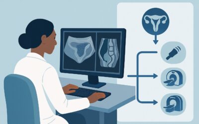
What Imaging Should You Order for Suspected Gyn Pelvic Pain with a Negative ß-hCG?

A 28-year-old female presents to the emergency department at 10 PM with acute-onset, severe right-sided pelvic pain. Her last menstrual period was two weeks ago, and her urine pregnancy test is negative. On exam, she has marked right adnexal tenderness. You suspect a gynecological etiology, with ovarian torsion high on your differential. You need to choose the right initial imaging study to confirm your suspicion and guide immediate management. This article provides a detailed clinical workflow for this specific scenario, grounded in the American College of Radiology (ACR) Appropriateness Criteria, which rates US duplex Doppler pelvis as Usually Appropriate.
Who Fits This Clinical Scenario for Acute Pelvic Pain?
This guidance applies to a specific and common clinical presentation: a patient in the reproductive age group with acute pelvic pain where the clinical suspicion points toward a gynecological source, and pregnancy has been ruled out. The key inclusion criteria are:
- Patient Demographics: A patient of reproductive age (typically post-menarche and pre-menopause).
- Clinical Presentation: Acute onset of pelvic pain.
- Suspected Etiology: History and physical exam findings suggest a gynecological origin (e.g., adnexal tenderness, pain related to menstrual cycle, abnormal vaginal bleeding, cervical motion tenderness).
- Pregnancy Status: A negative ß-hCG test, either from serum or urine, is confirmed.
It is critical to distinguish this scenario from similar presentations. This workflow does not apply if:
- The ß-hCG is positive. A positive pregnancy test fundamentally changes the differential, prioritizing the evaluation for ectopic pregnancy. This requires a different imaging pathway.
- A non-gynecological cause is more likely. If symptoms point strongly to appendicitis (migratory right lower quadrant pain, fever, leukocytosis), diverticulitis, or nephrolithiasis (flank pain, hematuria), the imaging strategy shifts accordingly.
- The patient is post-menopausal. While some overlap exists, the differential diagnosis and risk stratification for malignancy are different in this population.
What Diagnoses Are You Working Up in This Scenario?
With pregnancy excluded, the differential for acute gynecologic pelvic pain centers on adnexal and uterine pathologies that can present emergently. The choice of imaging is driven by the need to rapidly evaluate for these conditions.
Ovarian Torsion: This is the most time-sensitive diagnosis and a primary reason for urgent imaging. Torsion occurs when the ovary twists on its ligamentous support, compromising its blood supply. It causes sudden, severe, and often unrelenting pain. Prompt diagnosis via imaging is crucial to salvaging the ovary.
Hemorrhagic Ovarian Cyst or Ruptured Cyst: A very common cause of acute pelvic pain in this demographic. A functional cyst can bleed into itself or rupture, releasing blood and fluid into the pelvis, which causes peritoneal irritation and pain. While often self-limited, imaging is needed to confirm the diagnosis and exclude more sinister causes.
Pelvic Inflammatory Disease (PID) and Tubo-ovarian Abscess (TOA): An infection of the upper genital tract can lead to inflammation and, in severe cases, the formation of a complex inflammatory mass involving the fallopian tube and ovary. Patients may also present with fever, vaginal discharge, and cervical motion tenderness. Imaging helps assess the extent of disease and identify a drainable fluid collection.
Degenerating or Torsed Leiomyoma (Fibroid): Uterine fibroids are common, and they can outgrow their blood supply, leading to infarction and acute pain (degeneration). A pedunculated fibroid, which hangs off the uterus by a stalk, can also twist on itself (torsion), presenting similarly to ovarian torsion.
Why Is Pelvic Ultrasound with Doppler the Recommended First Study?
For a non-pregnant patient with suspected acute gynecologic pelvic pain, the ACR designates three ultrasound modalities as Usually Appropriate, with US duplex Doppler pelvis being the most comprehensive initial study. This recommendation is based on its high diagnostic yield, safety profile, and accessibility.
A complete pelvic ultrasound exam typically involves both transabdominal and transvaginal approaches. The transabdominal view provides a broad overview of the entire pelvis, identifying large masses or free fluid. The transvaginal approach uses a higher-frequency transducer placed closer to the adnexa, providing superior spatial resolution for detailed evaluation of the ovaries, fallopian tubes, and uterus.
The addition of color and spectral Duplex Doppler is what makes this study indispensable for this scenario. Doppler imaging assesses blood flow within the ovarian vessels. The absence or significant reduction of arterial and venous flow to an enlarged, edematous ovary is a key finding for diagnosing ovarian torsion. It can also demonstrate the characteristic “ring of fire” peripheral vascularity in a corpus luteum cyst, helping to distinguish it from other pathologies.
In contrast, other imaging modalities are rated lower for this initial workup:
- CT abdomen and pelvis with IV contrast is rated as May be appropriate. While CT can identify adnexal masses, free fluid, or complications like abscesses, it is less sensitive than ultrasound for characterizing ovarian parenchyma and detecting subtle signs of torsion. Crucially, it involves ionizing radiation (☢☢☢ 1-10 mSv), a significant consideration in young patients who may require future imaging. CT is best reserved for cases where the ultrasound is inconclusive or if a non-gynecologic diagnosis (like appendicitis) becomes more likely.
- CT abdomen and pelvis without IV contrast is rated as Usually not appropriate. Omitting intravenous contrast severely limits the evaluation of vascular compromise in torsion, abscess wall enhancement in a TOA, and the characterization of complex masses. It offers little diagnostic advantage over ultrasound while still exposing the patient to radiation.
What’s Next After Pelvic Ultrasound? Downstream Workflow
The results of the initial pelvic ultrasound will guide the subsequent clinical pathway. The next steps are determined by whether the findings are definitive, negative, or indeterminate.
If the study is positive for a surgical emergency:
A definitive finding of ovarian torsion (e.g., an enlarged ovary with absent Doppler flow) is a surgical emergency. The next step is an immediate consultation with gynecology for surgical detorsion. Similarly, a large, complex tubo-ovarian abscess may require gynecologic consultation for potential drainage and inpatient antibiotic therapy.
If the study is positive for a non-emergent diagnosis:
Findings of a simple or hemorrhagic ovarian cyst, or uncomplicated fibroids, can often be managed on an outpatient basis. The patient may be discharged with appropriate analgesia and instructions for follow-up with their gynecologist. A follow-up ultrasound may be recommended in several weeks to ensure resolution.
If the study is negative or indeterminate:
A completely normal pelvic ultrasound in a patient with persistent, severe pain is a diagnostic challenge. At this point, the clinical team must reconsider the differential diagnosis. The focus may shift to non-gynecological causes, such as appendicitis, diverticulitis, or ureteral stones. This is a key juncture where a different imaging study, such as CT abdomen and pelvis with IV contrast, may become appropriate to evaluate for these alternative diagnoses. If an adnexal mass is seen but cannot be fully characterized by ultrasound, MRI pelvis without and with IV contrast (rated May be appropriate) is the best problem-solving tool, offering superior soft-tissue contrast without ionizing radiation.
Pitfalls to Avoid (and When to Get Help)
Navigating this clinical scenario requires vigilance to avoid common diagnostic traps. Here are a few key pitfalls:
- Misinterpreting preserved Doppler flow: The presence of some blood flow on Doppler does not definitively exclude ovarian torsion. Torsion can be intermittent, or dual arterial supply may allow for some persistent flow early in the process. Correlate imaging with the high degree of clinical suspicion.
- Incomplete ultrasound technique: Failing to perform both transabdominal and transvaginal scans can lead to missed diagnoses. A large mass high in the pelvis may be missed on transvaginal imaging alone.
- Anchoring on a single diagnosis: If the ultrasound is negative but the patient’s pain is severe and persistent, do not stop the workup. Re-evaluate the patient and consider alternative, non-gynecologic etiologies.
- Delaying imaging: For suspected ovarian torsion, time is critical. Delays in obtaining an ultrasound can mean the difference between salvaging and losing an ovary.
If the clinical picture and imaging findings are discordant, or if a complex diagnosis like a TOA is identified, early consultation with gynecology and radiology is essential for optimal patient management.
Related ACR Topics and Tools
This article focuses on one specific clinical variant. For a comprehensive overview of all scenarios related to this topic, please consult our parent guide. The following GigHz tools can also support your clinical decision-making:
- For breadth across all scenarios in Acute Pelvic Pain in the Reproductive Age Group, see our parent guide: Acute Pelvic Pain in the Reproductive Age Group: ACR Appropriateness Decoded.
- Imaging Appropriateness Selector — for adjacent scenarios
- Imaging Protocol Library — for technique on the recommended study
- Radiation Dose Calculator — for cumulative dose conversations
Frequently Asked Questions
Why is ultrasound preferred over CT for initial imaging in a non-pregnant patient with suspected gynecologic pelvic pain?
Ultrasound is preferred because it provides excellent visualization of the ovaries and uterus without using ionizing radiation, which is a key safety consideration in the reproductive age group. The addition of Doppler is also highly sensitive for evaluating blood flow, which is critical for diagnosing ovarian torsion. CT is less sensitive for early or partial torsion and exposes the patient to radiation.
Does a normal pelvic ultrasound rule out all gynecologic causes of acute pain?
Not entirely. While a high-quality ultrasound is very sensitive for structural abnormalities like torsion, large cysts, or abscesses, it may not detect early pelvic inflammatory disease (PID) or small, non-hemorrhagic ruptured cysts. Furthermore, pain from a condition like endometriosis may not have obvious correlates on an emergent ultrasound. Clinical correlation is paramount.
What if the patient cannot tolerate a transvaginal ultrasound?
If a patient cannot tolerate or declines a transvaginal ultrasound, a transabdominal ultrasound can still be performed. While the image resolution of the adnexa will be lower, it can still identify large masses, free fluid, and in some cases, signs of torsion. The limitations of a transabdominal-only exam should be noted in the report, and if the study is non-diagnostic in a patient with high clinical suspicion, an alternative modality like MRI may be considered.
When should I order an MRI instead of an ultrasound for this presentation?
MRI is generally not a first-line imaging test for acute pelvic pain due to its cost, time, and limited availability in many emergency settings. However, it is an excellent problem-solving tool. According to the ACR, MRI (rated ‘May be appropriate’) should be considered when the ultrasound is indeterminate or equivocal, for example, to better characterize a complex adnexal mass or to confirm suspected torsion when Doppler findings are unclear.
If I suspect ovarian torsion but the Doppler flow appears normal, what should I do?
The presence of Doppler flow does not definitively exclude torsion, as it can be intermittent or incomplete. If your clinical suspicion for torsion remains high despite preserved flow on ultrasound, you should have an urgent discussion with both the radiologist and a gynecologic consultant. Secondary signs of torsion on ultrasound, such as ovarian enlargement, edema, and abnormal ovarian position, are very important. The clinical presentation is the most critical factor, and surgical exploration may still be warranted.
Reviewed by Pouyan Golshani, MD, Interventional Radiologist — May 26, 2026