What Imaging Should You Order for Suspected Pelvic-Origin Varicose Veins in Females?
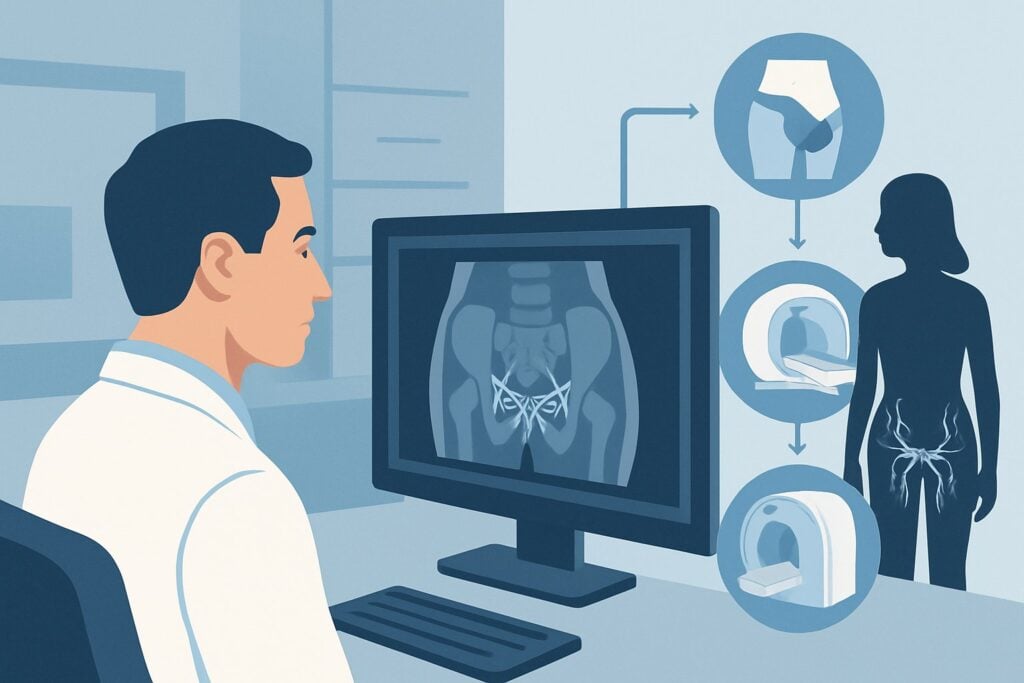
A 44-year-old multiparous woman presents to your clinic with painful, bulging veins in her left leg. Unlike typical varicose veins you’ve seen, these are most prominent on the upper medial thigh and she also reports associated perineal and gluteal varicosities. She describes a chronic, dull, aching pain in her pelvis that worsens with prolonged standing and at the end of the day. You suspect her lower extremity venous disease may originate from pelvic venous insufficiency. The immediate question is how to begin the diagnostic workup without ordering an invasive or high-radiation study prematurely. According to the American College of Radiology (ACR) Appropriateness Criteria, the initial imaging study for this presentation, US duplex Doppler lower extremity, is rated Usually Appropriate.
Who Fits This Clinical Scenario?
This guidance is for the initial diagnosis of a female patient presenting with lower extremity varicose veins where a pelvic origin is clinically suspected. The key indicators that should route a clinician to this specific workflow, rather than the standard varicose vein workup, are the location and pattern of the varicosities and associated symptoms.
Inclusion criteria for this scenario include:
- Female patient (this condition is rare in males).
- Varicose veins in atypical distributions, such as the upper medial thigh, perineum, vulva, or gluteal region.
- Lower extremity varicosities that appear to be extensions of perineal or gluteal veins.
- Co-existing symptoms of pelvic congestion syndrome (PCS), such as chronic pelvic pain, dyspareunia, or urinary urgency, though these are not required.
This workflow is NOT for:
- Standard Varicose Veins: Patients with varicosities clearly confined to the great saphenous vein (GSV) or small saphenous vein (SSV) distributions without perineal, vulvar, or gluteal involvement. This presentation fits the more common Varicose veins. Initial diagnosis. scenario.
- Post-Thrombotic Syndrome: Patients with a known history of deep vein thrombosis (DVT) and severe skin changes, edema, or ulceration. These patients fall under the Suspected iliocaval or lower extremity disease with severe post-thrombotic changes. scenario.
- Male Patients: While males can have pelvic venous disorders (e.g., varicoceles), the pathophysiology and presentation of pelvic-origin leg varicosities are distinctly different and not covered by this specific variant.
What Diagnoses Are You Working Up in This Scenario?
When you suspect a pelvic source for lower extremity varicosities, the differential diagnosis expands beyond simple saphenous vein reflux. The imaging strategy is designed to investigate several potential causes of pelvic venous hypertension that can manifest in the legs.
Pelvic Venous Insufficiency (PVI) is the most common underlying cause. This is often part of Pelvic Congestion Syndrome (PCS) and typically results from incompetent valves in the ovarian veins (most commonly the left) or internal iliac veins. Reflux from these pelvic veins allows blood to flow retrograde, pressurizing and dilating veins in the pelvis, perineum, and ultimately connecting to the medial thigh veins via perforators. This is especially common in multiparous women due to hormonal changes and increased venous capacitance during pregnancy.
A less common but consequential cause is extrinsic compression of major pelvic veins. May-Thurner Syndrome, or non-thrombotic iliac vein compression, involves the compression of the left common iliac vein by the overlying right common iliac artery. This increases pressure in the left iliac vein, which can lead to pelvic and left lower extremity venous hypertension and varicosities.
Another compression syndrome to consider is Nutcracker Syndrome. This involves the compression of the left renal vein between the superior mesenteric artery (SMA) and the aorta. This can cause left renal vein hypertension, leading to the development of collateral pathways, including reflux down the left ovarian vein, which then contributes to PVI and pelvic-origin leg varicosities.
Why Is US Duplex Doppler Lower Extremity the Recommended Initial Study?
While the clinical suspicion points toward the pelvis, the ACR designates US duplex Doppler lower extremity as a Usually Appropriate first step. This may seem counterintuitive, but the rationale is to first characterize the extent of the lower extremity disease and rule out or confirm co-existing saphenous incompetence before proceeding to more complex pelvic imaging.
The initial lower extremity ultrasound serves several key purposes:
- Rule out primary saphenous reflux: It confirms whether the patient’s varicosities are solely from a pelvic source or if there is a concurrent, and more common, issue with the great or small saphenous veins.
- Identify atypical reflux patterns: A skilled sonographer can identify reflux originating from perineal perforating veins that connect to the medial thigh veins. This finding is a strong indicator of a pelvic source.
- Provide a baseline: It maps the superficial venous system of the leg, which is essential for any future treatment planning, whether it involves pelvic vein embolization or saphenous vein ablation.
It is critical to note that several other studies are also rated Usually Appropriate, including US duplex Doppler pelvis, CTV abdomen and pelvis with IV contrast, and MRV abdomen and pelvis without and with IV contrast. These are typically considered the next step after the initial leg ultrasound confirms that the reflux pattern is atypical or that saphenous reflux cannot explain the full clinical picture. Starting with CTV (☢☢☢☢ 10-30 mSv) or MRV exposes the patient to radiation or contrast and higher costs unnecessarily if the issue is simple saphenous disease. Catheter venography, while a gold standard for confirming reflux, is invasive and rated May be appropriate, reserved for cases where non-invasive imaging is equivocal or when endovascular treatment is planned concurrently.
Once you’ve decided on the initial ultrasound, our protocol guide covers the essential technical details for a comprehensive exam. For a detailed review of the technique, contrast considerations, and reading principles, see our guide: US Lower Extremity Doppler (DVT).
What’s Next After US Duplex Doppler Lower Extremity? Downstream Workflow
The results of the initial lower extremity duplex ultrasound will guide your next steps. The downstream workflow is a critical decision point that hinges on whether the findings confirm a pelvic source, suggest a standard saphenous source, or are inconclusive.
If the study is positive for an atypical reflux pattern (e.g., reflux from perineal perforators into the medial thigh, with a competent saphenofemoral junction), this strongly supports the initial suspicion of a pelvic origin. The next step is dedicated pelvic venous imaging to identify the source of the reflux (e.g., incompetent ovarian or internal iliac veins) and rule out compression syndromes. Both MR Venography (MRV) of the abdomen and pelvis and CT Venography (CTV) of the abdomen and pelvis are rated Usually Appropriate for this purpose. MRV is often preferred as it avoids ionizing radiation, while CTV provides excellent anatomic detail for procedural planning.
If the study is negative for saphenous reflux and shows no clear source, but high clinical suspicion for a pelvic origin remains (based on symptoms and vein distribution), the next step is also dedicated pelvic imaging with MRV or CTV.
If the study is positive for significant great or small saphenous vein reflux that can explain the patient’s varicosities, the workflow shifts. The patient may have standard chronic venous insufficiency, and the pelvic symptoms may be unrelated or a secondary finding. The workup and treatment would then follow the standard Varicose veins. Treatment. pathway, which typically involves endovenous ablation or other treatments for the saphenous system.
Pitfalls to Avoid (and When to Get Help)
Navigating the workup for suspected pelvic-origin varicose veins requires careful interpretation to avoid common missteps. Be mindful of these potential pitfalls:
- Stopping the Workup Prematurely: A “normal” lower extremity venous duplex that only evaluates the saphenous systems is insufficient. If your clinical suspicion for a pelvic source is high, a negative leg ultrasound does not rule out the diagnosis and should prompt dedicated pelvic imaging.
- Ordering an Incomplete Ultrasound: When ordering the initial duplex, communicate your suspicion to the radiology department or vascular lab. The sonographer should be specifically instructed to evaluate for atypical reflux patterns and connections to perineal veins.
- Attribution Error: Do not automatically attribute all leg varicosities to saphenous incompetence, especially in a multiparous woman with pelvic pain and an atypical vein distribution. The two conditions can coexist.
- Prematurely Ordering Invasive Studies: Avoid jumping to catheter venography for initial diagnosis. This invasive procedure is best reserved for therapeutic intervention or when non-invasive imaging (US, MRV, CTV) is inconclusive.
If the non-invasive workup is complex or equivocal, this is the point to escalate. A consultation with an Interventional Radiologist or a vascular specialist experienced in treating pelvic venous disorders is the appropriate next step.
Related ACR Topics and Tools
This article covers one specific scenario in depth. For a broader view of imaging for all related clinical presentations, from venous ulcers to post-thrombotic syndrome, please refer to our comprehensive hub article. It provides a complete overview of the ratings for every variant within this topic.
- For breadth across all scenarios in Lower Extremity Chronic Venous Disease, see our parent guide: Lower Extremity Chronic Venous Disease: ACR Appropriateness Decoded.
- To explore other clinical scenarios or search the full ACR criteria, use the ACR Appropriateness Criteria Lookup.
- To review technical details for other imaging studies, visit the Imaging Protocol Library.
- To discuss radiation exposure with your patients, especially when considering CT, use the Radiation Dose Calculator.
Frequently Asked Questions
Why start with a lower extremity ultrasound if I suspect a pelvic problem?
Starting with a lower extremity duplex ultrasound is a strategic first step recommended by the ACR. It helps rule out or confirm co-existing saphenous vein insufficiency, which is a more common cause of varicose veins. It also helps identify atypical reflux patterns from perineal veins that strongly suggest a pelvic source, justifying the need for more advanced pelvic imaging like MRV or CTV.
What specific findings on a lower extremity US suggest a pelvic source?
The key finding is reflux in the upper medial thigh veins that does not originate from the saphenofemoral junction. A sonographer may identify large, incompetent perineal or gluteal perforating veins that connect the pelvic venous system to the leg veins. Seeing retrograde flow from these perforators during a Valsalva maneuver is a classic sign of a pelvic origin.
Is an MRV or CTV better for evaluating pelvic veins after the initial ultrasound?
Both MR Venography (MRV) and CT Venography (CTV) are rated ‘Usually Appropriate’ and are excellent for this purpose. MRV is often preferred because it provides detailed imaging of the pelvic veins, including ovarian and iliac veins, without using ionizing radiation. CTV is faster and can offer superior spatial resolution, which can be beneficial for pre-procedural planning, but it involves a significant radiation dose (10-30 mSv).
Can this condition occur in women who have not had children?
Yes, while pelvic venous insufficiency is most common in multiparous women due to hormonal changes and venous compression during pregnancy, it can also occur in nulliparous women. In these cases, it may be related to congenital valve absence or underlying venous compression syndromes like May-Thurner or Nutcracker syndrome.
What’s the main difference between this scenario and the standard ‘Varicose veins. Initial diagnosis.’ scenario?
The primary difference is the clinical suspicion of the origin. The standard ‘Varicose veins’ scenario assumes the problem originates in the leg’s saphenous system. This ‘Pelvic-origin’ scenario is triggered by atypical vein distribution (e.g., vulvar, perineal, upper medial thigh) and/or associated pelvic symptoms, which shifts the diagnostic focus to include the pelvic venous system as the potential primary source.
Reviewed by Pouyan Golshani, MD, Interventional Radiologist — May 30, 2026