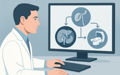
What Is the Best Drainage Strategy for a Loculated Empyema After Failed Antibiotics?

It’s day 21 of your patient’s admission for community-acquired pneumonia. Despite a full course of broad-spectrum antibiotics, their fever persists and their dyspnea is worsening. The latest contrast-enhanced chest CT confirms your suspicion: a large, loculated pleural collection with significant overlying pleural thickening and enhancement. The primary team is asking for the next step. Is simple needle aspiration enough, or does this complex, organized fluid collection require a more definitive intervention? This clinical scenario represents a critical decision point in managing complicated parapneumonic effusions, where medical therapy alone has failed. This article provides a workflow for this exact presentation, grounded in the American College of Radiology (ACR) Appropriateness Criteria. For this patient, the ACR rates ‘Percutaneous catheter drainage with administration of thrombolytic therapy’ as Usually Appropriate.
Who Fits This Clinical Scenario for Empyema Management?
This guidance is specifically for a patient with a complicated parapneumonic effusion that has progressed to an organized empyema. The key inclusion criteria are a clear history of pneumonia, clinical deterioration despite adequate antibiotic therapy, and specific imaging findings on CT scan—namely, a loculated pleural collection and associated pleural thickening. These features signify a transition from a simple, free-flowing effusion (exudative stage) to a more complex, fibrinopurulent, or organizing stage where the pus is thick and trapped within fibrinous septations.
This workflow does not apply to several similar-but-distinct clinical situations:
- Simple Parapneumonic Effusion: A patient with pneumonia and a small, free-flowing, non-loculated pleural effusion may respond to antibiotics alone or with a simple diagnostic/therapeutic thoracentesis. The presence of loculations and pleural thickening is the key differentiator.
- Lung Abscess: While also an infected collection, a lung abscess is located within the lung parenchyma itself, not the pleural space. It often appears as a thick-walled cavity on imaging and has a different management algorithm.
- Post-Operative Pleural Collection: An infected collection following thoracic surgery may have a different microbial profile and anatomical considerations, potentially altering the risk-benefit analysis of drainage procedures.
This article focuses squarely on the patient whose infection has become walled-off in the pleural space, demanding intervention beyond systemic antibiotics.
What Diagnoses Are You Working Up in This Scenario?
While the clinical picture strongly points to a single diagnosis, the intervention aims to both confirm this and rule out less common mimics. The primary goal is to obtain source control of the infection.
Empyema (Complicated Parapneumonic Effusion): This is overwhelmingly the most likely diagnosis. It represents the progression of a parapneumonic effusion to a frank, purulent infection of the pleural space. The loculations seen on CT are caused by fibrin septa that form as the infection organizes, trapping pus in pockets. Failure to drain this purulent material leads to uncontrolled sepsis, lung entrapment (fibrothorax), and poor clinical outcomes. The intervention is both diagnostic (confirming pus) and therapeutic (evacuating the infection).
Tuberculous Empyema: In patients with risk factors for tuberculosis (TB), such as immunosuppression or exposure history, a tuberculous pleural effusion can evolve into a chronic, loculated empyema. While less common than bacterial empyema in many regions, it remains a critical consideration. Fluid analysis from the drainage procedure, including acid-fast bacilli (AFB) stain and culture, is essential for diagnosis.
Malignant Pleural Effusion with Superinfection: A pre-existing malignant effusion can become secondarily infected, presenting identically to a primary empyema. This is a consequential diagnosis to make, as the patient’s underlying prognosis and long-term management strategy (e.g., potential for an indwelling pleural catheter) are significantly different. Cytology from the drained fluid is crucial in this context.
Why Is Percutaneous Drainage with Thrombolytics the Recommended Approach?
For a patient with a loculated empyema who has failed medical management, simply observing or repeating antibiotics is insufficient. The ACR evaluates several interventional options, highlighting the rationale for a minimally invasive, image-guided approach as a primary step.
The ACR rates Percutaneous catheter drainage with administration of thrombolytic therapy as Usually Appropriate. This procedure, typically performed by an interventional radiologist using ultrasound or CT guidance, involves placing one or more drainage catheters directly into the largest locules of the empyema. The key advantage over simple aspiration is the ability to provide continuous drainage. Furthermore, the administration of intrapleural thrombolytics (fibrinolytics) like alteplase (tPA) helps break down the fibrin septa that create the loculations, improving drainage efficacy and allowing a complex collection to be evacuated through a small-bore catheter. This approach is highly effective at achieving source control while avoiding the morbidity of a major surgical procedure.
In contrast, the ACR rates other options differently for this specific scenario:
- Needle aspiration alone is rated Usually not appropriate. While it can provide a diagnostic sample, a one-time aspiration is almost always insufficient to drain a thick, loculated, and voluminous collection. The fluid quickly reaccumulates, failing to provide adequate source control.
- Another course of antibiotics and postural drainage is also rated Usually not appropriate. At this stage, the infection is mechanically isolated from systemic circulation by the thickened pleural peel. Antibiotics cannot penetrate this space effectively, and the purulent material must be physically removed.
While Video-assisted thoracic surgery (VATS) decortication is also rated Usually Appropriate, percutaneous drainage is often considered first due to its lower invasiveness. VATS is an excellent and definitive therapy but requires general anesthesia and is a more significant physiologic insult. Many centers adopt a stepwise approach, starting with percutaneous drainage and escalating to VATS if the minimally invasive option fails.
What’s Next After Percutaneous Drainage? Downstream Workflow
Placing the drainage catheter is the beginning, not the end, of management. The post-procedure workflow is critical for success and involves close collaboration between interventional radiology and the primary clinical team.
- If the procedure is successful: The catheter is placed in the dominant collection, and purulent fluid is drained. The patient should show clinical improvement—defervescence, decreasing white blood cell count, and improved respiratory status—within 24 to 72 hours. The catheter is managed with regular flushes and intrapleural thrombolytic administration per institutional protocol. Daily chest radiographs monitor for lung re-expansion and resolution of the collection. The catheter is typically removed when drainage is minimal (<25 mL/day) and follow-up imaging confirms resolution.
- If the patient does not improve: If fever and leukocytosis persist after 48-72 hours of effective drainage, several possibilities must be considered. First, review the initial CT to assess for any undrained loculations. It is common for complex empyemas to require a second or even third catheter to address all infected pockets. If all collections appear adequately drained but the patient remains septic, a search for an alternative infectious source is warranted.
- If percutaneous drainage fails: Failure is defined as the inability to resolve the sepsis and effusion despite optimal catheter management. This may be due to extremely viscous pus or the development of a thick, inelastic visceral pleural peel that “traps” the lung, preventing re-expansion. In this case, the patient should be promptly evaluated by thoracic surgery for VATS or open decortication, which is rated May be appropriate as a next step.
Pitfalls to Avoid (and When to Get Help)
Navigating the management of a loculated empyema requires careful attention to detail to avoid common setbacks.
- Pitfall: Delaying Intervention. Once a patient with pneumonia has failed to improve on antibiotics and imaging confirms a complex, loculated collection, prompt source control is paramount. Delaying drainage allows the empyema to become more organized, increasing the likelihood that percutaneous measures will fail and major surgery will be required.
- Pitfall: Inadequate Catheter Management. A chest tube for empyema is not a passive device. It requires diligent nursing care, including regular flushing to maintain patency. Without this, the thick, fibrinous material will quickly clog the tube, rendering it ineffective.
- Pitfall: Stopping at One Catheter. A single drainage catheter can only access the locule it is placed in. Always review the pre-procedure CT and post-procedure imaging to ensure all significant collections are targeted, even if it requires placing multiple catheters.
If the patient’s clinical condition deteriorates rapidly or they develop signs of shock despite initial drainage, escalate immediately for a surgical consultation and critical care support.
Related ACR Topics and Tools
For a comprehensive overview of managing various infected fluid collections throughout the body, this article is best used alongside its parent topic guide. For additional decision support and technical details, the following GigHz resources are available:
- For breadth across all scenarios in Radiologic Management of Infected Fluid Collections, see our parent guide: Radiologic Management of Infected Fluid Collections: ACR Appropriateness Decoded.
- To look up appropriateness criteria for adjacent or alternative clinical scenarios, use the ACR Appropriateness Criteria Lookup tool.
- For detailed procedural techniques on image-guided interventions, explore the Imaging Protocol Library.
- To discuss cumulative radiation exposure from diagnostic and interventional procedures, our Radiation Dose Calculator can help frame conversations with patients.
Frequently Asked Questions
Why is CT guidance often preferred over ultrasound for placing the drainage catheter?
While ultrasound is excellent for guiding initial access into a pleural collection, especially for anterior or lateral fluid, CT provides a more comprehensive view of the entire thoracic cavity. CT guidance is particularly useful for placing catheters into posterior or unusually located loculations and for navigating around critical structures like the heart and great vessels. It also helps confirm that the catheter tip is in the optimal position within the largest pocket.
What is the role of intrapleural thrombolytics like tPA?
In a loculated empyema, the infected fluid is trapped by a meshwork of fibrin strands. Thrombolytics (also called fibrinolytics) like tissue plasminogen activator (tPA) are enzymes that dissolve these fibrin strands. When instilled into the pleural space via the drainage catheter, they break down the septations, allowing the thick, trapped pus to liquefy and drain more effectively. This can turn a complex, multiloculated cavity into a single, drainable space, often improving the success rate of percutaneous drainage.
How long should the drainage catheter remain in place?
The catheter should remain until the criteria for resolution are met: the patient is clinically improved (afebrile, with a normal white blood cell count), the drainage output is minimal (typically less than 25 mL over 24 hours), and follow-up imaging (usually a chest radiograph or CT) shows resolution of the collection and re-expansion of the lung. This process can take anywhere from a few days to two weeks.
Is Video-Assisted Thoracic Surgery (VATS) a better first option than percutaneous drainage?
Both percutaneous drainage with thrombolytics and VATS are rated ‘Usually Appropriate’ by the ACR, and both are excellent treatments. The choice often depends on patient factors and institutional preference. Percutaneous drainage is less invasive and avoids general anesthesia, making it a preferred first step for sicker, more fragile patients. VATS is more definitive and may be favored for very extensive or highly organized collections. Many centers use a stepwise approach, starting with the less invasive percutaneous option and escalating to VATS if it fails.
What if the fluid drained is not pus?
If the drained fluid is serous (straw-colored) or bloody rather than purulent, the diagnosis of empyema should be reconsidered. The sample must be sent for microbiology, chemistry (protein, LDH, glucose), and cytology. A non-purulent exudative effusion could be related to malignancy, tuberculosis, or rheumatologic disease. The clinical context and fluid analysis will guide the subsequent diagnostic workup.
Reviewed by Pouyan Golshani, MD, Interventional Radiologist — May 29, 2026