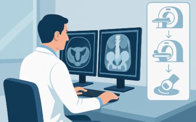
What Is the Best First Imaging Study for Suspected Nongynecological Pelvic Pain?

A 24-year-old female presents to the emergency department with two days of worsening right lower quadrant (RLQ) pain, anorexia, and subjective fever. Her last menstrual period was three weeks ago. On exam, she is tender at McBurney’s point with mild guarding. Her point-of-care urine ß-hCG is negative. The clinical picture strongly suggests a non-gynecological process like appendicitis, but the differential remains broad. You need to choose the initial imaging study that will provide a diagnosis quickly and safely. This article details the clinical workflow for this specific scenario, guiding you through the American College of Radiology (ACR) recommendations. For this patient, the ACR rates US abdomen and pelvis transabdominal as Usually Appropriate.
Who Fits This Clinical Scenario?
This guidance applies to a specific and common clinical presentation: a patient of reproductive age with acute pelvic pain where the initial history and physical exam point away from the reproductive organs. The two critical qualifiers for this workflow are a negative ß-hCG test (either urine or serum), which effectively rules out ectopic pregnancy, and a high clinical suspicion for a non-gynecological diagnosis.
Common non-gynecological culprits include appendicitis, diverticulitis, urolithiasis, or inflammatory bowel disease. The patient’s symptoms—such as pain migrating from the periumbilical region to the RLQ, gastrointestinal symptoms like nausea or diarrhea, or urinary symptoms like dysuria or hematuria—are what steer the workup down this path.
This workflow is distinct from other similar presentations:
- If the ß-hCG were positive: The entire workup would pivot to urgently rule out a life-threatening ectopic pregnancy, making it a different diagnostic algorithm.
- If a gynecological etiology were suspected: If the pain were cyclical, clearly localized to an adnexa, or associated with significant vaginal bleeding or discharge (despite a negative ß-hCG), the focus would shift to conditions like ovarian torsion, hemorrhagic cysts, or pelvic inflammatory disease.
This guidance is tailored for the non-pregnant patient where the primary question is related to the appendix, bowel, or urinary tract, while still acknowledging the need to evaluate the adjacent pelvic organs.
What Diagnoses Are You Working Up in This Scenario?
When a non-gynecological cause of acute pelvic pain is suspected, the differential diagnosis drives the choice of imaging. The goal is to select a study that can effectively evaluate for the most likely and most dangerous conditions.
Acute Appendicitis: This is the most common surgical emergency presenting with acute RLQ pain in this demographic. The classic presentation of migrating pain, anorexia, and focal tenderness makes it the leading consideration. Imaging is crucial to confirm the diagnosis, reduce the negative appendectomy rate, and identify potential complications like perforation or abscess.
Urolithiasis: An obstructing ureteral stone can cause severe, colicky flank pain that radiates to the pelvis and groin. While often associated with hematuria, its absence does not rule out the diagnosis. Imaging can identify the stone itself or secondary signs like hydronephrosis and hydroureter.
Diverticulitis: While more common in older populations, diverticulitis can occur in younger adults. It typically causes left lower quadrant pain but can affect the right colon or sigmoid colon, mimicking appendicitis. Imaging helps confirm inflammation, assess for complications like abscess or perforation, and differentiate it from other etiologies.
Inflammatory Bowel Disease (IBD): A flare of Crohn’s disease, particularly terminal ileitis, is a key consideration in the differential for RLQ pain in a young adult. Imaging can reveal bowel wall thickening, mesenteric inflammation (the “creeping fat” sign), and fistulae or abscesses.
Mesenteric Adenitis: This is an inflammatory condition of the mesenteric lymph nodes, often following a viral illness. It is a diagnosis of exclusion, typically made after imaging has confidently ruled out appendicitis. The nodes appear enlarged on imaging, but the appendix is normal.
Why Is Transabdominal Ultrasound the Recommended First Step for Suspected Nongynecological Pelvic Pain?
The ACR Appropriateness Criteria panel designates US abdomen and pelvis transabdominal as Usually Appropriate for this scenario, making it the preferred initial study. The rationale is rooted in diagnostic efficacy and patient safety, particularly the avoidance of ionizing radiation.
Ultrasound offers several key advantages as a first-line test. First and foremost, it uses no ionizing radiation (Relative Radiation Level: O 0 mSv), a critical consideration in reproductive-age patients who may undergo future imaging. Second, it has good diagnostic performance for the most common and urgent differential, acute appendicitis. A skilled sonographer can identify a non-compressible, dilated, blind-ending tubular structure consistent with an inflamed appendix. Ultrasound is also excellent for detecting hydronephrosis from an obstructing kidney stone and can identify free fluid or inflammatory changes in the pelvis. Finally, it provides a valuable, simultaneous survey of the uterus and ovaries, helping to rule out an unexpected gynecologic pathology that may have been missed on clinical exam.
While ultrasound is the best first step, other modalities are also rated for this scenario and serve important roles, often as the next step in the workup:
- CT abdomen and pelvis with IV contrast: This study is also rated Usually Appropriate. It is more sensitive and specific than ultrasound for appendicitis and other bowel pathology, especially in patients with a larger body habitus. However, it carries a radiation dose of ☢☢☢ 1-10 mSv and is therefore typically reserved as a second-line or problem-solving tool if the initial ultrasound is negative or equivocal but clinical suspicion remains high.
- CT pelvis with IV contrast: This study is rated Usually not appropriate. This is a critical distinction and a common ordering pitfall. Limiting the scan to the pelvis is insufficient because it can easily miss a high-riding appendix or other abdominal causes of pain. If CT is deemed necessary, the entire abdomen and pelvis must be included.
When ordering the initial ultrasound, it is helpful to specify the clinical concern (e.g., “Rule out appendicitis”) to guide the sonographer’s focused evaluation of the right lower quadrant in addition to the standard pelvic views.
What’s Next After the Ultrasound? Navigating the Downstream Workflow
The initial ultrasound result dictates the next step in patient management. The workflow branches based on whether the study is positive, negative, or indeterminate.
If the ultrasound is positive for appendicitis: The diagnostic workup is complete. The next step is a surgical consultation for appendectomy. No further imaging is typically required unless there is a concern for a complex complication like a large, drainable abscess that might be better visualized on CT.
If the ultrasound is negative or non-diagnostic: This is a frequent outcome, as bowel gas or patient body habitus can limit visualization of the appendix. If the clinical suspicion for appendicitis or another serious condition remains high despite a negative ultrasound, the workup must continue. The next logical step is a CT abdomen and pelvis with IV contrast. This study is also rated Usually Appropriate and serves as the definitive problem-solver to rule appendicitis in or out and evaluate for alternative diagnoses like diverticulitis or IBD.
If the ultrasound suggests urolithiasis (e.g., shows hydronephrosis): The next step is often a non-contrast CT of the abdomen and pelvis (a “stone protocol”) to locate the calculus, determine its size, and guide urologic management. This study, CT abdomen and pelvis without IV contrast, is also rated Usually Appropriate.
If the ultrasound reveals an unexpected gynecologic finding: Should the transabdominal scan identify a complex adnexal mass or other finding suspicious for a gynecologic issue, the workup pivots. The best next step is a US pelvis transvaginal, which provides superior resolution of the ovaries and uterus. This study is also rated Usually Appropriate and can be performed during the same imaging session.
Common Pitfalls to Avoid in This Acute Pelvic Pain Workup
Navigating this diagnostic pathway requires avoiding several common pitfalls that can delay diagnosis or lead to unnecessary radiation exposure.
- Stopping Too Soon: Do not accept a non-diagnostic or negative ultrasound as the final word in a patient with persistent, high-risk clinical signs for appendicitis. The “negative predictive value” of ultrasound is not perfect; escalation to CT is often necessary and appropriate.
- Ordering the Wrong CT Scan: A frequent error is ordering a “CT Pelvis” instead of a “CT Abdomen and Pelvis.” This limited field of view can miss a retrocecal or high-riding appendix, as well as other non-gynecological causes of pain originating in the abdomen.
- Ignoring Patient Preparation: For a transabdominal pelvic ultrasound, a full bladder is essential to create an acoustic window to visualize the uterus and ovaries. An empty bladder can render the pelvic portion of the study non-diagnostic.
- Anchoring on the Initial Suspicion: While the initial suspicion is non-gynecological, remain open to gynecologic possibilities. The initial ultrasound should always include a complete survey of the pelvic organs to avoid missing a concurrent or alternative diagnosis like an ovarian cyst or torsion. If clinical signs point to a time-sensitive emergency like ovarian torsion (e.g., sudden, severe, unilateral pain), escalate immediately for a dedicated pelvic ultrasound with Doppler.
Related ACR Topics and Tools
For a comprehensive overview of all clinical variants related to this topic, or to explore the technical details of the recommended imaging studies, the following resources are available.
- For breadth across all scenarios in Acute Pelvic Pain in the Reproductive Age Group, see our parent guide: Acute Pelvic Pain in the Reproductive Age Group: ACR Appropriateness Decoded.
- To explore adjacent clinical scenarios and their corresponding ACR ratings, use the Imaging Appropriateness Selector tool.
- For detailed procedural techniques on the recommended studies, consult the Imaging Protocol Library.
- To discuss cumulative radiation exposure with patients when considering CT, the Radiation Dose Calculator can help frame the conversation.
Frequently Asked Questions
Why not just order a CT scan first, since it’s also rated ‘Usually Appropriate’?
The principle of ALARA (As Low As Reasonably Achievable) guides this recommendation. In a reproductive-age patient, avoiding ionizing radiation is a priority to minimize lifetime cancer risk and radiation exposure to the gonads. Ultrasound provides no radiation and is often sufficient for diagnosis. CT is reserved as a highly effective second-line test for when ultrasound is inconclusive, balancing diagnostic need with radiation safety.
What if the patient is obese and ultrasound visualization is expected to be poor?
This is a critical clinical consideration. While the ACR guidelines recommend ultrasound as the first step for all patients in this group, the threshold to move to CT after a limited or non-diagnostic ultrasound should be very low in a patient with a high Body Mass Index (BMI). Some institutions may even have protocols to proceed directly to CT in patients with a high BMI where ultrasound is highly unlikely to be diagnostic, though this represents a deviation from the standard initial approach.
The scenario specifies a negative ß-hCG. Does it matter if it’s a urine or serum test?
For the purpose of this imaging algorithm, both a negative urine or serum ß-hCG test are generally considered sufficient to proceed. A serum test is more sensitive and can detect a very early pregnancy that a urine test might miss. However, a negative point-of-care urine test is widely accepted in the emergency setting to rule out ectopic pregnancy with a high degree of confidence and proceed with the non-gynecological workup, including CT if necessary.
When is MRI a good choice in this scenario?
MRI of the abdomen and pelvis without contrast is rated ‘May be appropriate.’ Its primary role is as a problem-solving tool in specific situations, such as in a young patient with an indeterminate ultrasound for whom the radiation from a CT scan is particularly undesirable. It is excellent for evaluating for inflammatory bowel disease or characterizing complex adnexal masses found incidentally on ultrasound. However, due to its higher cost, longer scan time, and limited availability in many emergency departments, it is not a first-line imaging test for acute, undifferentiated pelvic pain.
Does the patient need to have a full bladder for the transabdominal ultrasound?
Yes, a full or partially full bladder is crucial for the pelvic portion of the transabdominal ultrasound. The fluid-filled bladder acts as an ‘acoustic window,’ pushing bowel gas aside and providing a clear, unobstructed view of the uterus and ovaries. The sonographer will typically evaluate the pelvic organs first while the bladder is full, then may ask the patient to empty their bladder to improve visualization of the appendix and right lower quadrant.
Reviewed by Pouyan Golshani, MD, Interventional Radiologist — May 26, 2026