What Is the Best First-Line Intervention for Acute Variceal Bleeding in a High-Risk Cirrhotic Patient?
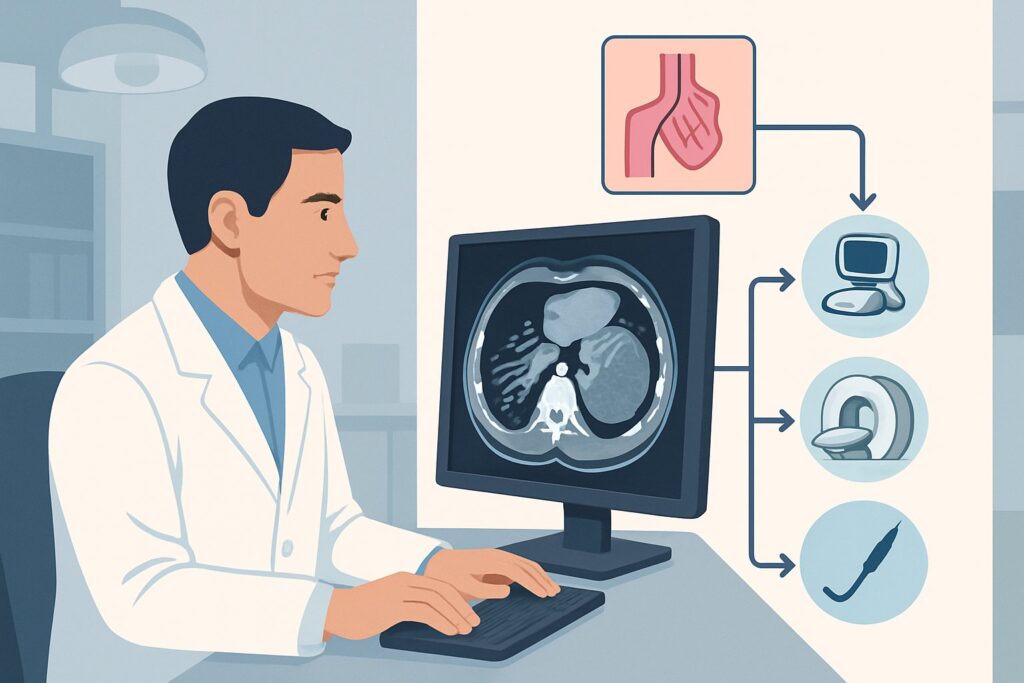
It’s 2 a.m., and the emergency department is calling about a patient with known alcohol-related cirrhosis, now presenting with massive hematemesis. He is hypotensive, tachycardic, and encephalopathic. His chart confirms a history of hepatocellular carcinoma with a branch portal vein tumor thrombus, a Child-Pugh class C score, and a Model for End-Stage Liver Disease (MELD) score of 24. You are facing a critically ill patient with active esophageal and gastroesophageal type 1 (GOV1) variceal hemorrhage. The immediate question is not just resuscitation, but which intervention offers the best chance of achieving hemostasis in this complex, high-risk scenario. According to the American College of Radiology (ACR) Appropriateness Criteria, the initial procedure of choice, Endoscopic management, is rated Usually appropriate.
Who Fits This Clinical Scenario?
This guidance is specifically for a high-acuity patient profile characterized by decompensated, end-stage liver disease and an active, life-threatening upper gastrointestinal bleed. The key inclusion criteria are:
- Acute Variceal Hemorrhage: Active, ongoing bleeding from known or suspected esophageal or gastroesophageal varices.
- Severe Liver Dysfunction: Child-Pugh class C status and a high MELD score (e.g., 24), indicating poor synthetic function, coagulopathy, and significant ascites or encephalopathy.
- Complex Portal Anatomy: The presence of hepatocellular carcinoma (HCC) and, critically, a branch portal vein tumor thrombus (PVTT), which complicates potential future interventions like a transjugular intrahepatic portosystemic shunt (TIPS).
This workflow is distinct from that for more stable patients. For example, a patient with a Child-Pugh class A score and an index variceal bleed has a different risk profile and potentially more therapeutic options. Similarly, this scenario differs from managing refractory ascites or a patient who has already failed prior endoscopic therapy and has a patent TIPS. This article addresses the crucial first-line decision in the sickest cohort, where the initial intervention is paramount.
What Diagnoses Are You Working Up in This Scenario?
In a patient with known cirrhosis and portal hypertension presenting with hematemesis, the differential is narrow but critical to confirm. The primary goal of the initial intervention is to both diagnose the precise bleeding source and treat it simultaneously.
Esophageal and/or Gastroesophageal Variceal Hemorrhage
This is the leading diagnosis and the most immediate life-threat. Portal hypertension causes portosystemic collaterals to form, most dangerously in the submucosa of the distal esophagus and gastric fundus. These varices are thin-walled and prone to rupture under high pressure, leading to massive, difficult-to-control hemorrhage. The patient’s presentation is classic for this etiology.
Portal Hypertensive Gastropathy (PHG)
While varices represent discrete collateral vessels, PHG is a more diffuse condition involving mucosal and submucosal capillary and venular dilation in the stomach. It creates a friable, “snakeskin” mucosal appearance that can ooze blood. While often causing chronic, occult bleeding, severe PHG can also contribute to an acute hemorrhage, though less commonly as the sole source of massive bleeding compared to a ruptured varix.
Peptic Ulcer Disease (PUD)
Patients with cirrhosis, particularly from alcohol use, have an increased risk of PUD. In the setting of profound coagulopathy (high INR) and thrombocytopenia common in Child-Pugh C cirrhosis, a bleeding ulcer can be just as catastrophic as a variceal bleed. Endoscopy is essential to differentiate this from a variceal source, as the management is entirely different.
Tumor-Related Bleeding
Less common, but a consequential consideration given the known diagnosis of hepatocellular carcinoma. The tumor itself could erode into a vessel, or the associated portal vein thrombus could exacerbate localized portal hypertension, leading to bleeding from ectopic varices near the tumor.
Why Is Endoscopic Management the Recommended First Step for This Presentation?
For a patient with active variceal hemorrhage, immediate intervention is required to control bleeding, and esophagogastroduodenoscopy (EGD) is the cornerstone of initial management. It is rated Usually appropriate because it uniquely combines diagnosis with therapy at the point of care. The endoscopist can directly visualize the bleeding source, confirm it is variceal, and immediately deploy therapeutic maneuvers like variceal band ligation or sclerotherapy. This approach directly targets the problem without the systemic risks or technical challenges of alternative procedures in this fragile patient.
Alongside endoscopy, Medical therapy with vasoactive drugs (e.g., octreotide, vasopressin) is also rated Usually appropriate and should be initiated immediately upon suspicion of variceal bleeding, even before endoscopy. These drugs reduce splanchnic blood flow and portal pressures, helping to control hemorrhage and improve the conditions for a successful endoscopic intervention.
Let’s consider the alternatives and why they are not the first choice in this specific scenario:
- Transjugular Intrahepatic Portosystemic Shunt (TIPS): This procedure is rated May be appropriate. While TIPS is highly effective at decompressing the portal system, it is a rescue therapy, not a first-line one. For this patient with a MELD of 24 and Child-Pugh C cirrhosis, the risk of precipitating severe, refractory hepatic encephalopathy or acute-on-chronic liver failure post-TIPS is substantial. Furthermore, the presence of a branch portal vein tumor thrombus can make the procedure technically difficult or impossible to perform safely. It is reserved for cases of endoscopic failure.
- Surgical Shunt: This option is rated Usually not appropriate. Creating a surgical portosystemic shunt is a major operation with prohibitive morbidity and mortality in a patient with decompensated Child-Pugh C cirrhosis and a MELD score in the mid-20s. It has no role in the acute management of this patient profile.
These interventions do not involve ionizing radiation. The priority is rapid stabilization and direct hemostatic control, which endoscopy provides most effectively.
What’s Next After Endoscopic Management? Downstream Workflow
The outcome of the initial EGD dictates the subsequent clinical pathway. The goal is to transition from acute resuscitation to secondary prevention and long-term management.
- If Endoscopy Achieves Hemostasis: The patient should be admitted to an intensive care unit for continued resuscitation, vasoactive drug infusion (typically for 2-5 days), and antibiotic prophylaxis to prevent spontaneous bacterial peritonitis. The immediate next steps involve initiating secondary prophylaxis with non-selective beta-blockers (e.g., nadolol, carvedilol) once hemodynamically stable. A plan for serial endoscopic variceal ligation (EVL) is made to obliterate the remaining varices. The patient’s HCC and PVTT require dedicated oncologic and transplant hepatology evaluation to determine eligibility for locoregional therapy, systemic therapy, or liver transplantation.
- If Endoscopy Fails to Control Bleeding: This constitutes a medical emergency requiring immediate escalation. A second-look EGD may be attempted, but if bleeding is torrential or re-bleeds occur, rescue therapy is necessary. The main options are a Coated esophageal self-expandable metal stent (May be appropriate) as a temporary bridge to tamponade the bleeding varices, or proceeding to TIPS (May be appropriate). The decision to pursue TIPS is a high-stakes, multidisciplinary discussion involving interventional radiology, hepatology, and the patient’s family, given the significant risks of liver failure and encephalopathy.
- If a Non-Variceal Source is Found: If endoscopy reveals a bleeding peptic ulcer, for example, the management shifts completely. Vasoactive drugs are discontinued, and therapy is focused on endoscopic treatment of the ulcer and initiation of high-dose proton pump inhibitors.
Pitfalls to Avoid (and When to Get Help)
In managing this critically ill patient, several common errors can lead to poor outcomes.
1. Delaying Endoscopy: Do not wait for cross-sectional imaging (e.g., CT scan) before proceeding to endoscopy. Time is critical, and EGD is the diagnostic and therapeutic priority.
2. Under-resuscitation: These patients require aggressive resuscitation with blood products to maintain hemodynamic stability before and during the procedure. A restrictive transfusion strategy (target hemoglobin 7-8 g/dL) is generally recommended, but the patient must be stable enough to tolerate endoscopy.
3. Forgetting Prophylactic Antibiotics: Bacterial infections are a major trigger for variceal bleeding and a common complication. Broad-spectrum antibiotics (e.g., ceftriaxone) should be started on admission.
4. Prematurely Offering TIPS: While a powerful tool, TIPS is not a benign procedure in a patient with a MELD of 24 and PVTT. It should be reserved for endoscopic failure after a careful risk-benefit analysis.
If bleeding cannot be controlled with initial endoscopic and medical therapy, this is a clear trigger for escalation. An urgent consultation with interventional radiology is required to discuss the feasibility and risks of rescue TIPS or other embolization techniques.
Related ACR Topics and Tools
This article focuses on a single, complex clinical scenario. For a comprehensive overview of all patient presentations and management options within this topic, please consult our parent guide.
- For breadth across all scenarios in Radiologic Management of Portal Hypertension, see our parent guide: Radiologic Management of Portal Hypertension: ACR Appropriateness Decoded.
For additional decision support and to explore related clinical questions, the following GigHz tools are available:
- ACR Appropriateness Criteria Lookup — for adjacent scenarios
- Imaging Protocol Library — for technique on the recommended study
- Radiation Dose Calculator — for cumulative dose conversations
Frequently Asked Questions
Why is TIPS not the first-line therapy if it’s so effective at lowering portal pressure?
TIPS is not first-line in this scenario due to the patient’s high risk of complications. With a MELD score of 24 and Child-Pugh C status, the risk of post-TIPS hepatic encephalopathy and acute-on-chronic liver failure is substantial. Additionally, the presence of a portal vein tumor thrombus can make the procedure technically challenging or impossible. It is reserved as a rescue therapy for when less invasive endoscopic management fails.
Does the presence of hepatocellular carcinoma (HCC) and portal vein thrombus change the immediate management of the bleed?
The immediate management of the acute hemorrhage remains focused on stabilization and endoscopic hemostasis. However, the HCC and portal vein tumor thrombus heavily influence the long-term plan and the choice of rescue therapies. They increase the risk of TIPS and may impact the patient’s candidacy for liver transplantation, making multidisciplinary consultation with hepatology and oncology essential after the acute bleed is controlled.
Should a CT scan be performed before endoscopy to confirm the bleeding source?
No. In a patient with known cirrhosis presenting with massive hematemesis, the pre-test probability of variceal hemorrhage is extremely high. A CT scan would delay the definitive, life-saving intervention. Endoscopy is both diagnostic and therapeutic and should be performed as emergently as possible after initial resuscitation.
What is the role of a balloon tamponade (e.g., Sengstaken-Blakemore tube) in 2026?
Balloon tamponade is a temporizing measure of last resort, used to control exsanguinating hemorrhage when endoscopy is not immediately available or has failed, and the patient is too unstable for transport to interventional radiology. It is a bridge to a more definitive therapy like TIPS or a coated stent. Its use is associated with significant complications, such as esophageal perforation and aspiration, and it should only be placed by experienced personnel.
If endoscopy is successful, what is the next most important step?
After achieving hemostasis, the next steps are to prevent re-bleeding and manage the underlying liver disease. This includes continuing vasoactive medications for 2-5 days, initiating secondary prophylaxis with non-selective beta-blockers, and starting a course of prophylactic antibiotics. A plan for serial endoscopic band ligation to eradicate residual varices should also be established.
Reviewed by Pouyan Golshani, MD, Interventional Radiologist — May 29, 2026