What Is the Best Follow-Up Imaging for a Small, Indeterminate Adrenal Mass?
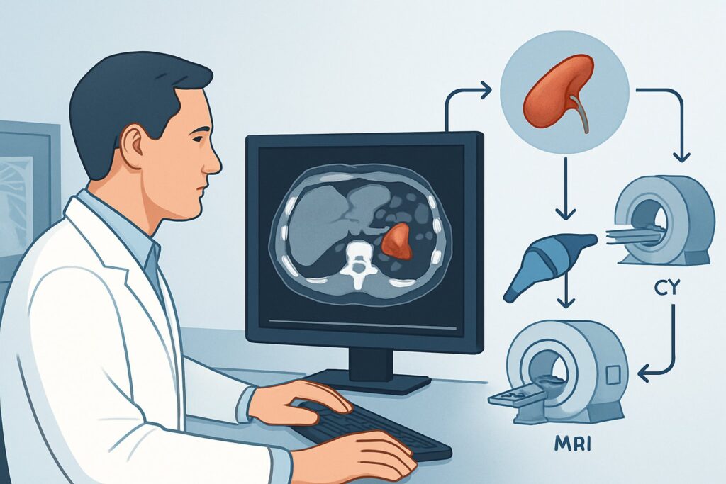
A 58-year-old patient returns to your clinic for a scheduled follow-up. One year ago, a computed tomography (CT) scan for an unrelated issue incidentally revealed a 1.5 cm adrenal nodule. The radiologist noted it was indeterminate, with an attenuation greater than 10 Hounsfield units on the unenhanced images, and recommended a 12-month follow-up to ensure stability. The patient has no history of cancer and is otherwise asymptomatic. Now, you must decide on the most appropriate imaging study to order, balancing diagnostic yield with radiation exposure. This article details the specific clinical workflow for this common scenario. According to the American College of Radiology (ACR) Appropriateness Criteria, an MRI abdomen without IV contrast is a Usually Appropriate choice for this 12-month follow-up.
Who Fits This Clinical Scenario?
This guidance applies specifically to patients undergoing a planned 12-month follow-up for a previously identified adrenal mass. The key inclusion criteria are:
- Size: The mass measured between 1 and 2 cm on the initial imaging study.
- Indeterminate Features: The initial study did not show classic benign features. For example, on an unenhanced CT, the mass measured >10 Hounsfield Units (HU), meaning it is not a lipid-rich adenoma.
- No Malignancy History: The patient has no known current or prior primary cancer that could metastasize to the adrenal gland.
- Timing: The patient is presenting for a scheduled 12-month follow-up exam to assess for stability.
It is crucial to distinguish this situation from similar but distinct clinical presentations that require a different diagnostic approach. This workflow does not apply to:
- Patients with a known primary malignancy: An adrenal mass in this context is highly suspicious for metastasis and is evaluated under a different ACR variant.
- Masses larger than 2 cm: Larger masses, particularly those approaching 4 cm, carry a higher risk of malignancy and warrant a more comprehensive or immediate workup.
- Newly discovered incidentalomas: The initial characterization of a newly found adrenal mass follows a different algorithm than a planned stability check.
- Masses with definitive benign features: A mass measuring <10 HU on unenhanced CT or containing macroscopic fat is a benign adenoma or myelolipoma, respectively, and typically requires no further imaging follow-up.
What Diagnoses Are You Evaluating at the 12-Month Follow-Up?
The primary goal of follow-up imaging is to confirm stability, which is the strongest indicator of benignity. However, the study also aims to further characterize the lesion and rule out concerning pathologies.
The most common diagnosis for an indeterminate adrenal mass is a lipid-poor benign adenoma. These non-cancerous tumors are extremely common but do not contain enough intracellular fat to meet the <10 HU density cutoff on unenhanced CT. Their defining characteristic is a lack of growth over time. The 12-month follow-up is designed to provide this evidence of stability, effectively confirming the benign nature of the lesion.
A much less common but critical diagnosis to exclude is adrenocortical carcinoma (ACC). While rare, especially for masses under 4 cm, any significant interval growth in a previously indeterminate nodule raises suspicion for this aggressive malignancy. The follow-up scan serves as a crucial safety check to detect this growth early.
Another consideration is a pheochromocytoma, a tumor of the adrenal medulla that can secrete catecholamines. While many are larger at diagnosis, they can present as small, indeterminate masses. Certain MRI features can suggest this diagnosis, which is critical to make before any potential biopsy, as that procedure could precipitate a life-threatening hypertensive crisis.
Why Is MRI Abdomen Without Contrast Usually Appropriate for This Follow-Up?
For the 12-month stability check of a 1-2 cm indeterminate adrenal mass, the ACR rates MRI abdomen without IV contrast as Usually Appropriate. This recommendation is based on its ability to both assess stability and provide definitive characterization without using ionizing radiation.
The key advantage of MRI in this scenario is its use of chemical shift imaging with in-phase and opposed-phase sequences. This technique can detect microscopic intracellular lipid that is invisible on CT. A lipid-poor adenoma will demonstrate a characteristic drop in signal on the opposed-phase images compared to the in-phase images. This finding is diagnostic for a benign adenoma, often ending the need for further imaging surveillance.
Furthermore, MRI provides excellent soft tissue resolution to accurately measure the mass and compare its size to the prior study. Crucially, it achieves this with no ionizing radiation (0 mSv), a significant benefit for follow-up imaging of a likely benign condition.
Alternative Imaging Options
- CT abdomen without IV contrast: This study is also rated Usually Appropriate. It is an excellent and widely available tool for assessing size stability. However, it cannot perform chemical shift analysis to definitively characterize a lipid-poor adenoma. It remains a strong alternative if MRI is unavailable, contraindicated, or not tolerated by the patient. It involves a moderate radiation dose (ACR RRL ☢☢☢, 1-10 mSv).
- Image-guided biopsy adrenal gland: This is rated Usually not appropriate. Biopsy of a small (1-2 cm) adrenal mass is technically challenging, carries risks of bleeding and pneumothorax, and has a significant rate of non-diagnostic results. Most importantly, it is dangerous if the mass is an undiagnosed pheochromocytoma. Biopsy is reserved for rare situations where imaging remains inconclusive after a full workup and confirming a diagnosis would directly alter urgent management.
What Are the Next Steps After the Follow-Up MRI?
The results of the 12-month follow-up MRI will guide the subsequent workflow. The decision tree is relatively straightforward and focuses on stability and characterization.
- If the mass is stable in size AND shows signal drop on opposed-phase imaging: The diagnosis of a benign, lipid-poor adenoma is confirmed. According to most societal guidelines, no further imaging follow-up is necessary for this lesion. The patient should still undergo a baseline biochemical evaluation for hormonal hypersecretion if not already performed.
- If the mass is stable in size but remains indeterminate on MRI (no signal drop): Stability is a reassuring feature that strongly suggests a benign etiology. While an adenoma is still most likely, other rare benign lesions remain on the differential. Depending on institutional practice and patient factors, a second follow-up scan at 24 months may be considered to ensure continued stability, after which surveillance is typically stopped.
- If the mass has grown significantly: Any definitive interval growth (e.g., >20% and at least 5 mm increase in the longest dimension) is a significant red flag. This patient’s condition has evolved, and they no longer fit this follow-up scenario. The workup should be escalated to evaluate for malignancy, which may involve a dedicated adrenal protocol CT or MRI with contrast, a full biochemical workup, and an urgent referral to an endocrinologist or endocrine surgeon.
Common Pitfalls to Avoid in 12-Month Adrenal Mass Follow-Up
Navigating this clinical scenario requires attention to detail to avoid common errors that can lead to unnecessary tests or missed diagnoses.
- Failing to provide prior imaging: The primary purpose of the exam is to assess for stability. The interpreting radiologist must have access to the initial study for a direct comparison. Always ensure priors are available.
- Assuming imaging stability negates the need for a functional workup: All adrenal incidentalomas, regardless of their imaging features or stability, warrant a biochemical evaluation to rule out hormonal hypersecretion (e.g., for Cushing’s syndrome, pheochromocytoma, or hyperaldosteronism).
- Ordering a contrast-enhanced study unnecessarily: For a simple stability check, a non-contrast MRI or CT is sufficient. Adding intravenous contrast increases cost, carries a small risk of reaction or nephrotoxicity, and is not needed unless growth is detected or specific characterization questions arise.
- Misinterpreting minor size variations: Small measurement discrepancies between studies or modalities can occur. Clinically significant growth is generally defined as an increase of >20% and >5 mm.
If significant growth is detected or the patient develops new symptoms concerning for hormonal excess, escalate care by consulting with an endocrinologist.
Related ACR Topics and Tools
This article focuses on a single, specific clinical scenario. For a comprehensive overview of all variants in this topic, or to explore related imaging resources, please use the tools below.
- For breadth across all scenarios in Adrenal Mass Evaluation, see our parent guide: Adrenal Mass Evaluation: ACR Appropriateness Decoded.
- ACR Appropriateness Criteria Lookup — for adjacent scenarios and different clinical questions.
- Imaging Protocol Library — for technical details on the recommended study.
- Radiation Dose Calculator — for discussing cumulative radiation exposure with patients.
Frequently Asked Questions
Why is MRI preferred over CT for a 12-month follow-up of a small adrenal mass?
MRI without contrast is preferred because it can both assess for size stability and definitively characterize many indeterminate masses as benign lipid-poor adenomas using chemical shift imaging. Crucially, it accomplishes this with no ionizing radiation, which is ideal for follow-up studies of a likely benign condition.
What if the patient has a contraindication to MRI, like a pacemaker?
If MRI is contraindicated, a CT abdomen without IV contrast is also rated ‘Usually Appropriate’ by the ACR. It is an excellent alternative for accurately assessing size stability, which is the primary goal of the follow-up. However, it cannot perform chemical shift analysis and involves a moderate radiation dose.
What constitutes significant growth on a follow-up scan?
While definitions can vary slightly, most guidelines consider significant growth to be an increase in the largest diameter of more than 20% AND at least 5 mm. This degree of growth is a red flag that warrants an escalated workup for potential malignancy.
If the 12-month follow-up MRI shows the mass is stable, is any further imaging ever needed?
If the MRI confirms the mass is a benign adenoma (by showing signal drop on opposed-phase imaging) or if it is stable after a 24-month scan, most guidelines recommend stopping imaging surveillance. The only other requirement is to ensure a one-time biochemical workup for hormonal activity has been completed.
Does this guidance apply if the initial mass was 3 cm?
No. This workflow is specifically for masses between 1 and 2 cm. An indeterminate mass that is 3 cm falls into a different ACR scenario (‘greater than 2 cm and less than 4 cm’). These larger masses may warrant a more comprehensive initial workup and have different follow-up recommendations due to a slightly higher baseline risk.
Reviewed by Pouyan Golshani, MD, Interventional Radiologist — May 29, 2026