What Is the Best Imaging for a Small Liver Lesion in a Patient with Cancer?

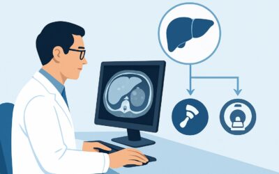
It’s 4:30 PM on a Tuesday. You’re reviewing the day’s imaging results and see an ultrasound report for a 68-year-old patient with a history of resected lung cancer. The report notes a new, indeterminate 8 mm hypoechoic lesion in the right hepatic lobe. The finding is too small to be confidently characterized by sonography alone. The patient’s oncologist needs to know if this represents metastatic disease, as it would significantly alter the patient’s management and prognosis. This clinical scenario—an indeterminate, sub-centimeter liver lesion on ultrasound in a patient with a known extrahepatic malignancy—requires a precise and high-yield next step.
This article provides a detailed clinical workflow for this specific presentation, grounded in the American College of Radiology (ACR) Appropriateness Criteria. For this scenario, the ACR panel rates MRI abdomen without and with IV contrast as Usually Appropriate.
Who Fits This Clinical Scenario?
This guidance is tailored for a very specific patient presentation. It is essential to confirm your patient matches these criteria before proceeding, as small changes can lead to a different recommended imaging pathway.
Inclusion Criteria:
- An indeterminate liver lesion was incidentally discovered on initial imaging.
- The initial imaging modality was ultrasound (US).
- The lesion measures less than 1 cm in its greatest dimension.
- The patient has a known, confirmed history of an extrahepatic malignancy (e.g., colon, breast, lung, melanoma). The risk of liver metastases makes this a high-stakes workup.
Exclusion Criteria (These patients require a different workflow):
- Lesion is larger than 1 cm: A lesion greater than 1 cm has a higher pre-test probability of being significant and may be more easily characterized. This falls under a different ACR variant.
- Initial imaging was CT or MRI: If the lesion was found on a cross-sectional study like a noncontrast CT or single-phase MRI, the diagnostic question and next steps are different.
- No history of malignancy: In a patient with a normal liver and no cancer history, the differential diagnosis for a small liver lesion shifts dramatically toward benign entities, and the urgency of the workup is lower.
What Diagnoses Are You Working Up in This Scenario?
In a patient with a known extrahepatic malignancy, any new, indeterminate liver lesion must be considered a metastasis until proven otherwise. The entire imaging workup is structured around confirming or refuting this primary concern. However, benign lesions are common and can be incidental findings even in this patient population.
Metastasis: This is the most consequential diagnosis to exclude. The liver is a common site for hematogenous spread from many primary cancers, particularly those of the gastrointestinal tract, lung, and breast. The enhancement characteristics of a lesion on dynamic, contrast-enhanced imaging are critical for identifying metastases, which are often hypervascular in the arterial phase with subsequent “washout” in later phases.
Hemangioma: The most common benign liver tumor. While often classic in appearance on ultrasound, small or atypical hemangiomas can appear indeterminate. On contrast-enhanced imaging, they typically demonstrate a characteristic pattern of peripheral, nodular enhancement that gradually fills in over time.
Simple or Complicated Cyst: A simple cyst is a very common, benign finding. However, on ultrasound, a tiny cyst may have some internal echoes or unclear margins, leading to an “indeterminate” classification. MRI is exceptionally good at confirming the simple fluid content of a cyst, effectively ruling it out as a concern.
Focal Nodular Hyperplasia (FNH): A less common benign lesion that represents a hyperplastic response of hepatocytes. While often found in younger women, it can occur in any patient. Its classic imaging feature is avid arterial enhancement and the presence of a central scar, though this scar can be absent in smaller lesions.
Why Is MRI Abdomen Without and With IV Contrast the Recommended Study?
The ACR designates MRI abdomen without and with IV contrast as Usually Appropriate for this scenario because it provides the highest diagnostic accuracy for characterizing small liver lesions without using ionizing radiation. The superior soft-tissue contrast resolution of MRI is the key advantage over other modalities.
The multiphasic, post-contrast imaging sequences are crucial. By acquiring images at different times after the injection of a gadolinium-based contrast agent (e.g., late arterial, portal venous, and delayed phases), the radiologist can assess the lesion’s vascularity and enhancement pattern over time. This dynamic information is often sufficient to distinguish a metastasis from a benign entity like a hemangioma or FNH. Furthermore, specific MRI sequences like T2-weighting are highly sensitive for identifying fluid-filled structures, making it simple to confirm a benign cyst.
Why are other studies rated lower for this specific scenario?
- CT abdomen with IV contrast multiphase: This study is rated May be appropriate. While it also uses dynamic contrast enhancement, its soft-tissue contrast resolution is lower than MRI’s, making it more difficult to confidently characterize a sub-centimeter lesion. It also exposes the patient to a significant level of ionizing radiation (ACR RRL® ☢☢☢☢, 10-30 mSv), which is a key consideration in patients who may require frequent surveillance imaging.
- Image-guided biopsy liver: This is rated Usually not appropriate. Attempting to biopsy a lesion smaller than 1 cm carries a high risk of sampling error (missing the lesion) and a non-diagnostic result. The potential complications of biopsy, such as bleeding or tumor seeding, generally outweigh the benefits when a non-invasive, high-accuracy imaging test like MRI is available.
What’s Next After MRI? Downstream Workflow
The results of the contrast-enhanced MRI will guide the subsequent clinical pathway. The goal is to achieve a definitive characterization of the lesion to inform oncologic management.
If the MRI is definitive for metastasis: The finding confirms metastatic disease. The next steps involve communication with the oncology team to integrate this information into the patient’s overall staging. This may trigger a change in systemic therapy, a consideration for local therapies like ablation, or a broader search for other metastatic sites if not already performed.
If the MRI is definitive for a benign lesion (e.g., cyst, hemangioma): The workup is complete. The lesion is considered an incidentaloma, and no further follow-up is required for the liver lesion itself. The patient can continue their standard oncologic surveillance as previously planned. This result provides significant reassurance and prevents unnecessary, costly, and potentially harmful further interventions.
If the MRI remains indeterminate: While MRI is highly accurate, some very small lesions may still lack classic features. In this situation, the decision becomes more complex and often involves a multidisciplinary discussion. Options include:
- Short-interval follow-up MRI: A repeat scan in 3-6 months can assess for stability or growth. Stability over time strongly suggests a benign etiology, while growth would increase suspicion for malignancy.
- Consider FDG-PET/CT: While rated Usually not appropriate for initial characterization, PET/CT may be used as a problem-solving tool in high-stakes cases where the MRI is equivocal. However, its spatial resolution is poor, and it may not detect metabolically active disease in a sub-centimeter lesion.
Pitfalls to Avoid (and When to Get Help)
Navigating this clinical scenario requires careful attention to detail to avoid common errors that can delay diagnosis or lead to incorrect conclusions.
- Accepting a technically limited study: Patient motion can severely degrade the quality of a liver MRI. If the report indicates the study is limited, consider repeating it rather than making a clinical decision based on suboptimal data.
- Not providing clinical history: The radiologist’s interpretation is heavily influenced by the provided history. Always specify the patient’s primary malignancy and the reason for the exam. This context is critical for an accurate differential.
- Misinterpreting a perfusion anomaly: The liver can have transient hepatic attenuation/enhancement differences (THADs/THEDs) that can mimic a true lesion. A multiphase study interpreted by an experienced radiologist is key to avoiding this pitfall.
If the imaging remains indeterminate after a high-quality MRI, or if the findings are discordant with the clinical picture, this is the time to escalate. A discussion at a multidisciplinary tumor board, including the oncologist, radiologist, and potentially a surgeon or interventional radiologist, is the best next step.
Related ACR Topics and Tools
For a comprehensive overview of all clinical variants related to the initial workup of a liver lesion, please see our parent guide. For other tools to assist in your clinical workflow, see the resources below.
- For breadth across all scenarios in Liver Lesion-Initial Characterization, see our parent guide: Liver Lesion-Initial Characterization: ACR Appropriateness Decoded.
- To look up other clinical scenarios, visit the Imaging Appropriateness Selector.
- To review institutional standards for the recommended study, see the Imaging Protocol Library.
- To discuss cumulative radiation exposure with your patients, use the Radiation Dose Calculator.
Frequently Asked Questions
Why not just go straight to a PET/CT for a new liver lesion in a cancer patient?
While FDG-PET/CT is excellent for staging, it is rated ‘Usually not appropriate’ by the ACR for initial characterization of a small liver lesion. Its spatial resolution is much lower than MRI or CT, meaning a sub-centimeter lesion may be too small to detect (a false negative). Furthermore, some non-cancerous inflammatory processes can be PET-avid (a false positive). MRI provides superior anatomical detail and tissue characterization, making it the more accurate first-line test in this specific scenario.
My patient has severe renal insufficiency. Can I still order an MRI with contrast?
This is an important consideration. For patients with an eGFR below 30 mL/min/1.73m², there is a risk of nephrogenic systemic fibrosis (NSF) with certain older gadolinium-based contrast agents (GBCAs). However, newer macrocyclic GBCAs have a much lower risk profile. A discussion with the radiology department is essential. They can advise on the specific risk with the agents they use or suggest alternative imaging, such as an MRI without contrast or a contrast-enhanced ultrasound, both of which are rated ‘May be appropriate’.
What if the ultrasound report called the lesion a ‘probable hemangioma’ but still recommended correlation?
Even when an ultrasound suggests a benign finding like a hemangioma, the high pre-test probability of metastasis in a patient with known cancer warrants definitive characterization. An 8 mm lesion is often too small for ultrasound to show the classic features of a hemangioma with high confidence. The MRI is performed to confirm the benign diagnosis and confidently exclude malignancy, which is the standard of care in this high-risk population.
Is contrast-enhanced ultrasound (CEUS) a good alternative to MRI?
Contrast-enhanced ultrasound (CEUS) is rated ‘May be appropriate’ and can be an excellent alternative, particularly if the patient has a contraindication to MRI (e.g., an incompatible implanted device) or gadolinium-based contrast. CEUS uses microbubble contrast agents that are not nephrotoxic and provides real-time dynamic enhancement information. However, its availability and the level of operator expertise can be variable between institutions, and it may be limited by patient body habitus or a lesion’s deep location.
Does the type of primary cancer change the recommendation?
No, the ACR recommendation for MRI remains the same regardless of the primary cancer type (e.g., colon, lung, breast). The core diagnostic challenge—differentiating a small metastasis from a small benign lesion—is best addressed by the superior soft-tissue contrast and dynamic enhancement assessment provided by MRI. The primary cancer type becomes more relevant when interpreting the results; for example, a lesion in a patient with neuroendocrine tumor might be evaluated with different imaging agents or techniques if it remains indeterminate.
Reviewed by Pouyan Golshani, MD, Interventional Radiologist — May 26, 2026