What Is the Best Imaging for a Suspected Extra-Axial Brain Tumor?
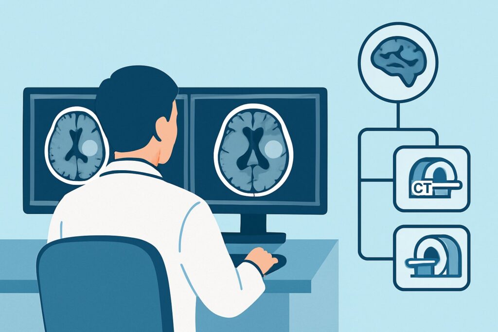
A 62-year-old patient with a history of breast cancer presents with persistent, localized headaches. An initial non-contrast head CT performed for another reason reveals a well-circumscribed, dural-based mass over the right cerebral convexity. The finding is incidental, but given the patient’s history and symptoms, you need to characterize the lesion to guide neurosurgical consultation and pretreatment planning. What is the most appropriate next imaging step to define the nature of this extra-axial mass and its relationship to adjacent structures? For this specific clinical scenario, the American College of Radiology (ACR) rates MRI head without and with IV contrast as Usually Appropriate.
## Who Fits This Clinical Scenario?
This guidance applies to adult patients where a prior imaging study, such as a CT or a non-contrast MRI, has already identified a mass suspected to be extra-axial. An extra-axial location means the tumor arises from structures outside the brain parenchyma itself, such as the meninges, cranial nerves, or skull. The primary goal of the imaging study is pretreatment evaluation—gathering the detailed anatomical information needed for surgical planning, radiation therapy targeting, or biopsy.
This workflow is distinct from several related clinical situations:
- Suspected Intra-Axial Tumors: If the mass appears to be located within the brain tissue (parenchyma), the imaging workup follows a different pathway. This article specifically addresses masses originating outside the brain.
- Initial Tumor Screening: This guidance is not for screening asymptomatic patients, even those with genetic risk factors for brain tumors. It is for patients with a known, visualized lesion.
- Post-treatment Surveillance: Patients with a known history of a brain tumor who are undergoing routine follow-up imaging fall under a separate set of ACR recommendations.
Correctly identifying your patient’s scenario is crucial for ordering the most effective and appropriate imaging study.
## What Diagnoses Are You Working Up in This Scenario?
When an extra-axial mass is discovered, the differential diagnosis is focused on tumors arising from the meninges and associated structures. The goal of advanced imaging is to narrow this differential, as the diagnosis dictates management, from observation to aggressive multimodal therapy.
The most common cause by a wide margin is a meningioma. These are typically slow-growing, benign (WHO Grade 1) tumors arising from the arachnoid cap cells of the meninges. Imaging often reveals a homogeneously enhancing, dural-based mass, sometimes with a characteristic “dural tail” sign. However, higher-grade (WHO Grade 2 and 3) meningiomas can appear more aggressive.
A critical consideration, especially in patients with a known extracranial malignancy, is a dural metastasis. Cancers of the breast, lung, and prostate are common primary sources. Metastases can closely mimic meningiomas on imaging, though they are often multiple and may lack a classic dural tail. Differentiating between a primary meningioma and a solitary metastasis is a key objective of the workup.
Less common but important possibilities include schwannomas, which are benign tumors arising from the sheath of cranial nerves. The most frequent location is the cerebellopontine angle, where they are known as vestibular schwannomas (or acoustic neuromas).
Other rarer entities on the differential include solitary fibrous tumor/hemangiopericytoma, a more aggressive vascular tumor of the dura, and primary dural lymphoma. These are often difficult to distinguish from meningioma based on imaging alone, but certain features on advanced MRI sequences can raise suspicion.
## Why MRI head without and with IV contrast Is the Recommended Study for This Presentation
The ACR designates MRI head without and with IV contrast as Usually Appropriate because it provides the most comprehensive, non-invasive evaluation of an extra-axial mass. Its superior soft-tissue resolution is unmatched for defining the tumor’s precise anatomy and relationship to critical neurovascular structures.
The rationale for this recommendation includes:
- Superior Anatomic Detail: MRI excels at delineating the tumor’s margins, its interface with the brain (identifying a cerebrospinal fluid cleft, a key sign of an extra-axial location), and any associated vasogenic edema in the adjacent brain parenchyma. This information is vital for neurosurgical planning.
- Contrast Enhancement Characteristics: The pattern of enhancement after gadolinium administration is crucial for diagnosis. A classic meningioma often shows avid, uniform enhancement. The “dural tail” sign, representing enhancing dura adjacent to the tumor, is best visualized on post-contrast MRI.
- Value of Non-Contrast Sequences: The “without and with” protocol is essential. Pre-contrast T1-weighted images can detect intrinsic hemorrhage or fatty components, while T2-weighted and FLAIR (Fluid-Attenuated Inversion Recovery) sequences are highly sensitive for edema. Diffusion-Weighted Imaging (DWI) provides information about cellularity, which can help differentiate between tumor types.
- Safety Profile: MRI involves no ionizing radiation (Relative Radiation Level: O 0 mSv), a significant advantage over CT, especially for patients who may require serial imaging.
Alternative studies are rated lower for this specific purpose. For example, CT head with IV contrast is rated Usually not appropriate. While CT is excellent for detecting calcification and bony involvement, its soft-tissue contrast is far inferior to MRI, limiting its ability to characterize the mass and its relationship to the brain, cranial nerves, and vessels. Similarly, FDG-PET/CT brain is Usually not appropriate for initial characterization because the high metabolic activity of the normal brain cortex can obscure the tumor, limiting its diagnostic utility in this context.
## What’s Next After MRI head without and with IV contrast? Downstream Workflow
The results of the pretreatment MRI directly guide the subsequent clinical pathway. The workflow branches based on the imaging findings and the patient’s clinical status.
- Classic, Asymptomatic Meningioma: If the MRI reveals a small mass with classic features of a benign meningioma in an asymptomatic patient, the next step is often active surveillance with serial MRI scans. This falls under the Posttreatment surveillance ACR scenario.
- Symptomatic or Atypical Mass: For a large, symptomatic, or radiologically atypical tumor, the MRI results are the roadmap for treatment. The next step is a neurosurgical consultation to discuss management options, which may include surgical resection, biopsy, or stereotactic radiosurgery. The detailed anatomy shown on the MRI—particularly the tumor’s relationship to dural venous sinuses, major arteries, and cranial nerves—is fundamental to this planning.
- Indeterminate Findings or High Suspicion for Meningioma: If the diagnosis remains uncertain but a meningioma is highly suspected, a more specialized imaging study may be considered. DOTATATE PET/CT brain or DOTATATE PET/MRI brain, both rated May be appropriate, can be highly valuable. These tracers target somatostatin receptors, which are densely expressed on most meningiomas but not on many mimics, helping to confirm the diagnosis non-invasively.
- Suspicion of Metastasis: If the findings are concerning for a dural metastasis in a patient without a known primary cancer, the MRI will prompt a systemic workup to identify the source.
## Pitfalls to Avoid (and When to Get Help)
In the workup of a suspected extra-axial tumor, several common pitfalls can lead to diagnostic delays or errors.
1. Omitting Pre-Contrast Sequences: Ordering an “MRI head with IV contrast” alone is a frequent mistake. This protocol is rated Usually not appropriate because the pre-contrast images are essential for detecting hemorrhage and accurately assessing the degree of enhancement. Always order the complete “without and with” study.
2. Over-interpreting the “Dural Tail”: While characteristic of meningioma, the dural tail sign is not pathognomonic. It can also be seen with dural metastases, lymphoma, and inflammatory conditions like sarcoidosis.
3. Mischaracterizing Location: A superficial, cortically based intra-axial tumor (like a glioma or metastasis) can sometimes be mistaken for an extra-axial mass on an initial non-contrast study. High-quality MRI is key to making this distinction.
If the MRI shows aggressive features such as significant brain invasion, bone destruction, or encasement of major vascular structures, it is critical to escalate care promptly to a multidisciplinary neuro-oncology tumor board for collaborative decision-making.
## Related ACR Topics and Tools
This article focuses on a single clinical variant. For a comprehensive overview of imaging for all brain tumor scenarios, from screening to post-treatment follow-up, please consult our parent guide.
- For breadth across all scenarios in Brain Tumors, see our parent guide: Brain Tumors: ACR Appropriateness Decoded.
For additional decision support and technical details, the following GigHz resources are available:
- ACR Appropriateness Criteria Lookup — for exploring adjacent scenarios and other clinical topics.
- Imaging Protocol Library — for detailed technical parameters of the recommended MRI study.
- Radiation Dose Calculator — for discussing cumulative radiation exposure with patients when considering CT or PET/CT.
Frequently Asked Questions
Why is an MRI needed if a CT scan already showed the extra-axial mass?
A CT scan, especially without contrast, is good at detecting a mass but provides limited detail. An MRI without and with IV contrast is essential for tissue characterization—distinguishing between a meningioma, metastasis, or other tumor types. It also provides superior detail of the tumor’s relationship to critical structures like blood vessels, nerves, and the brain itself, which is crucial for safe and effective treatment planning.
Is a ‘dural tail’ sign on MRI diagnostic for a meningioma?
The ‘dural tail’ sign, which is thickening and enhancement of the dura adjacent to the mass, is highly suggestive of a meningioma but is not 100% specific. Other conditions, including dural metastases, lymphoma, and inflammatory processes like sarcoidosis, can also exhibit this finding. It must be interpreted in the context of the tumor’s other imaging features and the patient’s clinical history.
When should I consider a DOTATATE PET/CT for an extra-axial tumor?
According to the ACR, a DOTATATE PET/CT or PET/MRI ‘May be appropriate.’ It is typically used as a problem-solving tool when the diagnosis is uncertain after MRI. Because most meningiomas (over 90%) express somatostatin receptors, they are avid on DOTATATE scans. This can help confirm a suspected meningioma and differentiate it from mimics like schwannomas or metastases, which are typically DOTATATE-negative.
What if the patient has a contraindication to gadolinium contrast, like severe renal failure?
If a patient cannot receive gadolinium, an ‘MRI head without IV contrast’ is rated as ‘May be appropriate.’ While suboptimal compared to a contrast-enhanced study, non-contrast sequences (T1, T2, FLAIR, DWI) can still provide significant information about the tumor’s size, location, mass effect, and edema. In some cases, advanced non-contrast techniques like Arterial Spin Labeling (ASL) perfusion may offer additional data, though this is not a routine part of the standard workup.
Does the imaging approach change if the suspected extra-axial tumor is in the spine instead of the head?
Yes, the approach is different. While the principles of using MRI with and without contrast are similar, the specific protocol and differential diagnosis change. For a suspected spinal extra-axial tumor (e.g., spinal meningioma or schwannoma), a dedicated MRI of the relevant spinal segment is required. The ACR has separate appropriateness criteria for spinal tumors.
Reviewed by Pouyan Golshani, MD, Interventional Radiologist — May 30, 2026