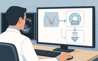
What Is the Best Imaging for Active Surveillance of Low-Risk Prostate Cancer?
A 68-year-old man with a history of Gleason 6 (3+3) prostate cancer returns for his annual follow-up. He opted for active surveillance two years ago and has been compliant with serial Prostate-Specific Antigen (PSA) testing. His latest PSA has risen from 4.5 ng/mL to 6.1 ng/mL over the past 12 months, representing a velocity that prompts re-evaluation. You need to determine if his low-risk disease has progressed or if a more clinically significant cancer was missed on his initial systematic biopsy. The central question is which imaging and biopsy strategy will most accurately re-stratify his risk without leading to overtreatment.
According to the American College of Radiology (ACR) Appropriateness Criteria, the next step in this active surveillance pathway involves imaging to guide tissue sampling. For this specific scenario, an MRI-targeted biopsy prostate is rated as Usually Appropriate. This article details the clinical workflow for this exact decision point.
Who Fits This Clinical Scenario?
This guidance applies specifically to patients with clinically established, low-risk prostate cancer who are currently undergoing active surveillance. The defining criteria for this cohort generally include:
- Gleason Score: 6 (Grade Group 1)
- PSA Level: Typically <10 ng/mL
- Clinical Stage: T1c or T2a
- Biopsy Findings: Low volume of cancer (e.g., few positive cores, low percentage of cancer in each core)
This workflow is intended for the surveillance phase, often triggered by a concerning change in clinical parameters like a rising PSA or a change on digital rectal exam.
It is crucial to distinguish this scenario from others that require a different diagnostic approach. This guidance does not apply to:
- Biopsy-Naïve Patients: Men with an elevated PSA but no prior biopsy fall under the initial diagnosis pathway.
- Patients with a Prior Negative Biopsy: If a patient has a persistently rising PSA despite a previous negative systematic biopsy, the workup is different.
- Intermediate- or High-Risk Prostate Cancer: Patients initially diagnosed with higher-grade (Gleason ≥7) or higher-stage disease require definitive staging, not surveillance imaging.
Applying the active surveillance protocol to a patient with intermediate- or high-risk disease would be a significant clinical error, potentially delaying necessary treatment.
What Diagnoses Are You Working Up in This Scenario?
When a patient on active surveillance presents with a rising PSA, the imaging and subsequent biopsy are designed to differentiate among several key possibilities. The goal is to confirm stability or accurately detect progression that warrants a change in management.
The primary concern is grade reclassification or disease progression. The patient’s known Gleason 6 cancer may have evolved into a more aggressive, clinically significant cancer (Gleason ≥7). Alternatively, a higher-grade component may have been present all along but was missed by the initial systematic biopsy. An MRI can identify suspicious areas that were not sampled previously, guiding a targeted biopsy to find this occult disease.
Another possibility is volume progression. Even if the grade remains low, a significant increase in the tumor’s size could change the risk calculation and prompt a discussion about definitive therapy. Multiparametric MRI is highly effective at estimating tumor volume and identifying anterior lesions often missed by traditional transrectal ultrasound (TRUS)-guided biopsies.
The most favorable outcome is confirmation of stable, low-risk disease. In many cases, PSA can fluctuate due to benign causes. The imaging and biopsy may confirm that the patient still has low-volume, low-grade cancer, reaffirming the decision to continue active surveillance and avoiding the morbidity of unnecessary treatment.
Finally, the workup must consider benign prostatic conditions. Benign Prostatic Hyperplasia (BPH) and prostatitis are common causes of PSA elevation. While MRI’s primary role here is cancer detection, it can also provide anatomical detail about the prostate’s size and zonal anatomy, helping to contextualize the clinical picture.
Why Is an MRI-Targeted Biopsy the Recommended Approach?
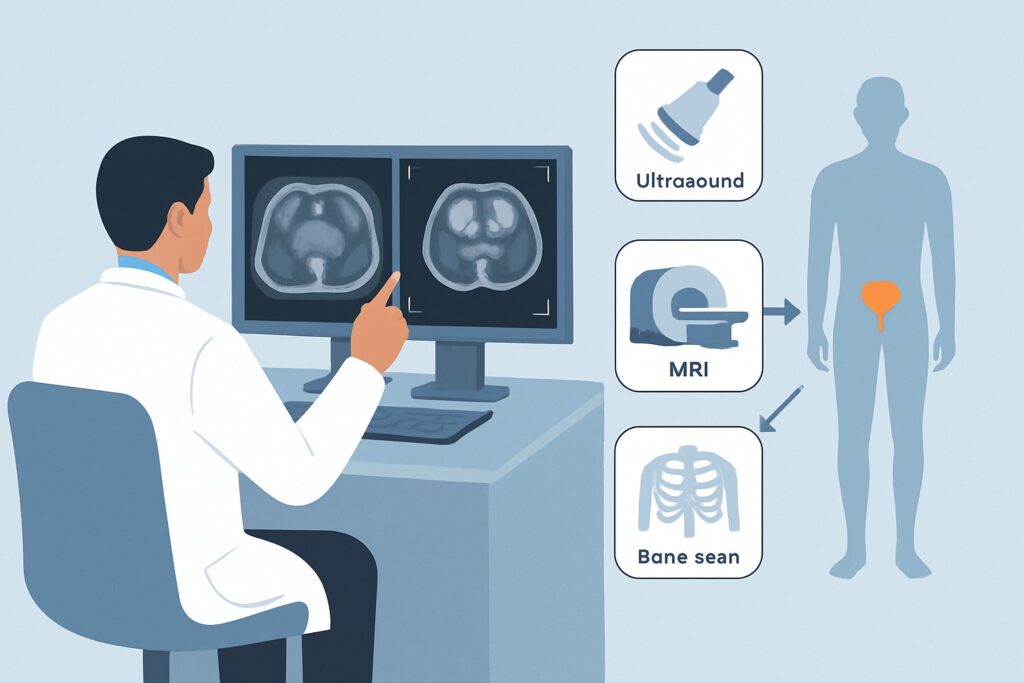
For a patient on active surveillance, the clinical need is to maximize the detection of clinically significant cancer while minimizing the detection of indolent disease. The ACR panel designates MRI-targeted biopsy prostate as Usually Appropriate because it directly addresses this need with high diagnostic precision and no ionizing radiation (0 mSv). This procedure typically involves a multiparametric MRI (mpMRI) of the pelvis followed by a biopsy that fuses the MRI images with real-time ultrasound to precisely sample suspicious lesions.
The rationale for this approach over alternatives includes:
- Superior Detection of Clinically Significant Cancer: mpMRI is highly sensitive for identifying aggressive tumors, which appear as distinct lesions on sequences like diffusion-weighted imaging (DWI) and dynamic contrast-enhanced (DCE) imaging. This allows for targeted sampling, substantially increasing the yield for Gleason score 7 or higher cancers compared to random, systematic biopsies alone.
- Risk Stratification: The Prostate Imaging Reporting and Data System (PI-RADS) score from the MRI provides a standardized way to estimate the likelihood of clinically significant cancer. A low PI-RADS score (1-2) is reassuring, while a high score (4-5) strongly indicates the need for a targeted biopsy.
- Avoiding Unnecessary Biopsies: In some protocols, a negative (PI-RADS 1-2) surveillance MRI may allow a patient to defer a scheduled biopsy, reducing the associated risks of infection, bleeding, and discomfort.
While an MRI-guided approach is preferred, other studies are also rated for this scenario:
- TRUS-guided biopsy prostate: This is also rated Usually Appropriate. It remains a valid standard of care. However, it is a “blind” systematic sampling technique that can miss anterior or small, aggressive tumors. Often, the best approach combines both: an MRI to identify targets, followed by a fusion biopsy that includes both targeted cores and a systematic template.
- Bone scan whole body or PSMA PET/CT skull base to mid-thigh: These are rated Usually Not Appropriate. For low-risk disease, even with a rising PSA, the pretest probability of metastatic disease is exceedingly low. Ordering these studies exposes the patient to unnecessary radiation (☢☢☢ to ☢☢☢☢, or 1-30 mSv) and cost, with a high risk of false-positive findings that can lead to further unnecessary workup and patient anxiety.
Once you’ve decided on an MRI-based strategy, ensuring a high-quality study is paramount for accurate diagnosis. For a detailed overview of the specific sequences, contrast administration, and interpretation framework, our protocol guide covers the technique, contrast, and reading principles: MRI Prostate (Multiparametric).
What’s Next After an MRI-Targeted Biopsy? Downstream Workflow
The results of the surveillance MRI and subsequent targeted biopsy will guide the next steps in management, creating a clear decision tree for you and your patient.
- If the biopsy confirms grade progression (e.g., Gleason 3+4=7 or higher): The patient no longer meets the criteria for low-risk disease or active surveillance. The conversation shifts to definitive treatment. The next step is a comprehensive discussion of the risks and benefits of options like radical prostatectomy or radiation therapy. Depending on the new risk stratification (intermediate or high), further staging studies, such as a bone scan or PSMA PET/CT, may now become appropriate to rule out metastatic disease before initiating treatment.
- If the biopsy is negative or confirms stable low-grade disease (Gleason 6): This is a reassuring result. It suggests the PSA rise may be due to benign causes or that the known cancer remains indolent. The patient can safely continue on the active surveillance protocol. You would schedule their next PSA test and follow-up appointment according to your institution’s or society’s guidelines (e.g., in 6-12 months).
- If the MRI shows an equivocal lesion (PI-RADS 3) and biopsy is negative: This scenario presents a clinical challenge. A PI-RADS 3 lesion has an indeterminate risk of being clinically significant cancer. Management may involve closer follow-up with more frequent PSA testing, incorporating other risk markers (e.g., genomic tests, PSA density), or planning for a repeat MRI and/or biopsy in a shorter interval (e.g., 12 months).
Pitfalls to Avoid (and When to Get Help)
Navigating active surveillance requires careful patient selection and adherence to established protocols. Common pitfalls in this specific scenario include:
- Misinterpreting PSA Velocity: Attributing every rise in PSA to cancer progression can lead to premature and unnecessary biopsies or treatment. PSA can be influenced by BPH, prostatitis, or even recent ejaculation.
- Ordering Systemic Staging Prematurely: Requesting a bone scan or PET/CT for a patient with confirmed low-risk disease is a frequent error that goes against ACR guidelines and adds no clinical value.
- Accepting a Low-Quality MRI: The diagnostic accuracy of mpMRI is highly dependent on technical quality and radiologist expertise. If the report is ambiguous or the images are suboptimal, consider seeking a second opinion from a center specializing in prostate imaging.
- Relying Solely on Targeted Cores: During a fusion biopsy, it is often prudent to perform systematic sampling in addition to targeting the MRI lesion, as multifocal disease is common.
If a patient’s case becomes complex—such as discordant MRI and biopsy results or a rapidly rising PSA despite negative findings—escalate by consulting with a multidisciplinary tumor board including urologists, radiologists, and radiation oncologists.
Related ACR Topics and Tools
For a comprehensive understanding of the imaging pathways for all prostate cancer scenarios and to access relevant clinical tools, please refer to the following resources:
- For breadth across all scenarios in Pretreatment Detection, Surveillance, and Staging of Prostate Cancer, see our parent guide: Pretreatment Detection, Surveillance, and Staging of Prostate Cancer: ACR Appropriateness Decoded.
- To explore other clinical presentations and their corresponding ACR-recommended imaging studies, use the Imaging Appropriateness Selector.
- For detailed technical specifications on hundreds of imaging studies, including the one recommended here, visit the Imaging Protocol Library.
- To discuss cumulative radiation exposure with patients when considering studies that are not appropriate for this scenario, our Radiation Dose Calculator can help frame the conversation.
Frequently Asked Questions
Is a prostate MRI necessary for every patient on active surveillance?
While not universally mandated at every single time point, a multiparametric MRI is considered ‘Usually Appropriate’ by the ACR, especially when there is clinical suspicion of progression, such as a rising PSA. It significantly improves the detection of clinically significant cancer that might be missed by systematic biopsy alone and helps in risk stratification.
What if the patient has a contraindication to MRI, like an incompatible pacemaker?
If an MRI is contraindicated, the next best option rated as ‘Usually Appropriate’ is a standard TRUS-guided systematic biopsy. While it lacks the targeting capability of an MRI-guided approach, it remains a valid method for re-sampling the prostate to assess for disease progression.
Does a ‘negative’ surveillance MRI (PI-RADS 1 or 2) mean we can skip the biopsy?
A negative MRI is highly reassuring and has a high negative predictive value for clinically significant (Gleason ≥7) cancer. In many clinical pathways, a negative surveillance MRI may allow for the deferral of a routine biopsy. However, this decision should be made in consultation with the patient, considering other factors like PSA density, PSA kinetics, and findings on digital rectal exam.
Should the prostate MRI be performed with or without IV contrast?
Both ‘MRI pelvis without IV contrast’ and ‘MRI pelvis without and with IV contrast’ are rated as ‘Usually Appropriate’. A full multiparametric MRI (mpMRI) protocol typically includes dynamic contrast-enhanced (DCE) sequences. However, some evidence suggests that a biparametric MRI (bpMRI), which omits the contrast agent, may have similar diagnostic performance, particularly for experienced readers. The decision often depends on institutional protocol and radiologist preference.
When should I order a PSMA PET/CT for a patient with prostate cancer?
PSMA PET/CT is ‘Usually Not Appropriate’ for patients with low-risk prostate cancer on active surveillance. Its role is primarily in the staging of high-risk disease before definitive treatment or in the setting of biochemical recurrence after treatment (e.g., a rising PSA after prostatectomy) to detect sites of metastasis.
Reviewed by Pouyan Golshani, MD, Interventional Radiologist — May 26, 2026