What Is the Best Imaging for Follow-Up After Thoracic Aortic Endovascular Repair (TEVAR)?
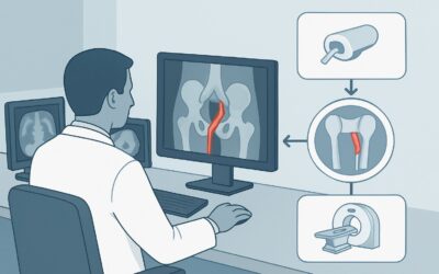
It’s Tuesday afternoon in the vascular surgery clinic. Your patient, a 68-year-old man, is six months out from a thoracic endovascular aortic repair (TEVAR) for a descending thoracic aortic aneurysm. He feels well and has no complaints, but today is his scheduled surveillance visit. The critical question is not whether to image, but how. You need to assess the stent-graft integrity, rule out complications, and ensure the aneurysm sac is regressing, all while considering the patient’s cumulative radiation exposure over a lifetime of follow-up. This article details the clinical workflow for this specific scenario, explaining why the American College of Radiology (ACR) finds one particular study to be the most effective choice. For routine post-TEVAR surveillance, a CTA chest abdomen pelvis with IV contrast is rated Usually Appropriate.
Who Fits This Clinical Scenario for Post-TEVAR Aortic Surveillance?
This guidance applies specifically to adults undergoing routine, scheduled imaging follow-up after a previously placed thoracic endovascular stent-graft. The initial procedure could have been for a thoracic aortic aneurysm or a dissection. The key element is that the patient is in a non-acute, surveillance phase of care, where the goal is to proactively detect potential long-term complications before they become symptomatic or life-threatening.
This workflow is distinct from several similar-sounding clinical situations:
- Patients with Post-Open Surgical Repair: If the patient underwent a traditional open surgical repair with a sewn-in graft, the potential complications and imaging appearance are different. This requires a separate workflow, detailed in the ACR variant for post-open repair follow-up.
- Patients with an Untreated Aneurysm or Dissection: An individual with a known thoracic aortic aneurysm or dissection who has not yet undergone repair falls under a different surveillance protocol. Their imaging is focused on native vessel changes, such as aneurysm growth rate, to determine the timing for intervention.
- Patients with Acute Symptoms: A patient presenting with acute chest or back pain, hypotension, or other signs of an aortic catastrophe—even with a known TEVAR graft—is not undergoing routine surveillance. This is an emergent workup where imaging must be performed immediately to rule out graft failure, new dissection, or rupture.
This article is for the stable, asymptomatic patient in your clinic for their planned check-up.
What Complications Are You Screening for After TEVAR?
Surveillance imaging after TEVAR is not a general screen; it is a focused search for a specific set of well-defined potential complications. The primary goal of the endograft is to exclude the aneurysm sac or false lumen from systemic blood pressure, leading to thrombosis and eventual shrinkage. Imaging is designed to confirm this process is occurring as expected and to detect failures.
The most common and clinically significant complication is an endoleak, defined as persistent blood flow into the aneurysm sac outside the stent-graft. There are several types, each with different implications. Type I (inadequate seal at the proximal or distal landing zones) and Type III (graft defect or component separation) are high-pressure leaks that require urgent intervention. Type II endoleaks, the most frequent type, involve retrograde flow from branch vessels (like intercostal or lumbar arteries) and are often managed conservatively unless they cause aneurysm sac expansion.
Beyond endoleaks, imaging assesses for stent-graft migration, where the device has moved from its intended position, potentially compromising the seal. The study also evaluates aneurysm sac dynamics. A stable or shrinking sac is the desired outcome. An enlarging sac is a red flag, indicating persistent pressurization, almost always from an endoleak (even one that is not immediately obvious). Less commonly, imaging can detect issues with device integrity, such as stent fractures, or identify new aortic pathology like a new dissection or aneurysm in an adjacent, untreated aortic segment.
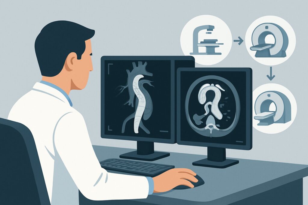
Why Is CTA of the Chest, Abdomen, and Pelvis the Recommended Study for Post-TEVAR Follow-up?
The ACR designates CTA chest abdomen pelvis with IV contrast as Usually Appropriate for post-TEVAR surveillance because it provides the most comprehensive and reliable assessment of the key potential complications. The high spatial and temporal resolution of modern multi-detector CT is ideal for visualizing the fine wires of the stent-graft, its position relative to key branch vessels, and, most importantly, the presence and source of contrast outside the graft within the aneurysm sac.
A complete study from the thoracic inlet through the pelvis is crucial. While the repair is in the chest, the distal landing zone and the access vessels (iliac and femoral arteries) must be evaluated. Furthermore, many Type II endoleaks originate from lumbar or mesenteric arteries in the abdomen, which would be missed on a chest-only scan. Multiphase imaging—including non-contrast, arterial, and delayed phases—is essential for detecting and characterizing endoleaks, distinguishing them from vessel wall calcification, and determining the timing of sac opacification.
Why are other studies rated lower for this specific scenario?
- MRA chest abdomen pelvis with IV contrast: This is also rated Usually Appropriate and is an excellent alternative, particularly for younger patients or those with renal insufficiency precluding iodinated contrast, as it involves no ionizing radiation. However, MRA can be limited by metallic artifact from the stent-graft, which may obscure small endoleaks. It is also less widely available, more time-consuming, and may be less sensitive for certain types of endoleaks compared to a multiphase CTA.
- Radiography chest: Rated Usually not appropriate, a plain chest X-ray is inadequate for surveillance. While it can grossly show the stent-graft position and detect wire fractures, it provides no information about endoleaks, aneurysm sac size, or perfusion—the most critical endpoints of follow-up.
The primary trade-off with serial CTA is the significant cumulative radiation dose (ACR Relative Radiation Level ☢☢☢☢☢). This is a critical consideration in patient counseling, as surveillance is often lifelong. The risk of repeated radiation exposure must be weighed against the life-threatening risk of a missed endoleak and subsequent aneurysm rupture.
Once you’ve decided on CTA chest abdomen pelvis with IV contrast, our protocol guide covers the technique, contrast, and reading principles: CT Chest/Abdomen/Pelvis with IV Contrast.
What Is the Downstream Workflow After a Post-TEVAR Surveillance CTA?
The radiologist’s report will guide your next steps, which diverge based on the findings.
- If the CTA is positive for a high-risk endoleak: Findings like a Type I or Type III endoleak, or a Type II endoleak associated with significant aneurysm sac expansion (typically >5 mm), warrant urgent consultation with the treating interventional radiologist or vascular surgeon. The next step is typically a catheter-based diagnostic aortogram to confirm the finding and plan for intervention, which may include deploying additional cuffs or extensions, or embolizing feeding vessels.
- If the CTA is negative: A report showing a stable or shrinking aneurysm sac with no endoleak and no device migration is the ideal outcome. The patient continues routine surveillance. The imaging interval is guided by societal guidelines and institutional protocols, but a common schedule is at 1, 6, and 12 months post-procedure, followed by annual scans thereafter, provided no adverse features develop.
- If the CTA is indeterminate: Sometimes, findings can be ambiguous. There might be a question of a low-flow endoleak or difficulty distinguishing thrombus from slow-flowing blood. In these cases, a problem-solving study like contrast-enhanced ultrasound (CEUS) or MRA may be considered. Alternatively, a shorter-interval follow-up CTA (e.g., in 3-6 months) may be recommended to assess for any change.
Pitfalls to Avoid (and When to Get Help)
Several common pitfalls can compromise the quality and safety of post-TEVAR surveillance.
- Ordering a Chest-Only CT: This is a frequent error that can miss distal endoleaks (e.g., from lumbar arteries) or issues at the distal landing zone. The standard of care is to image the entire aorta.
- Omitting Contrast or Phases: A non-contrast CT is insufficient as it cannot detect endoleaks. Similarly, a single-phase arterial study may miss delayed-filling endoleaks. Always specify a multiphase “endoleak protocol” when ordering.
- Ignoring Cumulative Radiation Dose: Failing to track a patient’s cumulative radiation exposure from serial scans is a significant oversight. For younger patients, actively consider alternating CTA with MRA to mitigate this long-term risk.
- Misinterpreting Sac Growth: Any confirmed increase in aneurysm sac diameter is a sign of treatment failure until proven otherwise and requires escalation. Do not dismiss small increases; they often precede more significant problems.
If you identify an enlarging aneurysm sac or a suspected Type I or III endoleak, escalate immediately to the proceduralist who performed the TEVAR.
Related ACR Topics and Tools
This article covers one specific clinical scenario. For a comprehensive overview of imaging for all related presentations, or for tools to help you implement this guidance, see the resources below.
- For breadth across all scenarios in Thoracic Aortic Aneurysm or Dissection-Treatment Planning and Follow-Up, see our parent guide: Thoracic Aortic Aneurysm or Dissection-Treatment Planning and Follow-Up: ACR Appropriateness Decoded.
- ACR Appropriateness Criteria Lookup: Look up ratings for thousands of clinical scenarios.
- Imaging Protocol Library: Find detailed, step-by-step techniques for ordering the recommended study.
- Radiation Dose Calculator: Estimate cumulative effective dose for patients undergoing serial imaging.
Frequently Asked Questions
How often should surveillance imaging be performed after TEVAR?
A common protocol is imaging at 1 month, 6 months, and 12 months after the initial procedure, followed by annual imaging for life. However, this can vary based on the initial pathology, specific device used, and institutional guidelines. If any adverse features like a small endoleak or minimal sac growth are detected, the surveillance interval may be shortened.
My patient has chronic kidney disease. Is CTA still the right test?
For patients with significant renal impairment (e.g., GFR < 30 mL/min/1.73 m²), the risk of contrast-induced nephropathy from CTA is high. In these cases, MRA with a gadolinium-based contrast agent (using a macrocyclic agent to minimize NSF risk) is an excellent alternative and is also rated 'Usually Appropriate' by the ACR. A non-contrast CT may also be performed to assess sac size and stent position, but it cannot rule out an endoleak.
What is the difference in follow-up for a TEVAR placed for an aneurysm versus a dissection?
The fundamental goals are the same: assess for endoleak, migration, and device integrity. However, in dissection cases, imaging also focuses on the behavior of the false lumen. The desired outcome is false lumen thrombosis and aortic remodeling. Persistent false lumen flow, especially with expansion, is an adverse finding that may require further intervention.
Can ultrasound be used for TEVAR surveillance?
Unlike with endovascular repair of the abdominal aorta (EVAR), standard duplex ultrasound is not useful for thoracic aortic surveillance due to the inability to visualize the thoracic aorta. However, Contrast-Enhanced Ultrasound (CEUS) is an emerging radiation-free option that can be highly sensitive for detecting endoleaks, though it is more operator-dependent and less standardized than CTA.
What if the aneurysm sac does not shrink but remains stable in size?
A stable aneurysm sac size without a detectable endoleak is a common finding. While sac shrinkage is the ideal outcome, a stable sac is generally considered an acceptable result, suggesting that the sac is depressurized. However, these patients still require continued annual surveillance, as delayed endoleaks or ‘endotension’ (sac pressurization without a visible leak) can still occur and lead to late sac expansion.
Reviewed by Pouyan Golshani, MD, Interventional Radiologist — May 30, 2026