What Is the Best Imaging for Follow-Up of Non-Liver Dominant Pancreatic Neuroendocrine Tumors?
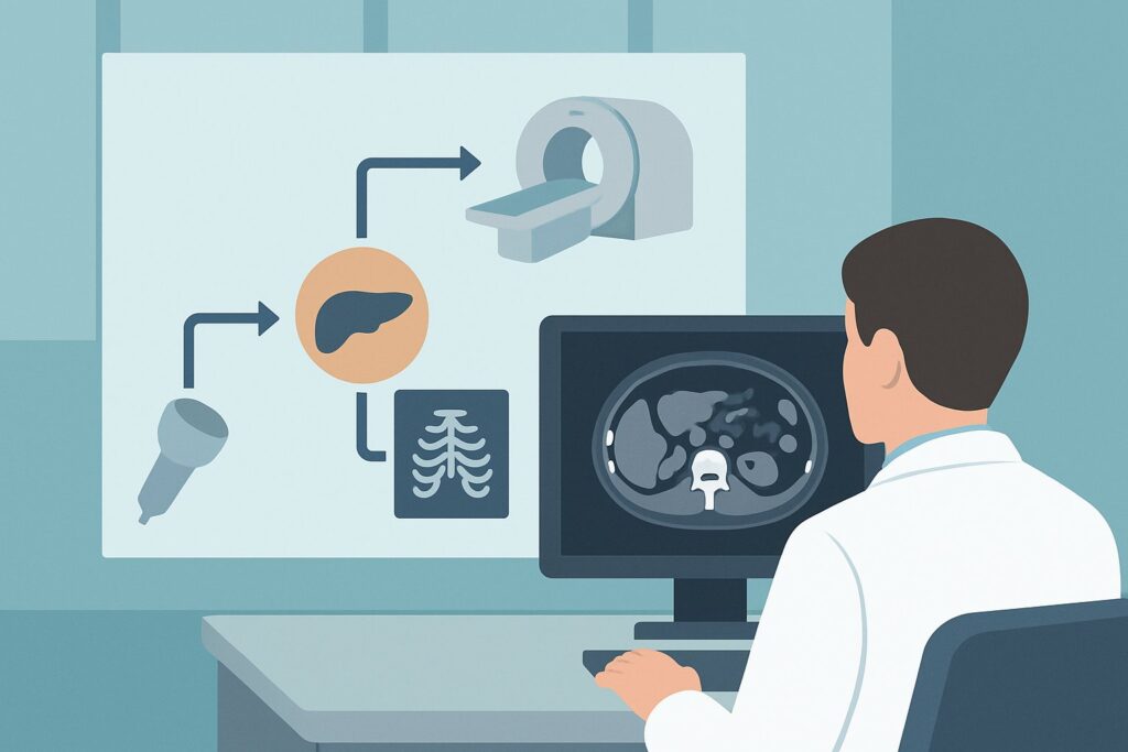
It’s Tuesday afternoon, and you are reviewing the follow-up plan for a 62-year-old patient with a well-differentiated pancreatic neuroendocrine tumor (pNET). Six months ago, they completed a course of peptide receptor radionuclide therapy (PRRT) for disease primarily involving the pancreatic tail and regional lymph nodes. Now, it’s time to assess their response. The patient feels well, and their chromogranin A levels are stable. You need to choose the optimal imaging study to detect any potential progression while minimizing cumulative radiation exposure over the long term. This article details the American College of Radiology (ACR) guideline-based workflow for this specific clinical question. For this scenario, the ACR rates `MRI abdomen and pelvis without and with IV contrast` as Usually Appropriate.
Who Fits This Clinical Scenario for pNET Follow-Up?
This guidance applies to adult patients with a histologically confirmed pancreatic neuroendocrine tumor who have already undergone treatment and are now being monitored. The key distinguishing feature of this scenario is that the disease is non-liver dominant. This means the primary tumor burden is located outside the liver—for instance, in the pancreas itself, regional lymph nodes, or peritoneum—even if small, non-dominant liver metastases are present.
This workflow is specifically for follow-up imaging to assess treatment response or monitor for recurrence. It is distinct from several similar, but separate, clinical situations:
- Initial Staging: This guidance does not apply to the initial workup of a newly diagnosed pNET to determine the extent of local or metastatic disease.
- Liver-Dominant Disease: Patients whose tumor burden is predominantly within the liver follow a different imaging pathway, as outlined in a separate ACR variant.
- Post-Resection Surveillance: This scenario is different from routine surveillance imaging for a patient who has undergone complete surgical resection and has no known or suspected residual disease.
- Untreated Disease: Patients with known, untreated disease who are undergoing active surveillance also represent a distinct clinical question with its own imaging considerations.
Correctly identifying your patient within this specific context—post-treatment, non-liver dominant disease—is crucial for applying the appropriate imaging strategy.
What Are You Assessing in Post-Treatment pNET Follow-Up?
In the follow-up setting for pNETs, imaging is not used to establish a new diagnosis but to evaluate the status of known disease after therapeutic intervention. The primary goals are to differentiate between treatment response, stable disease, and progression, each of which carries different implications for patient management.
Disease Progression is the most critical finding to identify. This can manifest as an increase in the size of known tumor deposits (in the pancreas, lymph nodes, or peritoneum) or the appearance of new lesions. Because pNETs are often slow-growing, even subtle changes can be significant over time, signaling that the current therapy is no longer effective and a change in strategy is needed.
Treatment Response is the desired outcome, characterized by a decrease in the size or enhancement of known tumors. In some cases, lesions may become cystic or necrotic. Quantifying this response using standardized criteria (like RECIST 1.1) is essential for determining whether to continue the current treatment regimen.
Stable Disease is a very common finding in the follow-up of well-differentiated, indolent pNETs. This indicates no significant change in tumor burden since the last imaging study. For slow-growing tumors, maintaining stable disease is often considered a successful therapeutic outcome.
Treatment-Related Changes must also be considered. Therapies like radiation or PRRT can cause inflammation and fibrosis, which can sometimes mimic tumor recurrence on imaging. Differentiating these benign post-treatment effects from true progression is a key task for the radiologist and requires careful comparison with prior studies.
Why Is Contrast-Enhanced MRI the Recommended Study for Non-Liver Dominant pNET Follow-Up?
The ACR designates `MRI abdomen and pelvis without and with IV contrast` as Usually Appropriate for this scenario, making it the primary recommended study. The rationale is based on its superior diagnostic capabilities and safety profile for long-term surveillance.
The principal advantage of MRI is its excellent soft-tissue contrast, which allows for detailed evaluation of the pancreatic parenchyma, surrounding soft tissues, and potential small peritoneal implants that can be difficult to visualize on other modalities. For pNETs, which are typically hypervascular, the dynamic enhancement pattern after administration of gadolinium-based contrast is crucial for identifying lesions and assessing their viability. A multiphasic acquisition protocol, including late arterial and portal venous phases, maximizes the conspicuity of these tumors against the background pancreatic and hepatic tissue.
A critical benefit of MRI in this setting is the complete absence of ionizing radiation (0 mSv). Patients with pNETs often require imaging every 6 to 12 months for many years. Choosing a radiation-free modality like MRI helps minimize the cumulative radiation dose and the associated long-term risks, a significant consideration in patient care.
While other studies can be used, they are often rated lower for specific reasons:
- CT abdomen and pelvis with IV contrast is also rated Usually Appropriate and is a valid alternative, particularly if MRI is contraindicated or unavailable. However, it involves ionizing radiation (☢☢☢ 1-10 mSv) and generally offers lower soft-tissue contrast compared to MRI for subtle peritoneal disease or complex post-treatment changes.
- Endoscopic ultrasound (US) is rated Usually not appropriate for this follow-up scenario. While invaluable for initial diagnosis and local staging of the primary pancreatic tumor, it is an invasive procedure and cannot assess for nodal, peritoneal, or distant metastatic disease, which is the primary goal of follow-up imaging.
What’s the Next Step After the Follow-Up MRI?
The results of the follow-up MRI will directly guide the subsequent clinical workflow and management decisions. The pathway diverges based on whether the findings show progression, stability, or an indeterminate result.
If the MRI shows clear evidence of disease progression (new lesions or unequivocal growth of existing ones), the standard next step is a discussion at a multidisciplinary neuroendocrine tumor board. This allows for integrated input from oncology, surgery, radiology, and nuclear medicine. Management changes may include switching to a different systemic therapy, considering targeted locoregional treatments, or exploring clinical trial options. A `DOTATATE PET/CT skull base to mid-thigh`, which is also Usually Appropriate, may be ordered to confirm somatostatin receptor expression in the progressive lesions, especially if another round of PRRT is being considered.
If the MRI demonstrates stable or responding disease, this is generally a favorable sign. The patient would typically continue on their current treatment plan, with the next surveillance scan scheduled at a clinically appropriate interval, often between 6 and 12 months.
If the MRI reveals an indeterminate finding, such as a tiny new nodule or an ambiguous area of enhancement, the decision is more nuanced. One option is a short-interval follow-up MRI in 3-4 months to assess for change. Alternatively, if the finding is concerning and could alter management, proceeding to functional imaging with DOTATATE PET/CT can provide crucial information. High uptake on a DOTATATE scan would confirm the finding as a focus of neuroendocrine tumor, prompting a change in management.
Pitfalls to Avoid (and When to Get Help)
Navigating post-treatment imaging for pNETs requires attention to several potential pitfalls to ensure accurate interpretation and optimal patient care.
- Not Providing Priors: Comparing the current scan to multiple previous studies is essential for distinguishing true progression from post-treatment inflammation or stable, long-standing findings. Always ensure the radiologist has access to all relevant prior imaging.
- Using a Generic Protocol: Ordering a “routine” abdomen MRI may not include the specific multiphasic contrast-enhanced sequences needed to optimally detect hypervascular pNETs. Specify a dedicated pancreas protocol.
- Ignoring Functional Correlation: In equivocal cases, relying solely on anatomic imaging can be misleading. Fibrosis and tumor can look similar. Don’t hesitate to use functional imaging like DOTATATE PET/CT to resolve ambiguity.
- Forgetting Extra-Abdominal Disease: While this scenario focuses on non-liver dominant disease in the abdomen, pNETs can metastasize elsewhere (e.g., bone, lungs). If a patient develops new, localized symptoms like bone pain, dedicated imaging of that area or a whole-body functional scan may be warranted.
If imaging results are complex, show unexpected patterns of progression, or contradict the clinical picture, escalate the case for review at a specialized neuroendocrine tumor board.
Related ACR Topics and Tools
For a comprehensive overview of imaging recommendations across all clinical presentations of pancreatic neuroendocrine tumors, please consult the parent topic article. Additional GigHz tools are available to support clinical decision-making for imaging orders.
- For breadth across all scenarios in Staging and Follow-up of Pancreatic Neuroendocrine Tumors, see our parent guide: Staging and Follow-up of Pancreatic Neuroendocrine Tumors: ACR Appropriateness Decoded.
- To explore imaging guidelines for other clinical presentations, use the ACR Appropriateness Criteria Lookup.
- For detailed technical parameters of the recommended study, refer to the Imaging Protocol Library.
- To discuss cumulative radiation exposure with patients when considering CT as an alternative, the Radiation Dose Calculator can be a helpful resource.
Frequently Asked Questions
Why is MRI preferred over CT for follow-up if both are rated ‘Usually Appropriate’?
MRI is often preferred for long-term follow-up of pNETs primarily because it does not use ionizing radiation. Since these patients may require scans every 6-12 months for many years, minimizing cumulative radiation exposure is a key consideration. Additionally, MRI offers superior soft-tissue contrast, which can be advantageous for detecting subtle peritoneal disease or evaluating the pancreas in a complex post-treatment setting.
What if my patient has a contraindication to MRI, like a non-compatible pacemaker?
If a patient cannot undergo an MRI, a multiphasic `CT abdomen and pelvis with IV contrast` is an excellent and appropriate alternative. It is also rated ‘Usually Appropriate’ by the ACR for this scenario and provides high-quality anatomic detail for assessing tumor burden and response to therapy.
When should I order a DOTATATE PET/CT instead of an MRI for follow-up?
While MRI is the recommended primary modality for anatomic follow-up, a DOTATATE PET/CT is invaluable for assessing functional status. It is often used when MRI findings are equivocal, to confirm that a new or growing lesion expresses somatostatin receptors before planning PRRT, or to provide a whole-body survey if widespread progression is suspected. Some centers alternate between anatomic (MRI/CT) and functional (DOTATATE) imaging for surveillance.
Does ‘non-liver dominant’ mean the patient has no liver metastases?
Not necessarily. A patient can have liver metastases and still be considered ‘non-liver dominant’. The term means that the bulk of the tumor volume and the primary driver of the patient’s clinical course is located outside the liver (e.g., a large primary pancreatic mass or extensive nodal/peritoneal disease). This is in contrast to ‘liver-dominant’ disease, where the majority of the tumor burden is within the liver.
How does the imaging strategy change if the patient had a complete surgical resection and is now in surveillance?
That is a different clinical scenario addressed by a separate ACR variant: ‘Imaging after surgical resection, no suspected or known recurrence. Surveillance.’ The imaging choice and frequency may differ, as the goal is to detect new recurrence in a patient presumed to be disease-free, rather than monitoring known disease after non-curative treatment.
Reviewed by Pouyan Golshani, MD, Interventional Radiologist — May 30, 2026