What Is the Best Initial Imaging for a Child with Back Pain and Suspected Idiopathic Arthritis?
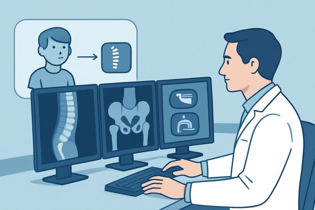
A 9-year-old presents with three months of worsening mid-back pain. There was no specific injury, the pain is worse in the morning, and it seems to improve with activity throughout the day. You note mild stiffness on examination. Inflammatory markers are slightly elevated. While juvenile idiopathic arthritis (JIA) is on your differential, so are more concerning diagnoses like infection or malignancy. You need to choose the initial imaging study that can best differentiate these possibilities without exposing the child to unnecessary radiation. This article details the American College of Radiology (ACR) workflow for this specific scenario. For a child with back pain and suspected idiopathic arthritis, the ACR rates an MRI complete spine without and with IV contrast as Usually appropriate.
Who Fits This Clinical Scenario?
This guidance applies to the initial imaging workup for a child or adolescent presenting with persistent, non-traumatic back pain where an inflammatory process, specifically a form of juvenile idiopathic arthritis like juvenile spondyloarthropathy, is a leading consideration. The key features include insidious onset of pain, morning stiffness, and the absence of a clear mechanical cause. The term “suspected” is critical; this workflow is for diagnostic clarification when the clinical picture is suggestive but not yet confirmed.
This pathway is distinct from several similar presentations:
- Acute Trauma: A child with back pain immediately following a fall or injury requires a different workup, often starting with radiographs to assess for fracture or malalignment.
- Focal Neurologic Deficits: If the child presents with weakness, sensory loss, or bowel/bladder changes, the imaging protocol becomes more urgent to evaluate for spinal cord or nerve root compression, though MRI remains the modality of choice.
- Pain Localized to the Pelvis: If the pain is clearly localized to the sacroiliac (SI) joints, a dedicated MRI of the SI joints is the preferred initial study, as detailed in the sibling ACR scenario for sacroiliac joint pain.
- Primary Appendicular Symptoms: A child whose primary complaint is swelling or pain in the knees, wrists, or ankles, even with minor back pain, falls under the appendicular joint pain scenario.
What Diagnoses Are You Working Up in This Scenario?
When ordering initial imaging for a child with suspected inflammatory back pain, the goal is to confirm or exclude several key diagnoses that can present similarly. The differential is broad, ranging from common inflammatory conditions to rare but critical pathologies.
Juvenile Spondyloarthropathy: This is a category of juvenile idiopathic arthritis that primarily affects the spine and sacroiliac joints. Imaging is crucial for detecting early signs of inflammation, such as bone marrow edema in the vertebral bodies (osteitis), inflammation of the facet joints (synovitis), or inflammation at the sites where ligaments and tendons attach to bone (enthesitis). These findings on MRI can secure the diagnosis and guide treatment with disease-modifying antirheumatic drugs (DMARDs) or biologics.
Infection (Discitis or Vertebral Osteomyelitis): Bacterial infection of the intervertebral disc or vertebral body is a critical mimic of inflammatory arthritis. Children may present with back pain, refusal to walk, and low-grade fever. MRI is highly sensitive for differentiating the characteristic enhancement patterns and associated abscesses of infection from the more diffuse edema of sterile inflammation.
Benign or Malignant Tumors: While less common, tumors must be considered. Benign lesions like an osteoid osteoma can cause significant nocturnal pain that responds well to NSAIDs. Malignant processes, including leukemia, lymphoma, or primary bone tumors like Ewing sarcoma, can also present with back pain. MRI is essential for characterizing mass lesions and determining their extent.
Scheuermann’s Disease: This structural condition, characterized by vertebral wedging and thoracic kyphosis, is a common cause of back pain in adolescents. While often diagnosed on radiographs, MRI can assess for associated disc herniations and rule out superimposed inflammatory or infectious processes.
Why Is MRI of the Complete Spine Without and With IV Contrast the Recommended Study?
The ACR designates an MRI complete spine without and with IV contrast as Usually appropriate because it provides the most comprehensive diagnostic information for this clinical question while avoiding ionizing radiation.
MRI’s superior soft-tissue contrast is its primary advantage. It is uniquely capable of detecting bone marrow edema, the earliest sign of active inflammation from either spondyloarthropathy or osteomyelitis, long before any structural changes become visible on other imaging modalities. This allows for early diagnosis and intervention.
The inclusion of intravenous (IV) gadolinium-based contrast is key. Post-contrast images highlight areas of active inflammation, helping to distinguish between sterile inflammatory synovitis, infectious phlegmon or abscess, and enhancing tumors. This is critical for narrowing the differential diagnosis. Ordering the study “without and with” contrast provides both the pre-contrast anatomical detail and the post-contrast physiological information needed for an accurate interpretation.
Let’s compare this to other modalities rated for this scenario:
- Radiography (X-rays): Rated as May be appropriate. While useful for assessing spinal alignment and detecting chronic changes like vertebral body squaring or erosions, radiographs are insensitive to early inflammation. A normal X-ray does not rule out active spondyloarthropathy or infection. Given its limitations in answering the primary clinical question, it is not the first-line study for suspected active inflammation.
- Computed Tomography (CT): Rated as Usually not appropriate. CT provides excellent bony detail but has poor soft-tissue contrast compared to MRI, making it unable to reliably detect bone marrow edema. Furthermore, it involves a significant pediatric radiation dose (3-10 mSv), which should be avoided whenever a non-ionizing modality like MRI can answer the clinical question.
The choice of “complete spine” over an “area of interest” is also deliberate. Children can have difficulty localizing spinal pain, and inflammatory conditions can be multifocal. Imaging the entire spine—cervical, thoracic, and lumbar—in a single session prevents missing pathology outside a limited field of view. Once you’ve decided on an MRI of the spine, our protocol guide covers the core technique, contrast considerations, and reading principles: MRI Lumbar Spine Without Contrast.
What’s Next After MRI? Downstream Workflow
The results of the spine MRI will guide your next steps, creating distinct clinical pathways.
If the MRI is positive for inflammatory spondyloarthropathy: Findings like multifocal vertebral bone marrow edema, facet joint synovitis and enhancement, or sacroiliitis confirm an active inflammatory process. This result is the trigger for a referral to a pediatric rheumatologist. The rheumatologist will correlate the imaging with clinical and laboratory findings (e.g., HLA-B27 status, ESR/CRP) to finalize the diagnosis and initiate appropriate systemic therapy, which may include NSAIDs, DMARDs, or biologic agents.
If the MRI is negative: A completely normal MRI makes active inflammatory spondylitis, infection, or a tumor highly unlikely. The diagnostic focus should shift toward mechanical or musculoskeletal causes of back pain. The next step is often a referral to physical therapy, a trial of conservative management, and clinical follow-up. If pain persists despite these measures, a repeat clinical evaluation is warranted.
If the MRI suggests infection or a tumor: Findings such as disc space enhancement with a paraspinal abscess (discitis/osteomyelitis) or a destructive, enhancing vertebral mass (tumor) are critical results. This requires immediate action. An urgent consultation with pediatric infectious disease and/or orthopedic surgery for suspected infection, or with pediatric oncology for a suspected tumor, is necessary. These pathways will likely lead to biopsy and specific antimicrobial or oncologic treatment.
If the MRI is indeterminate: Occasionally, findings can be ambiguous, such as mild, non-specific bone marrow edema. In these cases, a multidisciplinary discussion involving the ordering clinician, a pediatric radiologist, and a pediatric rheumatologist is invaluable to correlate the subtle imaging findings with the full clinical picture and decide on the next step, which could be close clinical follow-up or a short-interval repeat MRI.
Pitfalls to Avoid (and When to Get Help)
Navigating the workup for pediatric back pain requires avoiding several common pitfalls:
- Stopping at a Normal Radiograph: Do not be falsely reassured by normal X-rays. If your clinical suspicion for an inflammatory condition is high based on history and exam, proceed to MRI, as radiographs cannot detect the early changes of JIA or infection.
- Ordering a Limited Study: Unless the pain is precisely localized, ordering an MRI of only the lumbar spine risks missing thoracic or cervical pathology. The “complete spine” protocol is often the safer initial choice.
- Forgetting Sedation Needs: A high-quality MRI requires the patient to remain still for an extended period. Many younger children will require sedation or general anesthesia. Plan for this logistical step early to avoid delays in diagnosis.
- Underestimating the Differential: While JIA may be the leading suspicion, always maintain a broad differential. Review the MRI report carefully for any signs that point toward infection or malignancy, as these require immediate escalation.
If the MRI reveals findings suggestive of a tumor, abscess, or impending spinal cord compromise, escalate immediately to the appropriate surgical or medical subspecialty service.
Related ACR Topics and Tools
This article focuses on a single clinical scenario. For a comprehensive overview of all related presentations and for tools to help with ordering and patient communication, the following resources are available.
- For breadth across all scenarios in Joint Pain: Idiopathic Arthritis-Child, see our parent guide: Joint Pain: Idiopathic Arthritis-Child: ACR Appropriateness Decoded.
- Explore adjacent scenarios and their corresponding ACR recommendations using the ACR Appropriateness Criteria Lookup.
- Review detailed technical parameters for various studies in our Imaging Protocol Library.
- For discussions about radiation exposure from alternative studies like CT, use the Radiation Dose Calculator.
Frequently Asked Questions
Why is an MRI with and without contrast necessary instead of just a non-contrast study?
A non-contrast MRI is excellent for detecting bone marrow edema, but adding intravenous contrast significantly improves diagnostic specificity. Contrast helps differentiate active inflammation (which enhances) from chronic changes, and it is critical for distinguishing sterile inflammation from infection (e.g., an abscess) or an enhancing tumor. The ACR rates a non-contrast MRI as only ‘May be appropriate’ for this reason.
What if my patient has a contraindication to gadolinium contrast, like severe renal impairment?
In cases of a true contraindication to gadolinium, an MRI of the complete spine without IV contrast is still a valuable study and is rated ‘May be appropriate’ by the ACR. It can detect bone marrow edema and major structural abnormalities. However, its ability to differentiate between inflammation, infection, and some tumors is reduced. A discussion with the radiologist prior to the scan is recommended to optimize the non-contrast protocol.
My patient’s pain is mostly in the lower back. Can I just order an MRI of the lumbar spine?
While an MRI of the spine area of interest is also rated ‘Usually appropriate,’ children often have difficulty precisely localizing pain. Inflammatory spondyloarthropathies can be multifocal. Ordering a complete spine MRI is often the safer approach to ensure that pathology in the thoracic or cervical spine is not missed, which could lead to a delayed or incomplete diagnosis.
Is a whole-body bone scan a reasonable alternative to look for multifocal disease?
No, for this clinical scenario, a whole-body bone scan is rated ‘Usually not appropriate.’ While it can detect areas of increased bone turnover, it is not specific and cannot differentiate between inflammation, infection, trauma, or tumor. More importantly, MRI is far more sensitive for the early bone marrow edema characteristic of inflammatory arthritis. Bone scans also expose the child to significant radiation (3-10 mSv).
If the initial MRI is negative but the child’s back pain persists, what is the next step?
If the initial, high-quality MRI of the complete spine is negative, it strongly argues against an active inflammatory, infectious, or neoplastic process. The focus should shift to non-inflammatory causes (e.g., mechanical pain, stress injury). The next steps typically involve a trial of physical therapy and continued clinical observation. If symptoms worsen or new red flags develop, a repeat clinical evaluation is warranted, which may eventually lead to repeat imaging, but this would fall under a ‘follow-up’ imaging scenario.
Reviewed by Pouyan Golshani, MD, Interventional Radiologist — May 30, 2026