What Is the Best Initial Imaging for a Suspected Adnexal Mass in Pregnancy?
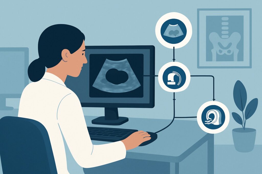
A 28-year-old patient, G1P0 at 14 weeks gestation, presents for a routine prenatal visit. On bimanual examination, you palpate a smooth, mobile, non-tender fullness in the left adnexa. The patient is asymptomatic, reporting no pain, fever, or bleeding. The clinical question is immediate: how do you evaluate this finding to characterize its nature and potential impact on the pregnancy, while ensuring the safety of the developing fetus? This article provides a focused clinical workflow for this exact scenario, guiding the choice of initial imaging. For this presentation, the American College of Radiology (ACR) Appropriateness Criteria rate US duplex Doppler pelvis as Usually Appropriate.
Who Fits This Clinical Scenario?
This guidance applies to a specific patient population: a pregnant female, at any gestational age, with a clinically suspected adnexal mass but no acute symptoms. The suspicion may arise from a physical examination finding, as in the vignette, or as an incidental finding on an early obstetric ultrasound performed for other reasons (e.g., dating, nuchal translucency).
Key inclusion criteria are:
- Patient is confirmed pregnant.
- An adnexal mass is suspected based on clinical exam or noted incidentally.
- The patient is asymptomatic, meaning no acute pelvic pain, fever, hemodynamic instability, or other signs suggesting an emergent condition like ovarian torsion, ectopic pregnancy rupture, or significant hemorrhage.
- This workflow is for the initial diagnostic imaging, not for follow-up of a previously characterized mass.
It is crucial to distinguish this scenario from others. This guidance does not apply to patients with acute pelvic pain, which requires an emergent workup for conditions like ovarian torsion or ruptured ectopic pregnancy. It also does not apply to non-pregnant patients (premenopausal or postmenopausal), whose evaluation follows a different diagnostic algorithm due to a different risk profile for malignancy.
What Diagnoses Are You Working Up in a Pregnant Patient with an Adnexal Mass?
When an adnexal mass is discovered during pregnancy, the differential diagnosis spans from benign, physiologic changes to rare but critical pathologies. The goal of initial imaging is to differentiate between these possibilities.
Corpus Luteum Cyst: This is the most common adnexal “mass” identified in the first trimester. It is a physiologic, functional cyst that supports the early pregnancy by producing progesterone. These cysts are typically simple or hemorrhagic in appearance on ultrasound, have a characteristic “ring of fire” on Doppler imaging, and usually resolve spontaneously by 14-16 weeks gestation as the placenta takes over progesterone production.
Dermoid Cyst (Mature Cystic Teratoma): These are common benign germ cell tumors that are often discovered incidentally during pregnancy. They can contain various tissues like fat, hair, and calcifications (teeth), which give them a characteristic appearance on ultrasound. While benign, they carry a small risk of torsion, which increases as the uterus grows and displaces the ovary.
Luteoma of Pregnancy: This is a rare, non-neoplastic, solid tumor-like mass that is hormone-dependent, developing during pregnancy and regressing spontaneously after delivery. Luteomas can be hormonally active, sometimes causing maternal virilization. Their solid appearance on ultrasound can mimic malignancy, making them a diagnostic challenge.
Ovarian Malignancy: Although the incidence of ovarian cancer in pregnancy is low (approximately 1 in 25,000 pregnancies), it remains the most critical diagnosis to exclude. Imaging seeks to identify suspicious features such as large solid components, thick septations, papillary projections, significant vascularity on Doppler, and associated findings like ascites.
Other Benign Neoplasms: Other possibilities include serous or mucinous cystadenomas. These are typically cystic masses that can grow to a large size during pregnancy. Ultrasound is key to assessing their size and internal complexity to gauge the likelihood of benignity.
Why Is Pelvic Ultrasound the Recommended Initial Study for a Suspected Adnexal Mass in Pregnancy?
The ACR designates several ultrasound techniques—including US duplex Doppler pelvis, US pelvis transabdominal, and US pelvis transvaginal—as Usually Appropriate for the initial evaluation of a suspected adnexal mass in a pregnant patient. This strong recommendation is rooted in the modality’s exceptional safety profile and high diagnostic efficacy in this specific clinical context.
The foremost reason for choosing ultrasound is safety. Ultrasound utilizes non-ionizing sound waves and has no known adverse effects on the developing fetus. Its radiation level is zero (O, 0 mSv), making it the ideal imaging tool during pregnancy, where avoiding fetal radiation exposure is a primary concern.
Beyond safety, pelvic ultrasound provides excellent anatomic detail of the adnexa. A combination of transabdominal and transvaginal approaches is often optimal. The transabdominal view gives a broader overview of the pelvis, assessing large masses and their relationship to the gravid uterus. The transvaginal approach, when feasible and safe depending on gestational age, offers higher-resolution images of the ovaries and the internal architecture of a mass, which is critical for characterization. It can reliably differentiate cystic from solid masses and identify internal features like septations, mural nodules, and debris.
The addition of Duplex Doppler is a crucial component of the evaluation. It assesses blood flow within the mass. Malignant tumors often induce angiogenesis, creating vessels with low-resistance flow patterns. In contrast, benign lesions and physiologic cysts typically show less prominent or higher-resistance flow. This information, combined with the morphologic features, helps risk-stratify the mass and guide further management.
Alternative imaging modalities are rated lower for initial evaluation:
- MRI pelvis without IV contrast is rated May be appropriate. It is not a first-line study but serves as a valuable problem-solving tool when ultrasound findings are indeterminate. It offers superior soft tissue contrast without using radiation but is more costly and less accessible. Gadolinium-based contrast is generally avoided during pregnancy unless the potential benefit unequivocally outweighs the potential fetal risk.
- CT pelvis with IV contrast is rated Usually not appropriate. CT exposes the fetus to ionizing radiation (☢☢☢ 1-10 mSv), which should be avoided whenever possible during pregnancy. It offers no significant diagnostic advantage over ultrasound or MRI for the primary characterization of an adnexal mass and is reserved for rare, specific indications where its benefits outweigh the radiation risk.
What’s Next After Pelvic Ultrasound? Downstream Workflow
The results of the initial pelvic ultrasound will dictate the subsequent clinical pathway, which balances maternal health with fetal well-being.
If the mass has classic benign features: For a simple cyst less than 5 cm or a mass with the classic appearance of a corpus luteum or dermoid cyst, the typical next step is conservative management. This involves serial ultrasound surveillance. The frequency of follow-up imaging depends on the specific findings and gestational age but may occur once per trimester to monitor for growth or changes in complexity. Most of these masses remain stable or resolve, requiring no intervention during pregnancy.
If the mass is indeterminate: When ultrasound cannot confidently classify a mass as benign (e.g., a complex cystic-solid mass without overtly malignant features), the next step is often a consultation with a maternal-fetal medicine (MFM) specialist and a gynecologic oncologist. The ACR rates MRI pelvis without IV contrast as May be appropriate in this situation. MRI can provide additional characterization of the mass’s tissue composition, helping to differentiate a benign entity like a fibroid or hemorrhagic cyst from a more suspicious lesion.
If the mass is suspicious for malignancy: If the ultrasound reveals features highly concerning for malignancy (e.g., large solid components, papillary projections, significant vascularity, ascites), immediate referral to a gynecologic oncologist is mandatory. Management becomes a complex, multidisciplinary decision involving the patient, MFM, oncology, and neonatology. The plan may involve further staging (often with MRI), consideration of surgical intervention during the second trimester (the safest window), or close monitoring with plans for postpartum surgery, depending on the specifics of the case and gestational age.
Pitfalls to Avoid (and When to Get Help)
Navigating an adnexal mass in pregnancy requires careful consideration to avoid common diagnostic and management errors.
- Misinterpreting a Corpus Luteum: Be cautious not to mistake a physiologic corpus luteum for a pathologic neoplasm in the first trimester. Its typical sonographic appearance and subsequent resolution are key differentiators.
- Delaying Follow-up: For a mass deemed likely benign, failing to schedule and perform recommended follow-up ultrasounds can lead to missing significant growth or the development of concerning features.
- Overlooking Torsion Risk: Remember that the risk of ovarian torsion increases during pregnancy, particularly for masses >5 cm. Counsel patients on the symptoms of acute pain and to seek immediate care if they occur.
- Inappropriate Use of CT: Avoid ordering CT as an initial or secondary imaging tool for mass characterization due to fetal radiation exposure. Reserve it for emergent, non-gynecologic indications where it is absolutely necessary.
If ultrasound findings are indeterminate or any features are suspicious for malignancy, escalate care by consulting with maternal-fetal medicine and gynecologic oncology specialists promptly.
Related ACR Topics and Tools
For a comprehensive overview of imaging for adnexal masses across all patient populations, or to explore the technical details of the recommended studies, the following resources are valuable. For breadth across all scenarios in Clinically Suspected Adnexal Mass, No Acute Symptoms, see our parent guide: Clinically Suspected Adnexal Mass, No Acute Symptoms: ACR Appropriateness Decoded.
- ACR Appropriateness Criteria Lookup — For adjacent scenarios and other clinical questions.
- Imaging Protocol Library — For technical guidance on performing imaging studies.
- Radiation Dose Calculator — For estimating cumulative dose and facilitating patient conversations.
Frequently Asked Questions
Is a transvaginal ultrasound safe to perform during pregnancy?
Yes, transvaginal ultrasound is considered safe throughout pregnancy when performed by a trained professional for an appropriate medical indication. In the first trimester, it provides superior image quality for evaluating the adnexa. In later pregnancy, its use may be guided by factors like cervical length and placental position, but it does not pose a direct risk to the fetus.
What if the patient is in her third trimester? Is ultrasound still the best initial test?
Yes, ultrasound remains the best initial imaging test regardless of gestational age. However, imaging in the third trimester can be technically challenging due to the large size of the gravid uterus, which can obscure the adnexa. A skilled sonographer may need to use various techniques, such as scanning the patient in a decubitus position, to visualize the ovaries. If ultrasound is non-diagnostic due to these limitations, an MRI without contrast may be considered.
Should tumor markers like CA-125 be ordered for an adnexal mass in pregnancy?
The utility of tumor markers like CA-125 is limited during pregnancy. CA-125 can be physiologically elevated in a normal pregnancy, especially in the first trimester, which significantly reduces its specificity for detecting ovarian malignancy. Therefore, management decisions are primarily based on imaging characteristics rather than serum tumor markers.
If an adnexal mass is found, does this automatically mean the patient needs a C-section?
No, the presence of an adnexal mass does not automatically necessitate a cesarean delivery. The mode of delivery is determined by standard obstetric indications. A very large mass that obstructs the birth canal or a mass at high risk of rupture or torsion during labor are rare exceptions where a planned C-section, possibly combined with removal of the mass, might be considered in consultation with a gynecologic oncologist.
Reviewed by Pouyan Golshani, MD, Interventional Radiologist — May 30, 2026