What Is the Best Initial Imaging for an Indeterminate Renal Mass Without Contrast Contraindications?
A 55-year-old patient undergoes a non-contrast computed tomography (CT) scan for suspected nephrolithiasis, which reveals a 2.5 cm solid-appearing mass in the right kidney. The density is too high for a simple cyst, but it cannot be definitively characterized without intravenous contrast. The patient has normal renal function and no history of allergies to imaging contrast agents. You are now faced with the decision of which dedicated imaging study to order next to characterize this indeterminate renal mass. This article provides a detailed clinical workflow for this specific scenario, guiding you through the differential diagnosis, study rationale, and downstream management steps. According to the American College of Radiology (ACR) Appropriateness Criteria, multiple studies are well-suited for this workup, with **US abdomen with IV contrast** rated as *Usually Appropriate*.
Who Qualifies for This Indeterminate Renal Mass Imaging Pathway?
This guidance is designed for the initial characterization of a renal mass that remains indeterminate after preliminary imaging, such as a non-contrast CT or a standard grayscale ultrasound. The key inclusion criteria for this specific workflow are:
- An incidentally discovered or suspected renal mass that lacks definitive features of a simple cyst or classic angiomyolipoma.
- The patient has no contraindications to iodinated intravenous contrast used in CT scans (e.g., no severe prior allergic-like reaction, adequate renal function).
- The patient has no contraindications to gadolinium-based intravenous contrast agents used in MRI (e.g., no history of nephrogenic systemic fibrosis, adequate renal function, no incompatible implanted devices).
It is critical to distinguish this scenario from others that require different imaging strategies. This workflow does not apply if the patient has a known contraindication to only iodinated CT contrast. In that case, MRI becomes the primary modality. Similarly, if a patient has contraindications to both CT and MR contrast agents, the diagnostic options are more constrained, often relying on non-contrast techniques or ultrasound. This article focuses exclusively on the patient for whom all standard contrast-enhanced imaging options are on the table.
What Are the Key Diagnoses in an Indeterminate Renal Mass Workup?
The primary goal of imaging in this scenario is to differentiate malignant tumors from benign lesions, which guides subsequent management, from active surveillance to surgical intervention. The differential diagnosis is broad, but several key entities are of primary concern.
The most pressing concern is Renal Cell Carcinoma (RCC), which accounts for the vast majority of malignant solid renal masses. Characterizing the enhancement pattern of a mass is crucial, as avid enhancement is a hallmark of clear cell RCC, the most common subtype. Other subtypes, like papillary and chromophobe RCC, may show less intense enhancement, making the diagnostic challenge more complex.
A variety of benign tumors can also present as indeterminate masses. Oncocytoma is a classic benign mimic of RCC and is often indistinguishable on imaging alone, sometimes exhibiting a central scar that can also be seen in some RCCs. Another common benign entity is the Angiomyolipoma (AML), particularly fat-poor AMLs. While fat-rich AMLs are easily diagnosed on non-contrast CT, those with minimal fat are a frequent cause of an indeterminate workup and can enhance avidly, mimicking RCC.
Finally, not all indeterminate masses are solid tumors. Complex cysts are a major consideration. The Bosniak classification system (from I to IV) is used to stratify the risk of malignancy in cystic renal lesions based on features like septations, wall thickening, and enhancing solid components. An indeterminate mass may prove to be a complex cyst (e.g., Bosniak III or IV) that carries a high probability of malignancy and often requires surgical management.
Why Are CT, MRI, and Contrast-Enhanced US Recommended for This Presentation?
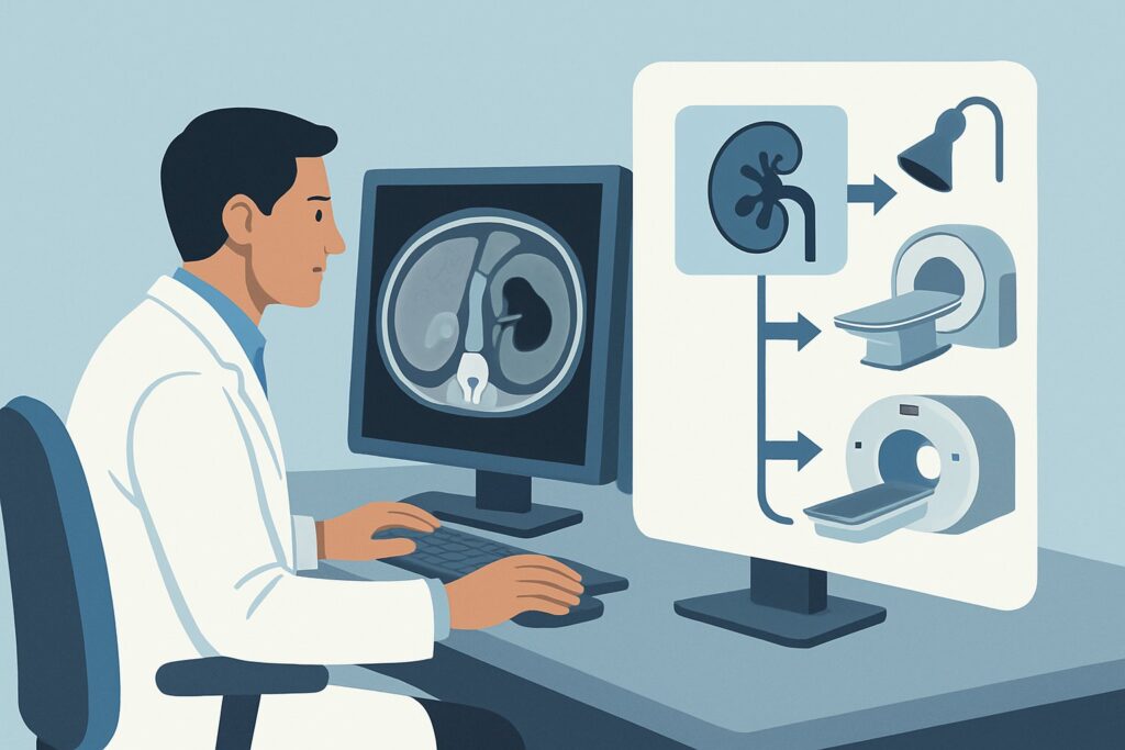
For a patient with an indeterminate renal mass and no contrast contraindications, the ACR rates three modalities as Usually Appropriate: CT abdomen without and with IV contrast, MRI abdomen without and with IV contrast, and US abdomen with IV contrast. Each has distinct advantages, and the optimal choice may depend on institutional expertise and patient-specific factors.
CT abdomen without and with IV contrast is often considered the workhorse for renal mass characterization. A multiphase “renal mass protocol” CT provides a non-contrast baseline, a corticomedullary phase, a nephrographic phase, and often a delayed/excretory phase. This allows for precise measurement of enhancement in Hounsfield Units (HU), which is the cornerstone of diagnosis; a change of >20 HU after contrast is considered definitive enhancement. CT offers rapid acquisition and excellent spatial resolution. Its main drawback is the use of ionizing radiation (ACR RRL ☢☢☢☢, 10-30 mSv), a key consideration in younger patients or those requiring serial imaging.
MRI abdomen without and with IV contrast is an equally powerful alternative that avoids ionizing radiation (ACR RRL O, 0 mSv). It provides superior soft-tissue contrast, which can be particularly helpful in differentiating tumor subtypes or evaluating for tumor thrombus in the renal vein. Specific sequences can detect intracellular lipid to help identify fat-poor AMLs. The primary limitations are longer scan times, higher cost, and contraindications related to metallic implants or claustrophobia.
US abdomen with IV contrast, also known as Contrast-Enhanced Ultrasound (CEUS), is another excellent radiation-free option (ACR RRL O, 0 mSv). It uses microbubble contrast agents that are not nephrotoxic, making it safe in patients with borderline renal function (though this scenario assumes no contraindications). CEUS provides a real-time, dynamic evaluation of vascularity and enhancement patterns, which is especially effective for differentiating complex cysts from solid masses and confirming or refuting enhancement in small lesions where CT density measurements can be equivocal due to partial volume averaging.
Lower-rated alternatives like a standard non-contrast renal ultrasound (May be appropriate) are insufficient for definitive characterization. Similarly, image-guided biopsy is rated Usually not appropriate for initial characterization, as it is typically reserved for cases where imaging remains indeterminate or when a non-surgical ablative therapy is planned.
What’s Next After Imaging? Downstream Workflow
The results of the characterization study will dictate the next steps in management, which typically involve a urologist or urologic oncologist.
- Clearly Malignant Features: If the study (CT, MRI, or CEUS) demonstrates features highly suspicious for RCC (e.g., a solid, avidly enhancing mass), the next step is typically consultation for management. Options include partial or radical nephrectomy, thermal ablation (cryoablation or radiofrequency ablation), or, for small masses in older or comorbid patients, active surveillance.
- Clearly Benign Features: If imaging confirms a benign diagnosis, such as a fat-rich AML or a simple cyst (Bosniak I), no further workup or follow-up is generally needed. The patient can be reassured.
- Indeterminate or Equivocal Result: If the initial dedicated study remains indeterminate (e.g., a possible fat-poor AML vs. RCC, or a Bosniak IIF/III cyst), several pathways exist. One option is to perform a second, complementary imaging study (e.g., an MRI if the initial study was a CT). Another option, particularly for solid masses where ablation is considered or the diagnosis is unclear, is a percutaneous renal mass biopsy. For indeterminate cystic lesions (Bosniak IIF), short-term imaging follow-up (e.g., in 6 months) is the standard recommendation.
The decision between these pathways is complex and depends on the specific imaging findings, patient age, comorbidities, and patient preference after a thorough discussion of the risks and benefits of each approach.
Pitfalls to Avoid (and When to Get Help)
Navigating the workup of an indeterminate renal mass requires careful attention to detail to avoid common errors. A primary pitfall is ordering an incomplete or non-dedicated study. A “CT abdomen with contrast” without specifying a multiphase renal mass protocol may not provide the necessary data to accurately measure enhancement. Another error is failing to review prior imaging; a mass that is new or has grown is more concerning than one that has been stable for many years. Finally, misinterpreting pseudolesions, such as a prominent column of Bertin or fetal lobulation, as a true mass can lead to unnecessary workups. If the imaging findings are complex or the diagnosis remains uncertain after a high-quality study, consultation with a radiologist specializing in genitourinary imaging or referral to a multidisciplinary urologic oncology team is the appropriate next step.
Related ACR Topics and Tools
For a comprehensive overview of all clinical variants related to this topic, please refer to our parent guide. The following GigHz resources can also help you navigate adjacent scenarios, understand imaging techniques, and discuss radiation safety with your patients.
- For breadth across all scenarios in Indeterminate Renal Mass, see our parent guide: Indeterminate Renal Mass: ACR Appropriateness Decoded.
- ACR Appropriateness Criteria Lookup — for adjacent scenarios
- Imaging Protocol Library — for technique on the recommended study
- Radiation Dose Calculator — for cumulative dose conversations
Frequently Asked Questions
If CT, MRI, and CEUS are all ‘Usually Appropriate’, how do I choose between them?
The choice often depends on local availability and radiologist expertise, particularly for CEUS. CT is fast and widely available, making it a common first choice. MRI is preferred for younger patients to avoid radiation or when there’s a specific question about tissue characterization (like identifying fat in a potential AML). CEUS is an excellent problem-solving tool, especially for cystic lesions or when CT findings are equivocal.
Is a CT Urogram (CTU) the same as a renal mass protocol CT?
No, they are different studies. A CT Urogram (CTU) is optimized to visualize the urothelium (renal pelves, ureters, bladder) and includes prominent excretory phase imaging. While it can characterize a renal mass, a dedicated renal mass protocol CT is specifically timed to evaluate parenchymal enhancement, which is superior for differentiating tumor types. For a parenchymal mass, order a renal mass protocol CT, not a CTU.
Why is biopsy ‘Usually Not Appropriate’ for the initial workup?
For most resectable solid renal masses, the pre-test probability of malignancy is high, and the treatment is surgical regardless of the specific subtype. Biopsy is reserved for situations where the diagnosis is uncertain after high-quality imaging, when the patient is a poor surgical candidate and an ablative therapy is planned (requiring tissue confirmation), or to confirm a diagnosis of lymphoma or metastatic disease.
Does the size of the indeterminate renal mass change the imaging recommendation?
For initial characterization, the recommended imaging modality (CT, MRI, or CEUS) does not change based on size. However, size is a critical factor in management. Small renal masses (<3-4 cm) are often candidates for active surveillance or minimally invasive treatments like thermal ablation, whereas larger, more complex masses are more likely to require surgery.
What if the patient has an allergy to iodinated contrast but not gadolinium?
That situation represents a different clinical scenario. In a patient with a contraindication to iodinated CT contrast but not gadolinium, MRI abdomen without and with IV contrast becomes the primary recommended study, as it avoids the risk of an allergic reaction while providing excellent diagnostic information.
Reviewed by Pouyan Golshani, MD, Interventional Radiologist — May 21, 2026