What Is the Best Initial Imaging for Female Infertility with Suspected PCOS?
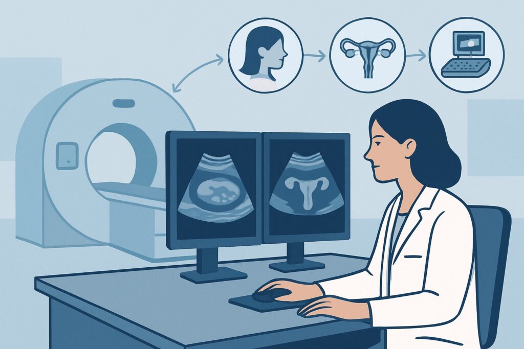
A 31-year-old woman presents to your clinic after one year of trying to conceive without success. She reports a history of irregular menstrual cycles, often skipping several months at a time, and notes mild hirsutism. Her clinical presentation raises suspicion for Polycystic Ovary Syndrome (PCOS) as the underlying cause of her infertility. You need to decide on the most appropriate initial imaging study to evaluate her pelvic anatomy, confirm ovarian morphology consistent with PCOS, and rule out other structural causes. This article details the clinical workflow for this specific scenario, explaining why the American College of Radiology (ACR) finds a transvaginal ultrasound to be `Usually appropriate` as the first imaging step.
Who Fits This Clinical Scenario?
This guidance applies to patients presenting with female infertility who also have clinical features or a known history suggestive of Polycystic Ovary Syndrome (PCOS). Infertility is typically defined as the inability to achieve pregnancy after 12 months of regular, unprotected intercourse (or 6 months for women over 35). The key feature anchoring this scenario is the concurrent suspicion of PCOS, based on signs of hyperandrogenism (e.g., hirsutism, severe acne) and/or a history of oligo-ovulation or anovulation (e.g., irregular menstrual cycles).
This workflow is distinct from other infertility workups. It is crucial to differentiate this presentation from nearby scenarios that require a different diagnostic approach:
- Suspicion of Tubal Occlusion: If the patient has a significant history of pelvic inflammatory disease (PID), prior pelvic surgery, or suspected tubal pathology, the primary question is different. That workup focuses on tubal patency, and the imaging pathway follows the ACR variant for suspected tubal occlusion.
- Known or Suspected Endometriosis: For a patient whose primary symptoms include significant dysmenorrhea, dyspareunia, and chronic pelvic pain suggestive of endometriosis, the imaging goals are to identify endometriomas and deep infiltrative disease. This follows a separate ACR workflow for suspected endometriosis.
- Recurrent Pregnancy Loss: If the patient has a history of two or more failed pregnancies, the investigation prioritizes uterine structural anomalies and other systemic causes, routing to the dedicated recurrent pregnancy loss variant.
What Diagnoses Are You Working Up in This Scenario?
When ordering initial imaging for infertility with suspected PCOS, the goal is to confirm specific morphologic features and exclude other structural abnormalities. The differential diagnosis guides the interpretation of the imaging findings.
Polycystic Ovary Syndrome (PCOS)
This is the primary diagnosis under consideration. PCOS is a diagnosis of exclusion based on the Rotterdam criteria, requiring two of the following three: oligo/anovulation, clinical or biochemical hyperandrogenism, and polycystic ovarian morphology on ultrasound. Imaging serves to establish that third criterion. The sonographic findings include an increased number of antral follicles (often described as a “string of pearls” appearance) and/or increased ovarian volume.
Diminished Ovarian Reserve
While evaluating for PCOS, the ultrasound provides a critical measure of ovarian reserve: the antral follicle count (AFC). A low AFC may suggest a diminished ovarian reserve, which is an important prognostic factor for fertility treatment success, independent of a PCOS diagnosis. This finding can significantly alter patient counseling and management, even if classic PCOS morphology is absent.
Other Anatomic Causes of Anovulation
Though less common, other ovarian pathologies can cause menstrual irregularities. The ultrasound helps exclude structural abnormalities such as benign or malignant ovarian neoplasms. Rarely, an androgen-secreting tumor (e.g., Sertoli-Leydig cell tumor) can mimic the hyperandrogenism seen in PCOS, and imaging is crucial for its detection.
Congenital or Acquired Uterine Abnormalities
The initial ultrasound also provides a detailed evaluation of the uterus. It can identify congenital Müllerian duct anomalies (such as a septate or bicornuate uterus) or acquired pathologies like endometrial polyps, submucosal fibroids, or adenomyosis. Any of these can impair implantation or contribute to infertility, and their identification is essential for a comprehensive workup.
Why Is Transvaginal Ultrasound the Recommended Study for This Presentation?
The ACR designates US pelvis transvaginal as `Usually appropriate` for the initial imaging of a patient with infertility and suspected PCOS. This recommendation is based on its high diagnostic accuracy, safety profile, and ability to answer the key clinical questions in this scenario.
A transvaginal ultrasound provides high-resolution images of the ovaries and uterus, which is essential for the detailed assessment required. It allows for precise measurement of ovarian volume and an accurate count of small (2-9 mm) antral follicles. According to current diagnostic criteria, the presence of 20 or more follicles per ovary and/or an ovarian volume greater than 10 mL is indicative of polycystic ovarian morphology. The transvaginal approach is superior to the transabdominal approach for this detailed evaluation.
Furthermore, this study involves no ionizing radiation (adult_rrl=O 0 mSv), a critical consideration in a patient population of reproductive age who may soon become pregnant. It is non-invasive, widely available, and cost-effective.
Why are other studies rated lower for this initial workup?
- US pelvis transabdominal is rated `May be appropriate`. While it can provide a general overview of the pelvis and is useful in patients who cannot undergo a transvaginal exam, its lower transducer frequency results in poorer spatial resolution. This makes it difficult to reliably count small antral follicles or assess ovarian stromal characteristics, limiting its utility for confirming PCOS morphology.
- MRI pelvis without IV contrast is also rated `May be appropriate`. MRI offers excellent soft tissue contrast and can be valuable for problem-solving. However, it is not the recommended initial study due to higher cost, longer examination time, and less accessibility compared to ultrasound. Its primary role is as a secondary test if the ultrasound is inconclusive or reveals a complex finding, such as a suspected congenital uterine anomaly or an indeterminate adnexal mass.
- MRI pelvis without and with IV contrast is rated `Usually not appropriate`. The addition of gadolinium-based contrast adds little to no diagnostic information for the primary questions of PCOS morphology or antral follicle counting and introduces the risks associated with contrast agents.
What’s Next After US pelvis transvaginal? Downstream Workflow
The results of the transvaginal ultrasound will guide the subsequent steps in the patient’s infertility management. The workflow branches based on whether the findings support a PCOS diagnosis, are normal, or reveal an unexpected abnormality.
If the study is positive for polycystic ovarian morphology:
When the ultrasound confirms PCO morphology (increased follicle count and/or volume) in a patient who already has oligo/anovulation or hyperandrogenism, the diagnosis of PCOS is solidified. The next steps are primarily clinical and biochemical, not radiological. Management will focus on lifestyle modifications (if applicable) and ovulation induction with agents like letrozole or clomiphene citrate. Further imaging is typically not needed unless treatment fails or other symptoms develop.
If the study is negative (normal ovarian morphology):
If the ultrasound shows normal ovaries and a normal uterus, the focus of the infertility investigation shifts away from an ovulatory disorder of the PCOS type. The next logical step is to evaluate for other causes of infertility. This often involves assessing tubal patency, which moves the patient into the ACR workflow for suspected tubal occlusion, where a hysterosalpingogram (HSG) is the next indicated study.
If the study is indeterminate or shows an unexpected finding:
If the ultrasound identifies a complex adnexal mass, a suspected congenital uterine anomaly that cannot be fully characterized (e.g., differentiating a septate from a bicornuate uterus), or extensive fibroids, further characterization is necessary. In these cases, an MRI pelvis without IV contrast, rated `May be appropriate`, becomes the best next step to clarify the anatomy before proceeding with treatment or potential surgery.
Pitfalls to Avoid (and When to Get Help)
Navigating the imaging workup for suspected PCOS-related infertility requires attention to several potential pitfalls to ensure an accurate diagnosis and appropriate management.
- Over-relying on imaging for diagnosis: Remember that polycystic ovarian morphology is just one of three potential criteria for PCOS. A patient can have polycystic-appearing ovaries and be perfectly fertile with regular cycles. The diagnosis requires a full clinical picture.
- Misinterpreting multifollicular ovaries (MFO): MFO can be seen in young women or those with hypothalamic amenorrhea and can be mistaken for PCOS. Unlike PCOS, MFO typically presents with normal ovarian volume and stromal echogenicity.
- Incorrect timing of the scan: The antral follicle count and endometrial assessment are most accurate when performed during the early follicular phase of the menstrual cycle (days 2-5). A scan performed mid-cycle may show a dominant follicle or corpus luteum, which can obscure the underlying morphology and alter ovarian volume measurements.
Escalation is warranted if the ultrasound reveals a suspicious adnexal mass with solid components, septations, or vascularity. In such cases, immediate referral to a gynecologic subspecialist is crucial.
Related ACR Topics and Tools
This article covers one specific scenario within the broader topic of female infertility. For a comprehensive overview of all related clinical variants, from tubal occlusion to recurrent pregnancy loss, please see our parent guide. The following GigHz tools can also support your clinical decision-making:
- Parent Topic Hub: For breadth across all scenarios in Female Infertility, see our parent guide: Female Infertility: ACR Appropriateness Decoded.
- Imaging Appropriateness Selector — for quickly finding guidance on adjacent or alternative clinical scenarios.
- Imaging Protocol Library — for detailed technical parameters of the recommended imaging studies.
- Radiation Dose Calculator — for discussing cumulative radiation exposure with patients when considering multiple studies.
Frequently Asked Questions
Does a normal transvaginal ultrasound rule out PCOS?
No. A normal ultrasound means the patient does not meet the ‘polycystic ovarian morphology’ criterion for PCOS. However, a diagnosis of PCOS can still be made if the patient meets the other two Rotterdam criteria: oligo- or anovulation and clinical or biochemical signs of hyperandrogenism.
Is a transabdominal ultrasound ever sufficient for this workup?
A transabdominal ultrasound is rated ‘May be appropriate’ and can be used if a patient cannot tolerate a transvaginal exam (e.g., is not yet sexually active). However, its resolution is significantly lower, making it less reliable for accurately counting small antral follicles and measuring ovarian volume, which are key to the diagnosis. It is considered a second-line option.
Why isn’t MRI the first choice if it provides more detailed images?
While MRI provides excellent anatomical detail, it is not the initial study of choice for suspected PCOS due to its higher cost, lower availability, and longer scan time compared to ultrasound. Transvaginal ultrasound provides sufficient information to diagnose polycystic ovarian morphology in the vast majority of cases. MRI is reserved for problem-solving when ultrasound findings are inconclusive or reveal a complex abnormality.
What is the difference between polycystic ovaries (PCO) and polycystic ovary syndrome (PCOS)?
Polycystic ovaries (PCO) is a sonographic finding describing the appearance of the ovaries (increased follicle number and/or volume). Polycystic ovary syndrome (PCOS) is a complex endocrine disorder and a clinical diagnosis. A person can have PCO on ultrasound without having the syndrome, and conversely, a person can have PCOS without having the classic ovarian morphology on ultrasound.
Should I order a Doppler ultrasound for this scenario?
US color Doppler of the pelvis is rated ‘May be appropriate’ by the ACR. While not essential for the initial diagnosis of PCOS morphology, it can be a useful adjunct performed at the time of the grayscale ultrasound to assess blood flow to the ovaries or to help characterize any unexpected adnexal mass by evaluating its vascularity.
Reviewed by Pouyan Golshani, MD, Interventional Radiologist — August 4, 2026