What Is the Best Initial Imaging for Suspected Hiatal Hernia? An ACR-Guided Workflow
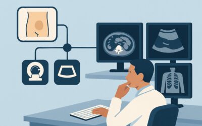
A 58-year-old patient presents to your clinic with several months of worsening epigastric pain, particularly after meals. He describes a burning sensation that rises into his chest, frequent regurgitation of food, and an uncomfortable feeling of fullness even after small meals. You suspect a hiatal hernia is contributing to, or is the primary cause of, his significant gastroesophageal reflux symptoms. The immediate clinical question is which imaging study will most effectively and safely confirm the diagnosis and guide the next steps in management. For this specific presentation, the American College of Radiology (ACR) Appropriateness Criteria rate a Fluoroscopy biphasic esophagram as Usually Appropriate for initial evaluation.
Who Fits This Clinical Scenario?
This guidance applies to adult patients presenting with epigastric pain accompanied by a constellation of symptoms strongly suggestive of a hiatal hernia. The key clinical indicators include:
- Chronic or worsening gastroesophageal reflux disease (GERD), often refractory to initial medical therapy.
- Postprandial fullness or early satiety.
- Regurgitation of undigested food.
- Intermittent dysphagia or a sensation of food “sticking” in the chest.
- Belching, bloating, or non-cardiac chest pain.
This workflow is specifically for the initial imaging workup where a structural cause like a hernia is high on the differential. It is crucial to distinguish this presentation from related but distinct clinical scenarios that require a different diagnostic approach:
- Exclusion 1: Predominant Dyspepsia or Ulcer Symptoms. If the patient’s symptoms are primarily burning epigastric pain without significant reflux, regurgitation, or dysphagia, the workup should align with the ACR variant for suspected peptic ulcer disease or gastritis, which may prioritize endoscopy over fluoroscopy.
- Exclusion 2: Alarm Features for Malignancy. Patients presenting with alarm features such as unintentional weight loss, iron deficiency anemia, progressive dysphagia, or odynophagia require a different, more urgent evaluation, often starting with endoscopy to rule out gastric or esophageal cancer.
What Diagnoses Are You Working Up in This Scenario?
When ordering imaging for suspected hiatal hernia, you are investigating a differential that centers on the anatomy and function of the esophagogastric junction. The primary goal is to identify a structural abnormality that explains the patient’s symptoms.
Hiatal Hernia
This is the principal diagnosis under consideration. A hiatal hernia occurs when the upper part of the stomach bulges through the diaphragm into the chest. The most common type is a sliding hernia (Type I), where the gastroesophageal junction itself slides above the diaphragm. Less common but more clinically significant are paraesophageal hernias (Types II-IV), where the junction remains in place but a portion of the gastric fundus herniates alongside the esophagus. These can lead to complications like incarceration or volvulus.
Gastroesophageal Reflux Disease (GERD)
While a clinical diagnosis, imaging can provide objective evidence of reflux and its anatomical substrate. A hiatal hernia is a major risk factor for GERD because it impairs the function of the lower esophageal sphincter (LES). An esophagram can directly visualize the retrograde flow of barium from the stomach into the esophagus, confirming reflux.
Esophageal Motility Disorder
Symptoms like dysphagia and regurgitation can overlap with primary motility disorders such as achalasia or diffuse esophageal spasm. A fluoroscopic esophagram is an excellent initial test to evaluate esophageal peristalsis and emptying, potentially revealing these less common but important diagnoses.
Esophageal Stricture or Web
Chronic, severe reflux can lead to inflammation (esophagitis) and subsequent scarring, resulting in a benign peptic stricture. These structural abnormalities can be a direct cause of dysphagia and are well-visualized on an esophagram, which can define their location and severity.
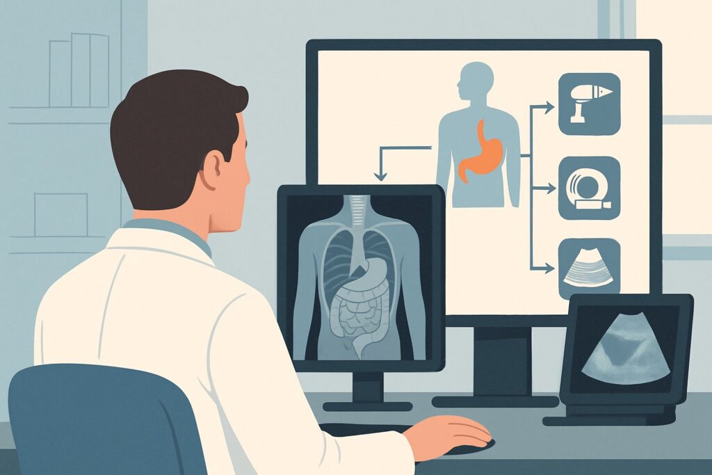
Why Is a Fluoroscopy Biphasic Esophagram the Recommended Study for Suspected Hiatal Hernia?
The ACR designates a fluoroscopy biphasic esophagram as Usually Appropriate because it uniquely provides both anatomical and functional information in a single, dynamic examination. This is the key advantage for this clinical scenario. The study involves the patient swallowing barium contrast of different consistencies while the radiologist observes its transit in real-time using fluoroscopy.
The biphasic technique—using both high-density barium (double-contrast) to coat the mucosa and low-density barium (single-contrast) to distend the lumen—is highly effective for this workup. The double-contrast phase provides exquisite detail of the mucosal lining to detect subtle inflammation, ulcers, or webs. The single-contrast phase is superior for evaluating motility and outlining the anatomy of a hernia.
This dynamic assessment allows the radiologist to employ provocative maneuvers, such as placing the patient in a Trendelenburg position or having them perform a Valsalva maneuver. These actions increase intra-abdominal pressure and are crucial for demonstrating an intermittent sliding hiatal hernia or eliciting gastroesophageal reflux that may not be present at rest.
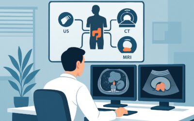
Why Alternatives Are Rated Lower
- CT Abdomen with IV Contrast: This study is rated Usually not appropriate. While a large hiatal hernia may be incidentally seen on a CT scan, it is a static imaging modality. It cannot assess esophageal motility, evaluate for reflux, or provide the same level of mucosal detail as an esophagram. Furthermore, it delivers a higher radiation dose (ACR relative radiation level ☢☢☢ 1-10 mSv) without answering the primary functional questions.
- MRI Abdomen: All MRI variants are rated Usually not appropriate. MRI offers no radiation exposure (ACR RRL of O), but it has poor temporal resolution for evaluating the dynamic process of swallowing and peristalsis. It is more expensive, less available, and provides no diagnostic advantage over fluoroscopy for assessing hiatal hernia or reflux.
The recommended esophagram involves a moderate radiation dose (ACR RRL ☢☢☢ 1-10 mSv), which is considered a reasonable trade-off for the high diagnostic yield in this specific clinical context.
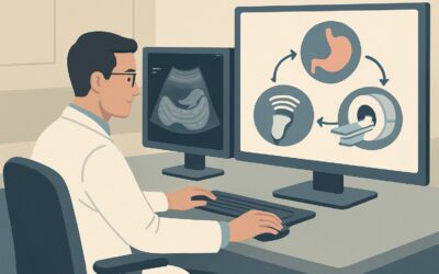
What’s Next After the Esophagram? Downstream Workflow
The results of the fluoroscopy biphasic esophagram will directly inform your next steps. The post-imaging workflow depends on whether the findings confirm the clinical suspicion, are negative, or reveal an alternative diagnosis.
If the study is positive for a hiatal hernia:
The management depends on the type and size of the hernia and the severity of symptoms. For a small, sliding hiatal hernia with reflux, the next step is typically medical management with proton pump inhibitors (PPIs) and lifestyle modifications. For a large hernia or any paraesophageal hernia, referral to a gastroenterologist and a thoracic or general surgeon is warranted. Further workup often includes upper endoscopy (EGD) to assess for esophagitis and Barrett’s esophagus, followed by esophageal manometry and pH testing to quantify reflux before considering surgical repair.
If the study is negative for a hernia but shows significant reflux:
The diagnosis is GERD. The workflow proceeds with medical management. If symptoms are refractory to high-dose PPIs, referral to gastroenterology for EGD and further functional testing is appropriate.
If the study is entirely negative:
If no hernia, reflux, or motility disorder is identified, the patient’s epigastric pain is likely due to another cause. The workup should be redirected. This may involve investigating for peptic ulcer disease or gastritis (often with EGD) or considering non-GI causes of epigastric pain, such as pancreatic or biliary pathology.
If the study suggests a motility disorder:
Findings like a “bird’s beak” appearance in achalasia or uncoordinated contractions (“corkscrew esophagus”) point toward a primary motility disorder. The essential next step is a referral to a gastroenterologist for high-resolution esophageal manometry, which is the gold standard for diagnosing these conditions.
Pitfalls to Avoid (and When to Get Help)
Navigating the workup for suspected hiatal hernia requires avoiding several common pitfalls to ensure an accurate and efficient diagnosis.
- Not Providing Clinical Context: Failing to specify “rule out hiatal hernia and reflux” on the imaging order can lead to a generic study without the necessary provocative maneuvers, potentially missing an intermittent hernia.
- Stopping at a Negative Study: A negative esophagram in a patient with persistent, classic reflux symptoms does not rule out GERD. The test has limited sensitivity for reflux, and the patient may still require empiric medical therapy or further testing like 24-hour pH monitoring.
- Over-relying on CT: Do not order a CT scan as the initial test for this specific question. It provides the wrong type of information (static anatomy vs. dynamic function) at a higher radiation cost.
- Ignoring Paraesophageal Hernias: If the report describes a paraesophageal hernia, this is a significant finding that requires prompt surgical consultation, even if symptoms seem manageable, due to the risk of strangulation and volvulus.
If a patient presents with acute, severe chest or epigastric pain, vomiting, and an inability to pass a nasogastric tube in the known context of a large hiatal hernia, escalate immediately for surgical evaluation to rule out acute gastric volvulus.
Related ACR Topics and Tools
For a comprehensive overview of imaging for all types of epigastric pain, or to explore the tools used to develop this workflow, the following resources are available:
- Parent Topic Hub Article: For breadth across all scenarios in Epigastric Pain, see our parent guide: Epigastric Pain: ACR Appropriateness Decoded.
- ACR Appropriateness Criteria Lookup — for adjacent scenarios
- Imaging Protocol Library — for technique on the recommended study
- Radiation Dose Calculator — for cumulative dose conversations
Frequently Asked Questions
Why is an esophagram preferred over an upper endoscopy (EGD) for the initial diagnosis of a hiatal hernia?
While an EGD is excellent for evaluating the mucosa for esophagitis or Barrett’s esophagus, it can miss or underestimate the size of a hiatal hernia, especially a sliding hernia that reduces when the esophagus is insufflated. The fluoroscopic esophagram is a dynamic study that uses gravity and provocative maneuvers to distend the esophagogastric junction fully, making it the superior initial test for defining the anatomy of a suspected hernia.
Can I just order a ‘barium swallow’?
It is better to be specific. A ‘barium swallow’ can sometimes be interpreted as a limited, pharyngeal-focused study. Ordering a ‘fluoroscopy biphasic esophagram’ or ‘upper GI series’ (which includes the stomach and duodenum) and stating the clinical indication (e.g., ‘epigastric pain, rule out hiatal hernia and reflux’) ensures the radiologist performs the comprehensive examination needed to answer the clinical question.
What if my patient is pregnant and has symptoms of a hiatal hernia?
In a pregnant patient, non-ionizing radiation studies are strongly preferred. While an esophagram is relatively low dose, it should be avoided if possible. The first step would be empiric medical management. If imaging is absolutely necessary due to severe, refractory symptoms, an upper endoscopy would be a safer alternative to a fluoroscopic study. Consultation with a radiologist and gastroenterologist is recommended.
Does the esophagram show other causes of epigastric pain, like gallstones or pancreatitis?
No. The esophagram is a focused examination of the esophagus, stomach, and sometimes the duodenum. It provides no information about the gallbladder, pancreas, liver, or other solid organs. If your clinical suspicion for these conditions is high, a different imaging modality, such as an abdominal ultrasound or CT scan, would be appropriate.
Is there any special preparation required for a fluoroscopy biphasic esophagram?
Yes, the patient is typically required to be NPO (nothing by mouth) for several hours before the study, usually 6 to 8 hours. This ensures the stomach is empty, allowing for optimal mucosal coating with the barium and preventing food from obscuring the anatomy or being mistaken for pathology.
Reviewed by Pouyan Golshani, MD, Interventional Radiologist — May 29, 2026