What Is the Best Initial Imaging for Suspected Pediatric Cerebral Venous Thrombosis?
A 9-year-old with a recent history of severe sinusitis presents to the emergency department with a thunderclap headache, vomiting, and a new-onset focal seizure. You are concerned for an intracranial complication, and your leading differential diagnosis is cerebral venous sinus thrombosis (CVST). The child is now postictal but stable. You need to decide on the most appropriate initial imaging study to confirm or exclude this time-sensitive diagnosis. This is a critical decision point where the right study provides a definitive answer, while a suboptimal choice could lead to diagnostic delay.
This article provides a detailed workflow for this specific clinical scenario, based on the American College of Radiology (ACR) Appropriateness Criteria. For a child with a clinical presentation suggestive of acute stroke due to suspected cortical vein or dural venous sinus thrombosis, the ACR rates MRI head without and with IV contrast as Usually Appropriate.
Who Fits This Clinical Scenario?
This guidance applies specifically to a pediatric patient presenting with signs and symptoms suggestive of an acute stroke where the underlying suspicion is for thrombosis of the cerebral venous system, not the arterial system. This includes thrombosis of the major dural venous sinuses (e.g., superior sagittal, transverse, sigmoid) or the smaller cortical veins.
Presentations may include severe headache, seizures (often focal), focal neurologic deficits that do not conform to a classic arterial territory, papilledema, or altered mental status. Risk factors in children often include prothrombotic states, severe dehydration, systemic infection or malignancy, and local infections like mastoiditis or sinusitis.
This workflow is distinct from other pediatric stroke scenarios. Exclude patients from this pathway if:
- The presentation is classic for an acute arterial ischemic stroke in a child older than 6 months, where emergent intervention might be considered. That scenario involves a different imaging algorithm focused on identifying large vessel occlusion.
- There is a known or suspected arteriopathy, such as moyamoya disease or arterial dissection. These conditions require dedicated arterial imaging, often with MR or CT angiography of the head and neck.
- The primary finding is a nontraumatic intraparenchymal hemorrhage of unknown etiology. While venous thrombosis can cause hemorrhagic infarcts, the workup for an unexplained primary bleed follows a separate diagnostic path.
What Diagnoses Are You Working Up in This Scenario?
When ordering imaging for suspected pediatric CVST, you are evaluating for a specific set of primary and secondary pathologies. The differential diagnosis guides the choice of imaging modality and protocol.
Cerebral Venous Sinus Thrombosis (CVST)
This is the primary diagnosis of concern. CVST is a rare but serious cause of stroke in children. It involves the formation of a blood clot in the dural venous sinuses, which drain blood from the brain. This obstruction increases venous and capillary pressure, which can lead to cerebral edema and parenchymal injury. Imaging aims to directly visualize the thrombus as a filling defect or abnormal signal within a sinus or cortical vein.
Venous Infarction and Edema
A direct consequence of CVST is venous congestion leading to vasogenic edema and, if severe, cytotoxic edema and infarction. Unlike arterial infarcts, venous infarcts do not typically respect arterial territories. They are often parasagittal or temporal and can be hemorrhagic due to venous hypertension and capillary rupture. Imaging must be sensitive to these parenchymal changes, which are often the first clue to the underlying venous pathology.
Meningitis or Encephalitis
Infectious or inflammatory processes can mimic the symptoms of CVST, causing headache, seizures, and altered consciousness. Furthermore, intracranial or adjacent infections (e.g., mastoiditis, orbital cellulitis) are significant risk factors for developing secondary septic CVST. Imaging helps differentiate primary infection from venous thrombosis and can identify both conditions if they coexist.
Idiopathic Intracranial Hypertension (IIH)
Also known as pseudotumor cerebri, IIH presents with headaches, papilledema, and vision changes due to elevated intracranial pressure without an identifiable cause. However, CVST is a well-known secondary cause of intracranial hypertension. Therefore, imaging in any new diagnosis of suspected IIH must first exclude an underlying venous sinus stenosis or thrombosis as the root cause.
Why Is MRI Head Without and With IV Contrast the Recommended Study?
The ACR designates MRI head without and with IV contrast as Usually Appropriate because it provides the most comprehensive evaluation for both the primary vascular event and its secondary parenchymal consequences, all without using ionizing radiation.
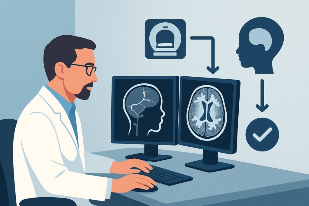
An optimal MRI protocol is a multi-tool examination. Non-contrast sequences like Diffusion-Weighted Imaging (DWI) are highly sensitive for detecting acute cytotoxic edema from venous infarction. Fluid-Attenuated Inversion Recovery (FLAIR) sequences excel at identifying vasogenic edema. Susceptibility-Weighted Imaging (SWI) or Gradient-Echo (GRE) sequences are exquisitely sensitive for detecting hemorrhage associated with venous infarcts. These sequences provide critical information about the brain tissue itself.
The addition of dedicated MR venography (MRV) sequences, either without contrast (time-of-flight) or with contrast, directly visualizes blood flow within the dural sinuses. A thrombosed segment will appear as an absence of flow signal. Post-contrast T1-weighted images can show enhancement of the dura surrounding a thrombus (the “empty delta sign”) or abnormal enhancement of congested veins.
Comparison to Alternative Studies
- CT head without IV contrast: While also rated Usually Appropriate and often more readily available, non-contrast CT is less sensitive than MRI. It may show a hyperdense (“dense cord”) sign in an acutely thrombosed sinus or cortical vein, but this finding is often absent. It can detect large hemorrhagic infarcts but may miss non-hemorrhagic infarcts or subtle edema. It is a reasonable first step in an unstable patient to quickly rule out a large hemorrhage but is not sufficient to rule out CVST.
- CTV head with IV contrast: This study is also Usually Appropriate and provides excellent visualization of filling defects within the dural sinuses. However, it delivers a significant radiation dose (Pediatric RRL: ☢☢☢☢ 3-10 mSv) and offers less detailed information about the brain parenchyma compared to the multiple sequences of an MRI. It is a strong alternative when MRI is contraindicated or unavailable.
The primary advantage of MRI is its superior soft tissue contrast and its ability to characterize tissue pathology in multiple ways, all with a pediatric radiation dose of 0 mSv. This combination makes it the definitive initial study for this clinical question.
What’s Next After MRI? Downstream Workflow
The results of the MRI will guide your immediate next steps and downstream management. The workflow branches based on whether the findings are positive, negative, or indeterminate for venous thrombosis.
If the study is positive for CVST:
A confirmed diagnosis of cerebral venous sinus thrombosis is a neurologic emergency. The immediate next steps involve a multi-disciplinary approach. You should urgently consult pediatric neurology and pediatric hematology. The primary treatment is typically anticoagulation to prevent thrombus propagation and promote recanalization. The patient will require admission for initiation of therapy, neurologic monitoring, and a diagnostic workup to identify any underlying prothrombotic disorders.
If the study is negative for CVST:
If the MRI and MRV are entirely normal, CVST is effectively ruled out. The clinical focus must return to the other items on the differential. If symptoms persist, particularly headache and papilledema, a lumbar puncture may be indicated to measure opening pressure (for IIH) and analyze cerebrospinal fluid (for meningitis/encephalitis). If focal deficits persist without an imaging correlate, the differential may broaden to include complex migraine, postictal state (Todd’s paralysis), or metabolic derangements.
If the study is indeterminate or shows other pathology:
Occasionally, MRI findings can be ambiguous (e.g., a hypoplastic sinus mimicking thrombosis). In these cases, CTV can be a valuable problem-solving tool. If the MRI reveals an unexpected finding, such as evidence of vasculitis or a tumor, the diagnostic pathway shifts entirely, often requiring further specialized imaging or biopsy and consultation with the appropriate subspecialists (e.g., rheumatology, neuro-oncology).
Pitfalls to Avoid (and When to Get Help)
Navigating a suspected pediatric CVST case requires vigilance to avoid common diagnostic traps. Be mindful of these potential pitfalls:
- Relying on a negative non-contrast CT: A normal non-contrast head CT does not exclude CVST. If clinical suspicion remains high, you must proceed to definitive venous imaging with MRV or CTV.
- Mistaking anatomic variants for pathology: Asymmetric or hypoplastic transverse sinuses are common anatomic variants that can be mistaken for thrombosis on MRV. Correlating with post-contrast images and parenchymal findings is key.
- Delaying the diagnosis: The symptoms of CVST can be nonspecific. Maintain a high index of suspicion in children with risk factors who present with severe headache, seizures, or unexplained neurologic deficits.
- Incomplete imaging protocol: When ordering the MRI, explicitly request a dedicated MR venogram (MRV) sequence. A standard brain MRI protocol may not be sufficient to confidently assess the venous sinuses.
If the diagnosis of CVST is confirmed or strongly suspected, escalate care immediately by consulting pediatric neurology and hematology for guidance on anticoagulation and further management.
Related ACR Topics and Tools
The ACR Appropriateness Criteria are a powerful resource for evidence-based imaging decisions. For a comprehensive overview of all pediatric stroke scenarios, this article is best used alongside its parent topic guide. The following tools can help you apply these guidelines in your daily practice.
- For breadth across all scenarios in Cerebrovascular Disease-Child, see our parent guide: Cerebrovascular Disease-Child: ACR Appropriateness Decoded.
- To explore adjacent clinical scenarios and their corresponding imaging recommendations, use the ACR Appropriateness Criteria Lookup.
- For detailed technical specifications on the recommended study, consult the Imaging Protocol Library.
- To discuss cumulative radiation exposure with families when considering CT, the Radiation Dose Calculator can be a useful aid.
Frequently Asked Questions
Why is MRI preferred over CT/CTV for suspected pediatric CVST?
MRI is preferred primarily because it involves no ionizing radiation (0 mSv), which is a critical consideration in children. Additionally, it provides superior detail of the brain parenchyma, allowing for better detection of subtle edema, non-hemorrhagic venous infarcts, and other soft tissue abnormalities that can be missed on CT. While CTV is excellent for visualizing the sinuses, MRI offers a more comprehensive diagnostic picture of both the vessels and the brain tissue.
Is intravenous contrast always necessary for an MRV to diagnose CVST?
Not always, but it is highly recommended. MR venography can be performed without contrast using a technique called 2D time-of-flight (TOF). However, TOF MRV is prone to artifacts, especially in areas of slow or complex flow, which can mimic thrombosis. Contrast-enhanced MRV is generally faster, less prone to these artifacts, and provides a more confident and definitive assessment of the venous sinuses. The combination of pre- and post-contrast imaging provides the most diagnostic information.
What should I order if my patient has a contraindication to MRI?
If a child has a contraindication to MRI (e.g., an incompatible implanted medical device, extreme claustrophobia, or instability precluding a longer scan), the best alternative is a CT head without contrast followed by a CTV head with IV contrast. This combination allows for assessment of hemorrhage and provides direct visualization of filling defects in the dural venous sinuses. It is a highly effective alternative, with the main drawback being the radiation exposure.
How urgently should this imaging be performed?
A workup for suspected CVST should be considered urgent. While it may not always require the same minute-by-minute speed as an arterial stroke workup for thrombectomy, a delay in diagnosis can lead to significant morbidity. The imaging should be performed as soon as is feasible, typically within hours of presentation, to allow for prompt initiation of anticoagulation therapy if the diagnosis is confirmed.
What specific MRI sequences are most important for this diagnosis?
A complete protocol should include multi-planar T1- and T2-weighted images, FLAIR (for edema), DWI (for acute infarct), and a susceptibility-weighted sequence like SWI or GRE (for hemorrhage). Crucially, it must also include a dedicated MR venography (MRV) sequence, either 2D TOF or contrast-enhanced 3D, as well as post-contrast T1-weighted images to assess for abnormal enhancement.
Reviewed by Pouyan Golshani, MD, Interventional Radiologist — June 27, 2026