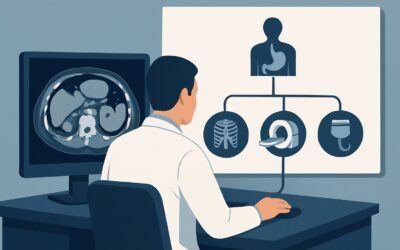
What Is the Best Initial Imaging for Undifferentiated Left Lower Quadrant Pain?

A 58-year-old male presents to the emergency department on a Tuesday evening with two days of worsening, steady left lower quadrant (LLQ) pain, low-grade fever, and mild nausea. His physical exam reveals focal tenderness in the LLQ without rebound or guarding. His white blood cell count is moderately elevated. While acute diverticulitis is high on the differential, other etiologies like colitis, renal colic, or a less common inflammatory process remain possibilities. You need to select the single best initial imaging study to confirm the diagnosis, assess for complications, and guide immediate management. This article details the clinical workflow for this exact scenario, where the American College of Radiology (ACR) has determined that a CT abdomen and pelvis with IV contrast is Usually appropriate.
## Who Fits This Clinical Scenario?
This guidance applies to adult patients presenting with new-onset, undifferentiated left lower quadrant pain requiring initial imaging. The key feature is the lack of a clear, pre-existing diagnosis or a highly specific clinical suspicion that would narrow the differential significantly. These are patients for whom you need a comprehensive first look to distinguish between several potential causes, both common and rare.
This workflow is specifically for the initial imaging step. It is not intended for:
- Patients with suspected uncomplicated diverticulitis: A patient with a known history of diverticulitis presenting with a typical, mild flare may not require imaging, or may fall under the more specific ACR variant for that condition. This scenario is for when the diagnosis is still in question.
- Patients with suspected complications of diverticulitis: If the patient presents with signs of sepsis, peritonitis (rebound, guarding), or a palpable mass suggesting an abscess, they fit a different, more urgent clinical scenario focused on identifying complications like perforation or fistula.
- Pregnant patients or young children: Imaging decisions in these populations require special consideration for radiation dose and alternative modalities, often prioritizing ultrasound or MRI.
The goal here is to provide a diagnostic roadmap when the clinical picture is suggestive but not definitive, requiring a robust imaging modality to clarify the underlying pathology.
## What Diagnoses Are You Working Up in This Scenario?
When ordering initial imaging for undifferentiated LLQ pain, you are evaluating a broad differential diagnosis. The chosen study must be capable of identifying or excluding several distinct pathologies located in this crowded anatomical region.
The most common cause is acute diverticulitis. This is an inflammation of diverticula, small pouches that can form in the wall of the colon, most often the sigmoid colon. Imaging is crucial to confirm inflammation (e.g., colonic wall thickening, surrounding fat stranding) and, importantly, to rule out complications like abscess formation or perforation, which dramatically alter management.
Another significant consideration is colitis, which can be infectious, ischemic, or inflammatory (e.g., a flare of inflammatory bowel disease). While clinically similar to diverticulitis, the imaging findings and underlying causes differ. CT can reveal the characteristic wall thickening and enhancement patterns that help distinguish between these conditions.
In both male and female patients, a left-sided ureteral stone can cause renal colic that refers pain to the LLQ. While a non-contrast CT is the gold standard for stone detection, a contrast-enhanced study can still identify most stones while also evaluating for secondary signs like hydronephrosis and providing information on other non-urologic causes.
In female patients, gynecologic and adnexal pathology is a critical part of the differential. This includes ovarian torsion, a ruptured ovarian cyst, pelvic inflammatory disease (PID), or a tubo-ovarian abscess. These conditions can mimic gastrointestinal pain, and a comprehensive imaging study of the pelvis is essential.
Less common but consequential diagnoses include epiploic appendagitis or omental infarction, which are self-limiting inflammatory conditions of mesenteric fat that can precisely mimic diverticulitis. CT is excellent at identifying these mimics, preventing unnecessary antibiotic treatment or hospital admission.
## Why Is CT Abdomen and Pelvis with IV Contrast the Recommended Study for This Presentation?
The ACR rates CT abdomen and pelvis with IV contrast as Usually appropriate because it offers the highest diagnostic yield across the wide range of potential causes for undifferentiated LLQ pain. Its excellent spatial resolution and tissue contrast provide a comprehensive evaluation of the colon, solid organs, vasculature, and pelvic structures in a single, rapid acquisition.
The use of intravenous (IV) contrast is key. It enhances the bowel wall, allowing radiologists to identify and characterize inflammation, ischemia, or masses. It opacifies blood vessels to rule out vascular causes and highlights abscess walls, making them easier to detect. This capability is what makes it superior to a non-contrast study for this particular clinical question.
Let’s compare this to other modalities rated for this scenario:
- CT abdomen and pelvis without IV contrast: This study is rated May be appropriate. While it is the ideal test for detecting kidney stones and can identify free air from a perforation, it is significantly less sensitive for detecting early or subtle inflammation, differentiating phlegmon from abscess, or evaluating for ischemic colitis. You risk missing the diagnosis if you omit contrast.
- Ultrasound (US abdomen transabdominal / US pelvis transvaginal): Both are rated May be appropriate. Ultrasound is valuable in young or pregnant patients to avoid radiation and is excellent for evaluating gynecologic pathology or the kidneys. However, its utility for assessing the sigmoid colon is often severely limited by overlying bowel gas and patient body habitus, making it an unreliable first-line test for suspected diverticulitis in the general adult population.
The primary trade-off with CT is the use of ionizing radiation. This study carries a relative radiation level of ☢☢☢ (1-10 mSv), a moderate dose that is justified by its high diagnostic accuracy in an acute setting where a missed diagnosis could lead to significant morbidity. The clinical benefit of obtaining a definitive diagnosis quickly and accurately is deemed to outweigh the radiation risk in most adult patients.
Once you’ve decided on CT abdomen and pelvis with IV contrast, our protocol guide covers the technique, contrast, and reading principles: CT Chest/Abdomen/Pelvis with IV Contrast.
## What’s Next After CT Abdomen and Pelvis with IV Contrast? Downstream Workflow
The radiologist’s report will guide your next steps, branching your workflow based on the findings.
- If the study is positive for uncomplicated diverticulitis: The CT will show sigmoid colonic wall thickening and pericolic fat stranding without abscess, fistula, or free air. This confirms the diagnosis and allows for confident outpatient management with oral antibiotics (or observation in select cases) and follow-up.
- If the study is positive for complicated diverticulitis: Findings like a contained fluid collection (abscess), extraluminal air (perforation), or fistulous tracts necessitate a change in management. This typically requires hospital admission, IV antibiotics, and consultation with general surgery and/or interventional radiology for potential percutaneous drain placement. The patient’s clinical scenario now shifts to the “Suspected complication(s) of diverticulitis” workflow.
- If the study is negative or normal: A normal CT scan effectively rules out most acute intra-abdominal pathologies. The next step is to reconsider the clinical picture. Could this be a non-intra-abdominal cause of pain (e.g., musculoskeletal, shingles)? Does the patient need further evaluation with a different modality (e.g., colonoscopy for subacute symptoms) or simply observation?
- If the study identifies an alternative diagnosis: The workflow follows the specific finding. A ureteral stone prompts urology consultation. An ovarian cyst rupture may require only pain management, while ovarian torsion is a surgical emergency. Epiploic appendagitis is managed conservatively with anti-inflammatory medication. The CT provides the clarity needed to route the patient to the correct care pathway.
## Pitfalls to Avoid (and When to Get Help)
When managing a patient with undifferentiated LLQ pain, be mindful of these common pitfalls:
- Omitting IV contrast: Ordering a non-contrast CT to “save time” or avoid a creatinine check can lead to a non-diagnostic study for inflammatory or ischemic conditions, potentially delaying the correct diagnosis.
- Anchoring on diverticulitis: While it is the most common cause, do not disregard other possibilities, especially gynecologic emergencies in female patients of reproductive age.
- Ignoring the appendix: Although appendicitis typically causes right-sided pain, a long, redundant appendix can occasionally present with LLQ pain. Ensure the entire appendix is visualized and normal.
- Misinterpreting the report: Understand the difference between a phlegmon (inflammation) and a drainable abscess, as their management is different.
If the patient shows signs of hemodynamic instability, peritonitis, or sepsis, escalate immediately to a surgical consultation, often in parallel with obtaining the CT scan.
## Related ACR Topics and Tools
This article covers one specific scenario within the broader topic of Left Lower Quadrant Pain. For a comprehensive overview of all related variants and their recommended imaging pathways, consult our parent guide. The tools below can help you apply these criteria in your daily practice.
- For breadth across all scenarios in Left Lower Quadrant Pain, see our parent guide: Left Lower Quadrant Pain: ACR Appropriateness Decoded.
- To explore other clinical scenarios, use the ACR Appropriateness Criteria Lookup.
- For detailed procedural techniques, browse the Imaging Protocol Library.
- To discuss radiation exposure with patients, use the Radiation Dose Calculator.
Frequently Asked Questions
Is oral contrast necessary for a CT scan for left lower quadrant pain?
In the acute setting, oral contrast is generally not required and can delay diagnosis. Modern multidetector CT scanners provide excellent bowel wall visualization with IV contrast alone. Oral contrast is typically reserved for more specific, non-emergent indications like evaluating for a subtle fistula or complex abscess.
What if my patient has a contrast allergy or renal insufficiency?
If a patient has a severe allergy to iodinated contrast, an alternative modality like MRI may be considered, though it is less available and takes longer. For mild allergies, premedication with steroids and antihistamines is an option. For renal insufficiency, the decision to use IV contrast depends on the severity of the kidney disease and the urgency of the clinical question, often involving a risk-benefit discussion and hydration protocols. A CT without contrast may be performed if the risk is too high, accepting its diagnostic limitations.
Can I start with an abdominal X-ray instead of a CT?
Radiography (X-ray) of the abdomen and pelvis is rated as *May be appropriate* by the ACR. While it can identify free air from a bowel perforation or signs of a high-grade bowel obstruction, it has very low sensitivity for most causes of LLQ pain, including diverticulitis and colitis. In most cases, it is a low-yield study that should be skipped in favor of the more definitive cross-sectional imaging provided by CT.
In a young female patient, should I order an ultrasound first?
Yes, in a young female patient, starting with a pelvic ultrasound (transabdominal and transvaginal) is often a very reasonable approach. It is rated *May be appropriate* and is excellent for evaluating for gynecologic causes of pain (like ovarian cysts, torsion, or PID) without using radiation. If the ultrasound is negative or non-diagnostic and clinical suspicion for a gastrointestinal cause remains high, you would then proceed to CT.
How does this guidance change if I strongly suspect diverticulitis based on history and exam?
If you have a high pre-test probability for uncomplicated diverticulitis (e.g., a patient with a known history and a typical flare), you may be in a different clinical scenario. The ACR has a separate variant for “Left lower quadrant pain. Suspected diverticulitis.” In some mild, recurrent cases, imaging may not be necessary at all. However, for a first presentation or if there is any concern for complication, CT remains the standard to confirm the diagnosis and rule out more serious pathology.
Reviewed by Pouyan Golshani, MD, Interventional Radiologist — May 30, 2026