What Is the Best Initial Imaging for Unilateral Isolated Tongue Weakness (CN XII Palsy)?
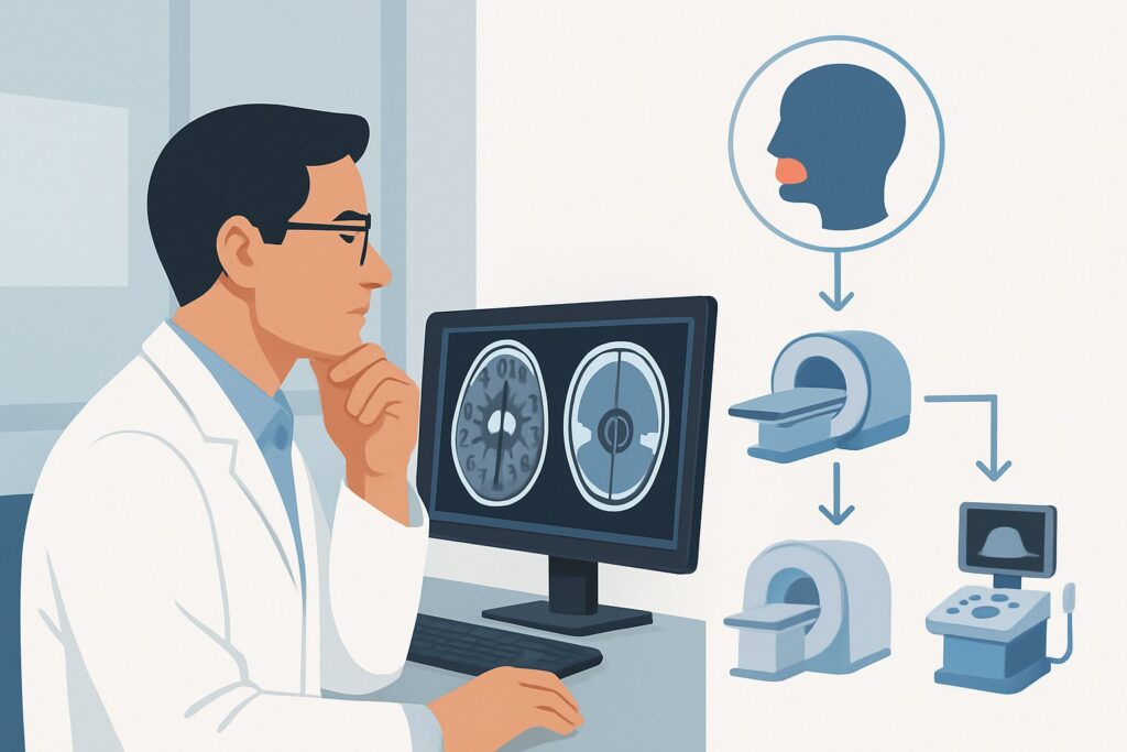
A 62-year-old man presents to your neurology clinic with a two-month history of slurred speech and difficulty moving food in his mouth. On examination, you observe his tongue deviates to the right upon protrusion, with visible atrophy and fasciculations on the right side. The remainder of his cranial nerve and neurologic exam is unremarkable. This isolated finding points toward a right hypoglossal nerve palsy, but the critical question is what lies along the nerve’s path causing the dysfunction. This article provides a focused, evidence-based workflow for the initial imaging workup of isolated unilateral hypoglossal nerve (cranial nerve XII) paralysis. According to the American College of Radiology (ACR) Appropriateness Criteria, the definitive first step is an MRI head without and with IV contrast, a study rated “Usually Appropriate” for this specific clinical scenario.
Who Fits This Clinical Scenario for Isolated Tongue Weakness?
This imaging guidance is intended for patients presenting with new-onset, unilateral signs of hypoglossal nerve dysfunction. The key clinical features include weakness, paralysis, atrophy, or fasciculations of one side of the tongue. A classic examination finding is deviation of the tongue toward the side of the lesion when the patient attempts to protrude it. The crucial qualifier for this specific workflow is that the finding must be isolated. A thorough neurologic examination should reveal no other cranial nerve deficits or long-tract signs.
This pathway is not appropriate for patients with more complex presentations. If the patient’s tongue weakness is accompanied by other neurologic signs, a different diagnostic algorithm is required. Key exclusions include:
- Associated Palatal or Vocal Cord Paralysis: If the patient also has a hoarse voice, dysphagia, or an asymmetric soft palate, this suggests involvement of the vagus nerve (CN X) and points to a different ACR scenario for combined lower cranial neuropathies.
- Concurrent Facial Weakness: Weakness of the muscles of facial expression indicates a facial nerve (CN VII) deficit, which has its own distinct imaging workup.
- Multiple Cranial Neuropathies: The presence of deficits involving several cranial nerves (e.g., CN V, VI, and VII) suggests a broader process at the skull base, cavernous sinus, or brainstem, requiring a more extensive evaluation than that for an isolated CN XII palsy.
–
This guidance also assumes the absence of acute, high-energy trauma to the head or neck, which would trigger a trauma-specific imaging protocol.
What Diagnoses Are You Working Up with Imaging for Hypoglossal Nerve Palsy?
An isolated hypoglossal nerve palsy is an alarming clinical sign that requires a diligent search for an underlying structural cause. The nerve has a long and complex course, originating in the medulla, exiting the skull through the hypoglossal canal, and traveling through the neck to innervate the tongue. A lesion anywhere along this path can produce symptoms. Imaging is essential to evaluate for several diagnostic possibilities.
The most pressing concern is often malignancy. Head and neck squamous cell carcinoma is a primary consideration, as tumors of the oropharynx, nasopharynx, or tonsil can directly invade or compress the nerve. Metastatic disease, particularly to the skull base or cervical lymph nodes, is another important cause. Primary tumors of the skull base, such as chordomas, chondrosarcomas, or meningiomas near the hypoglossal canal, can also present with this finding.
Vascular pathology is a less common but critical differential. Spontaneous dissection of the internal carotid artery, which runs in close proximity to the nerve in the upper neck, can cause a compressive or ischemic neuropathy. While often associated with pain or other neurologic symptoms (Horner syndrome), it can rarely present as an isolated CN XII palsy. Other vascular lesions like aneurysms or dural arteriovenous fistulas are also on the differential.
Inflammatory and infectious processes must also be considered. Skull base osteomyelitis, particularly in diabetic or immunocompromised patients, can affect the nerve as it exits the cranium. Systemic inflammatory conditions like sarcoidosis can cause cranial neuropathies, though an isolated presentation is less typical. Rarely, a primary nerve sheath tumor like a hypoglossal schwannoma can arise directly from the nerve itself.
Why Is MRI of the Head with and without Contrast the Recommended First Study?
The American College of Radiology rates MRI head without and with IV contrast as “Usually Appropriate” for this presentation because it provides the most comprehensive evaluation of the hypoglossal nerve’s entire course. The superior soft-tissue resolution of MRI is unmatched for visualizing the nerve from its nucleus in the brainstem, through the cisternal space, into the hypoglossal canal, and along its path in the neck to the tongue.
The inclusion of both pre- and post-contrast sequences is essential. Pre-contrast images are sensitive for detecting hemorrhage or intrinsic signal abnormalities within the brainstem. The administration of gadolinium-based IV contrast is critical for identifying pathologic enhancement, which is a key feature of tumors, perineural spread of malignancy, and inflammatory or infectious processes. Contrast enhancement can pinpoint the exact location of the lesion, whether it’s an enhancing mass at the skull base, abnormal thickening of the nerve itself, or dural-based disease.
Several alternative studies are rated lower for this specific initial workup:
- CT neck with IV contrast is also rated “Usually Appropriate” but is generally considered a secondary option to MRI. While CT provides excellent detail of the bony skull base and hypoglossal canal, its ability to visualize the nerve itself and subtle soft-tissue pathology is inferior to MRI. It also exposes the patient to ionizing radiation (ACR Relative Radiation Level ☢☢☢). It may be a reasonable alternative if MRI is contraindicated or unavailable.
- CT head without IV contrast is rated “Usually not appropriate.” This study would miss the vast majority of potential causes, such as enhancing tumors or inflammatory changes, and provides poor visualization of the nerve in the neck. Its utility is limited to detecting large bony destructive lesions or acute intracranial hemorrhage, which are less likely etiologies for this isolated presentation.
When ordering the recommended MRI, it is helpful to provide the radiologist with the specific clinical concern of an “isolated CN XII palsy.” This allows them to tailor the protocol with thin-slice, high-resolution sequences through the skull base and upper neck, ensuring the entire nerve path is scrutinized for subtle abnormalities.
What’s Next After MRI? Downstream Workflow
The results of the initial MRI will dictate the subsequent clinical pathway. The goal is to move from a nonspecific neurologic sign to a specific diagnosis that can be managed appropriately.
If the MRI is positive for a discrete mass or lesion: The next steps depend on the location and appearance of the finding. A lesion suggestive of a primary head and neck cancer will prompt an urgent referral to an Otolaryngology–Head and Neck Surgery (ENT) specialist for biopsy and staging. A finding consistent with a meningioma or schwannoma warrants a neurosurgical consultation. If metastatic disease is suspected, the workup will shift to identifying the primary malignancy.
If the MRI is negative: A high-quality, technically adequate MRI that shows no causative lesion is a significant finding. In this case, the differential shifts toward causes not always visible on conventional imaging. Further workup may include laboratory testing for inflammatory or infectious etiologies (e.g., ACE level for sarcoidosis, Lyme serology in endemic areas). If a vascular cause like a carotid dissection is still suspected despite a negative MRI (which is highly sensitive but not perfect), a dedicated vascular study like MRA head and neck or CTA head and neck (both rated “May be appropriate”) could be considered.
If the MRI is indeterminate: Occasionally, the MRI may show nonspecific findings, such as subtle nerve enhancement without a clear mass. In these cases, correlation with clinical symptoms and laboratory data is key. A short-term follow-up MRI may be recommended to assess for any change. If malignancy remains a high concern, FDG-PET/CT skull base to mid-thigh (rated “Usually not appropriate” for initial imaging) may be used as a problem-solving tool to search for an occult primary tumor or metastatic disease.
Pitfalls to Avoid (and When to Get Help)
Navigating the workup for isolated tongue weakness requires careful attention to detail to avoid common diagnostic errors. One major pitfall is an incomplete neurologic exam, potentially misclassifying a multi-nerve palsy as an isolated deficit and leading to an inadequate imaging search. Another is ordering an imaging study without intravenous contrast; for this indication, a non-contrast study has a very low diagnostic yield and often necessitates a second, contrast-enhanced scan, delaying diagnosis.
Do not underestimate the significance of this finding, even if the onset is gradual. Attributing isolated tongue atrophy in an older adult to “just aging” without a proper workup can lead to a missed diagnosis of an underlying malignancy. Finally, be aware that a “negative” MRI is only as good as its quality. If the clinical suspicion remains high after a non-diagnostic study from a general-purpose scan, consider requesting a dedicated review or repeat imaging at a center with neuroradiology expertise. If the initial imaging is negative and symptoms progress, escalation to a neurologist or neurosurgeon for consideration of further, more specialized testing is warranted.
Related ACR Topics and Tools
This article focuses on a single, specific clinical scenario. For a comprehensive overview of imaging for all twelve cranial nerves, refer to the parent topic article. For additional resources to aid in imaging selection and interpretation, the following tools are available.
- For breadth across all scenarios in Cranial Neuropathy, see our parent guide: Cranial Neuropathy: ACR Appropriateness Decoded.
- ACR Appropriateness Criteria Lookup — for adjacent and alternative clinical scenarios.
- Imaging Protocol Library — for detailed technical parameters of the recommended study.
- Radiation Dose Calculator — for discussing cumulative radiation exposure with patients when considering CT-based options.
Frequently Asked Questions
Why is MRI preferred over CT for an initial workup of isolated tongue weakness?
MRI is preferred because of its superior soft-tissue contrast, which allows for detailed visualization of the hypoglossal nerve itself, its brainstem nucleus, and surrounding soft tissues. It is far more sensitive than CT for detecting subtle nerve enhancement, perineural tumor spread, and small masses that are common causes of this condition. While CT excels at showing bone, most causes of CN XII palsy are soft-tissue abnormalities.
Is a non-contrast MRI of the head sufficient for this workup?
No, a non-contrast MRI is rated ‘May be appropriate’ but is generally insufficient. Intravenous contrast is critical because many of the potential causes, including tumors, schwannomas, and inflammatory lesions, are primarily detected by their pattern of gadolinium enhancement. Ordering a study without contrast significantly reduces its diagnostic yield and may lead to a missed diagnosis.
What if the patient has a contraindication to MRI, like a non-compatible pacemaker?
In cases where MRI is contraindicated, ‘CT neck with IV contrast’ is the best alternative and is also rated ‘Usually Appropriate’ by the ACR. While less sensitive for soft-tissue pathology, it provides excellent detail of the bony skull base and can identify destructive lesions, large masses, and some vascular abnormalities. It is important to use IV contrast to maximize the diagnostic information obtained from the CT.
Does the imaging protocol need to include the neck as well as the head?
Yes, a comprehensive evaluation must cover the entire path of the nerve. The ACR-recommended ‘MRI head without and with IV contrast’ protocol for this indication should include thin-slice sequences that extend from the medulla in the brainstem down through the skull base and into the high neck, where the nerve travels before reaching the tongue. An alternative ‘Usually Appropriate’ study is ‘MRI orbits face neck without and with IV contrast’, which explicitly covers this full range.
If the MRI is completely normal, what is the most likely diagnosis?
If a high-quality, contrast-enhanced MRI of the entire nerve course is negative, the likelihood of a structural lesion like a tumor is significantly reduced. In this situation, the diagnosis may be an idiopathic hypoglossal nerve palsy, sometimes referred to as ‘hypoglossal nerve neuritis.’ However, this is a diagnosis of exclusion, and other non-structural causes like a subtle vascular insult or an atypical inflammatory process should still be considered before concluding the workup.
Reviewed by Pouyan Golshani, MD, Interventional Radiologist — May 29, 2026