What Is the Best Initial Therapy for Symptomatic Pedunculated Submucosal Fibroids?
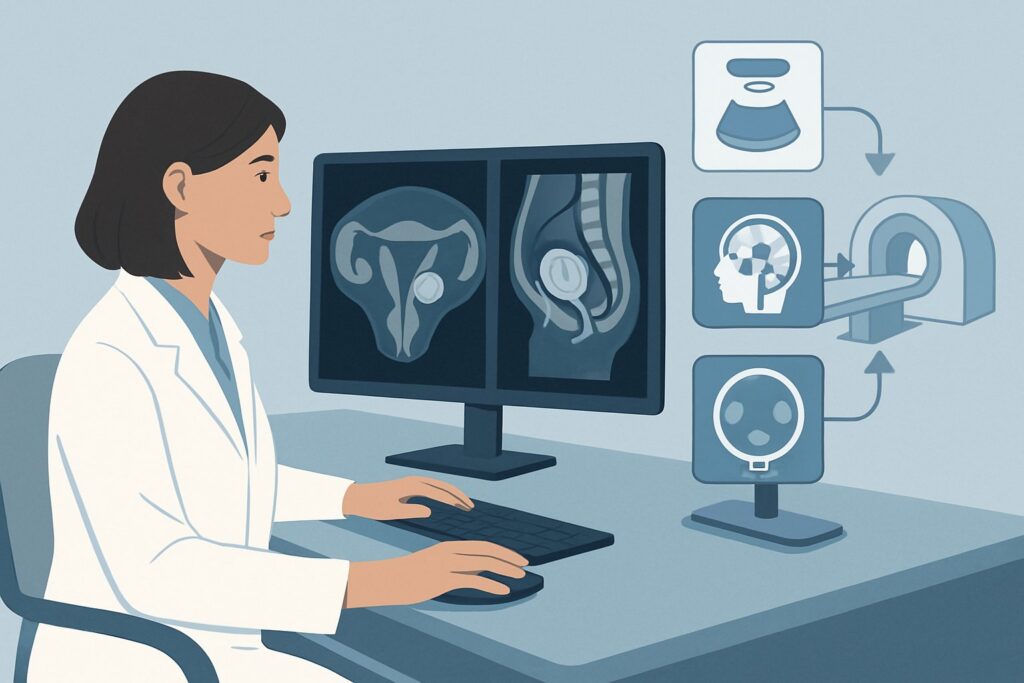
A 34-year-old patient presents to your clinic with a six-month history of progressively heavier menstrual bleeding, significantly impacting her quality of life. She is otherwise healthy and hopes to conceive in the next few years. A recent transvaginal ultrasound confirmed the suspected diagnosis: a 3 cm pedunculated submucosal uterine fibroid. You are now faced with the decision of initial therapy. The goal is to resolve her symptoms effectively while preserving her uterus and fertility potential. This article provides a focused clinical workflow for this exact scenario, guiding you through the American College of Radiology (ACR) Appropriateness Criteria. For this specific presentation, the ACR panel on Interventional Radiology rates Hysteroscopic myomectomy as a “Usually Appropriate” initial therapeutic procedure.
Who Fits This Clinical Scenario?
This guidance is specifically for a reproductive-age patient presenting with symptomatic, pedunculated submucosal uterine fibroids who is seeking initial therapy. The key inclusion criteria are:
- Patient Age: Reproductive age, where uterine preservation and potential future fertility are primary considerations.
- Fibroid Type and Location: Confirmed pedunculated submucosal leiomyoma (FIGO type 0 or 1). This means the fibroid is located on a stalk within the uterine cavity. The diagnosis is typically made via transvaginal ultrasound or saline-infusion sonohysterography.
- Primary Symptom: Heavy uterine bleeding (menorrhagia) is the dominant symptom driving the need for intervention.
- Treatment Stage: This is the initial therapy decision point, not a workup for failed prior treatments.
It is critical to distinguish this presentation from similar but distinct clinical scenarios that follow different management pathways. This guidance does not apply if:
- Bulk symptoms are primary: If the patient’s main complaints are pelvic pressure, pain, or urinary frequency from large intramural or subserosal fibroids, the therapeutic options and their appropriateness ratings change significantly.
- Concurrent adenomyosis is suspected: Patients with co-existing adenomyosis often have a more complex symptom profile and may respond differently to fibroid-directed therapies.
- The patient is postmenopausal: Management in postmenopausal women involves different considerations, including a lower threshold for considering endometrial biopsy and less emphasis on fertility preservation.
- Infertility is the primary complaint: While submucosal fibroids can cause reproductive dysfunction, the workup and management priorities are different when infertility, rather than bleeding, is the presenting problem.
What Diagnoses Are You Working Up in This Scenario?
While a pedunculated submucosal fibroid has been identified, the therapeutic intervention also serves to confirm it as the source of bleeding and to rule out other contributing pathologies. The key considerations include:
Pedunculated Submucosal Leiomyoma This is the primary therapeutic target. A fibroid on a stalk within the uterine cavity acts as a foreign body, disrupting the normal endometrial lining. This disruption can lead to ulceration, abnormal vascular development, and an inability of the endometrium to properly respond to hormonal cues, resulting in heavy and prolonged bleeding. Its intracavitary location makes it an ideal target for a hysteroscopic approach.
Endometrial Polyp Clinically, an endometrial polyp can present with identical symptoms of heavy or intermenstrual bleeding. While ultrasound can often differentiate a polyp from a fibroid, there can be overlap in appearance. A key advantage of hysteroscopy is that it provides direct visualization of the uterine cavity, allowing for definitive diagnosis and simultaneous resection of polyps if they are found instead of, or in addition to, the fibroid.
Adenomyosis This condition, where endometrial tissue grows into the uterine muscle wall, is a common cause of heavy menstrual bleeding and can coexist with fibroids. While hysteroscopy does not treat diffuse adenomyosis, it can rule out an intracavitary cause for the patient’s bleeding. If bleeding persists after myomectomy, underlying adenomyosis becomes a more likely diagnosis, guiding subsequent management decisions toward medical therapy or uterine artery embolization.
Anovulatory or Dysfunctional Uterine Bleeding Hormonal imbalances can also cause heavy bleeding. However, in the presence of a known structural abnormality like a submucosal fibroid, the fibroid is the most probable cause. If symptoms persist after the fibroid is removed, a workup for an underlying endocrinopathy or coagulopathy may be warranted.
Why Is Hysteroscopic Myomectomy a Recommended Initial Therapy for This Presentation?
The ACR Appropriateness Criteria designate Hysteroscopic myomectomy as “Usually Appropriate” for this scenario because it directly addresses the intracavitary pathology in a minimally invasive, uterine-sparing manner. This procedure involves inserting a hysteroscope through the cervix into the uterine cavity, allowing the surgeon to visualize the fibroid on its stalk and resect it directly. The rationale is compelling: it is both diagnostic and therapeutic, has a high success rate for controlling bleeding from this specific type of fibroid, and is associated with a rapid recovery and preservation of fertility.
Medical management is also rated “Usually Appropriate” and represents a valid non-surgical first step. Options like hormonal intrauterine devices (IUDs), oral contraceptives, or tranexamic acid can be effective in reducing bleeding. This is an excellent choice for patients who wish to avoid a procedure or for whom surgery poses a higher risk. However, it manages symptoms without removing the underlying structural cause.
Conversely, other interventions are rated lower for this specific patient presentation:
- Uterine Artery Embolization (UAE): Rated “May be appropriate.” UAE works by blocking the blood supply to the fibroids, causing them to shrink. While highly effective for intramural fibroids, its utility for a pedunculated submucosal fibroid is less established. There is a risk that the devascularized fibroid could detach and be expelled (slough), which can be a prolonged and painful process requiring further intervention. The long-term effects on fertility and pregnancy outcomes are also a subject of ongoing research, making it a secondary option for a patient desiring conception.
- Laparoscopic or Open Myomectomy: Rated “Usually not appropriate.” These surgical approaches are designed to remove fibroids located within the uterine wall (intramural) or on the outer surface (subserosal). Accessing a pedunculated fibroid inside the uterine cavity via an abdominal approach would require a significant incision into the uterus (hysterotomy), which is far more invasive than necessary and carries a higher risk of uterine scarring, adhesion formation, and complications in future pregnancies.
Since these are therapeutic procedures, not diagnostic imaging studies, considerations of ionizing radiation do not apply.
What’s Next After Hysteroscopic Myomectomy? Downstream Workflow
The post-procedure workflow depends on the resolution of symptoms and the patient’s reproductive goals.
- If symptoms resolve: The patient can be monitored annually. If she desires pregnancy, she can typically begin trying to conceive within a few months, following her surgeon’s specific guidance. The procedure is considered curative for the bleeding caused by that specific fibroid.
- If symptoms persist or recur: This is an important clinical branch point. The first step is to confirm that the myomectomy was complete and that no new intracavitary lesions have developed, often with a follow-up ultrasound or office hysteroscopy. If no structural cause is found, the workup should pivot to other causes of abnormal uterine bleeding. This may involve investigating for underlying adenomyosis (often with MRI) or evaluating for hormonal or coagulation disorders. The patient’s presentation may now align with a different ACR scenario, such as “uterine fibroids and concurrent adenomyosis.”
- If pathology is unexpected: The resected tissue is sent for histopathologic analysis. In the rare event of an unexpected finding, such as a uterine sarcoma, the patient requires immediate escalation to a gynecologic oncologist for further staging and management.
Pitfalls to Avoid (and When to Get Help)
Several potential pitfalls exist in managing this specific scenario. First, mischaracterizing the fibroid’s location is critical; a hysteroscopic approach is not suitable for deeply intramural or subserosal fibroids. Accurate pre-procedural imaging, sometimes including saline-infusion sonohysterography, is key. Second, failing to consider and discuss medical management as an equally appropriate first-line option can lead to unnecessary procedures for patients who might prefer a non-invasive approach. Finally, attributing all bleeding to the known fibroid without considering co-existing pathology can lead to persistent symptoms post-procedure. If heavy bleeding continues after a technically successful hysteroscopic myomectomy, it is crucial to escalate the workup to investigate for adenomyosis, polyps, or non-structural causes.
Related ACR Topics and Tools
For a comprehensive overview of all clinical variants related to uterine fibroids, please see our parent guide. The resources below can help you apply ACR guidance to other scenarios, understand procedural techniques, and discuss radiation safety with your patients.
- For breadth across all scenarios in Management of Uterine Fibroids, see our parent guide: Management of Uterine Fibroids: ACR Appropriateness Decoded.
- ACR Appropriateness Criteria Lookup — for adjacent scenarios
- Imaging Protocol Library — for technique on the recommended study
- Radiation Dose Calculator — for cumulative dose conversations
Frequently Asked Questions
Why isn’t hysterectomy considered appropriate for this patient?
Hysterectomy is rated ‘Usually not appropriate’ because the patient is of reproductive age and desires uterine preservation. Hysterectomy is a definitive treatment but is reserved for cases where less invasive, fertility-sparing options have failed or are not desired by a patient who has completed childbearing.
Can I offer an endometrial ablation instead of a myomectomy?
Endometrial ablation is rated ‘Usually not appropriate’ for this scenario. While it can reduce heavy bleeding, it does not address the underlying structural problem of the fibroid. Furthermore, ablation destroys the uterine lining, rendering the patient sterile, which is contraindicated in someone desiring future fertility. It can also cause scarring that may obscure the uterine cavity and complicate future surveillance.
What if the ultrasound shows the fibroid is partially in the uterine wall (intramural)?
If a significant portion of the fibroid is intramural (e.g., FIGO type 2), the feasibility and safety of a purely hysteroscopic approach become questionable. These cases often require a more complex surgical technique or a different approach, such as a laparoscopic or open myomectomy. Accurate pre-operative imaging and assessment by an experienced gynecologic surgeon are essential to determine the best approach.
How soon after a hysteroscopic myomectomy can a patient try to conceive?
The recommended waiting period before attempting conception varies but is typically between one to three menstrual cycles. This allows the endometrium to heal completely. Patients should follow the specific advice of their surgeon, as the recommendation may depend on the size and location of the resected fibroid.
Is an MRI necessary before proceeding with hysteroscopic myomectomy?
In most straightforward cases of a pedunculated submucosal fibroid identified on high-quality transvaginal ultrasound or sonohysterography, an MRI is not necessary. However, an MRI may be valuable if there is uncertainty about the degree of intramural extension, if multiple fibroids are present, or if there is a strong suspicion of co-existing adenomyosis that might alter the treatment plan.
Reviewed by Pouyan Golshani, MD, Interventional Radiologist — May 30, 2026